Bronchography
Historical document, translated for reference. It reflects medical knowledge of the 1920s–30s and is not medical advice.
Summary
This article from the 1928–1936 Soviet medical encyclopedia describes the diagnostic technique of bronchography, which involves using contrast media to visualize the bronchial tree via X-ray. It details the historical development of the method, various introduction techniques, clinical applications for diagnosing conditions like bronchiectasis and tumors, and potential complications.
Encyclopedia article (1928–1936)
BRONCHOGRAPHY (from Greek bronchos—bronchus and grapho—I write), a diagnostic method in radiology consisting of obtaining a photographic image of the bronchi in a living person by introducing into the bronchial system, which is normally transparent to X-rays, a contrast medium, i.e., a substance opaque to rays, which casts a shadow of the bronchi on a photographic plate. The first such radiograph was produced by Jackson in 1907 by insufflating dry bismuth powder into the bronchi. In 1918, Stewart used an emulsion of bismuth with oil, and at the present time, according to
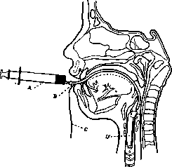
Introduction of a contrast medium into the bronchi via a cannula: A—syringe containing the contrast medium; B—rubber tube; C—thread for extracting the cannula; D—intubation cannula.
the proposal of Sicard and Forestier, vegetable oils containing 40% iodine are used, which represent a chemical compound with it, rather than a solution or mixture. These preparations are called Lipiodol (French preparation), Iodipin (German), Iodumbrin (Danish), and the newest preparation by Dyroff—Contrastol (contains bromine instead of iodine). All these preparations, especially the latter, irritate the mucous membrane little or not at all, which is why their use and the method of bronchography itself can be considered a relatively safe method of examination, as the greater part of the introduced substance is coughed back up by the patient. The substance is introduced into the bronchus after preliminary cocainization of the pharynx and upper respiratory tract, because without this, the patient would prematurely cough it back up, and it would not flow deeper into the bronchus. It can be introduced through a special laryngological (laryngeal) syringe with a cannula or with the help of a previously inserted bronchoscope, which allows for more precise direction of the liquid into the desired section. Introduction through a needle inserted into the larynx, in the space between the thyroid and cricoid cartilages, is also used. Sgalitzer suggested injecting the liquid through a Nelaton catheter passed into the larynx through the mouth, and Iglauer uses intubation of the larynx with a special cannula with a soldered tube for the oil and a thin rubber tube attached to the latter. The introduced liquid flows deeper by gravity to the small branches of the bronchi. By giving the patient's body the appropriate position, one can direct the Iodipin into the desired section of the lung and obtain a radiograph of the necessary part of the bronchi. The amount of liquid introduced at one time is, for obvious reasons, limited to 20-40 cubic cm. The figure (p. 106) shows a cross-section of the pharynx and larynx with the cannula inserted according to Iglauer and the introduction of the mass with a syringe. Nather found that if, after cocainization of the pharynx and larynx, the patient is made to swallow the Iodipin, the latter will enter the trachea. This simple method of introduction, however, suffers from a lack of asepsis: with it, at least theoretically, one can fear purulent pneumonia from the entry of infected material into the lung. The radiographs themselves must be taken quickly, before the mass is expectorated. They are taken with the patient in a lying or standing position and preferably after preliminary fluoroscopy on a screen, where the filled bronchi are perfectly visible. In this way, the most advantageous position for the radiograph can be determined. The trachea, the walls of which are coated with iodized oil, clearly stands out on the radiograph (see table, fig. 3). The entire branching system is clearly visible, and often the filling goes to the smallest bronchi, approaching the pleura pulmonalis itself. In the trachea and bronchi, one can establish displacement, obstruction, and changes in shape and lumen. The method is of particular value for the diagnosis of bronchiectasis, which sometimes presents difficulties for the clinician. Bronchiectasis and bronchiectatic cavities appear on the radiograph (see table, fig. 4) in the form of lumpy, spindle-shaped, or grape-cluster-like thickenings in the bronchial system. In this way, the size and position of lung abscesses having a connection with a bronchus can be determined, as well as tuberculous cavities, which are sometimes also difficult to recognize (see table, fig. 2). Bronchography is of great importance for the recognition of lung tumors: here the bronchus terminates abruptly due to compression or deviates to the side of the tumor, which is not observed with a lung tissue infiltrate. Among the complications described with bronchography, one should note a few cases of dyspnea and difficulty in breathing; furthermore, after bronchography, febrile phenomena are noted—a rise in temperature, sometimes lasting several days (no serious consequences have been described); in tuberculous patients, the appearance of some hemoptysis after injections has been described. The most serious phenomenon must be considered the very long retention of oil in the lung, where insignificant portions in the deep lobes remained from 5 to 16 weeks after the injection. Iodine is usually no longer detected in the urine from the 6th-7th day, which indicates that the main mass of the residue is absorbed by this time. Clinically, no phenomena from the lungs, either subjective or objective, have been observed from the retention of small portions of these iodine preparations; histological studies after death, apparently, do not yet exist. Contraindications to bronchography are: 1) severe cardiac compensation disorders, 2) very significantly pronounced weakness and exhaustion of the patient, 3) very extensive lung damage, as well as more active forms of tuberculosis.
A. Gagish
Related articles
Mentioned in
Cite this page
“Bronchography.” Soviet Medical Encyclopedia. English translation of Bolshaya Meditsinskaya Entsiklopediya, 1st ed. (Moscow, 1928–1936), ed. N. A. Semashko. https://sovietmedicalencyclopedia.pages.dev/article/bronchography/