Pyelography
Historical document, translated for reference. It reflects medical knowledge of the 1920s–30s and is not medical advice.
Summary
Pyelography is a radiological method for examining the urinary system by introducing contrast substances to visualize the renal pelvis and ureters. The article details various techniques including retrograde and excretory pyelography, historical developments, and technical considerations for the procedure.
Encyclopedia article (1928–1936)
PYELOGRAPHY (from Greek pyelos--pelvis and grapho--I draw), a radiological method of examining the urinary system, which makes it possible to obtain on an X-ray plate after filling the renal pelvis with a contrast substance an image of the contours of the latter. It would be more correct to call this method pyeloureterography, since usually an image is sought not only of the pelvis but also of the ureter. Depending on the route of introduction of the contrast substance into the pelvis, ascending or retrograde P. and excretory P. are distinguished. In retrograde P., the contrast substance is introduced through a ureteral catheter, in excretory P. through the blood (intravenous P.), through the rectum (rectal) or through the mouth (oral). If in retrograde P. gases are used as the contrast substance, it is called pneumopyelography. Pyeloscopy is the method of examining the pelvis by X-ray transillumination. History. In view of the fact that on ordinary X-ray films the hollow organs of the urinary system (pelvis, ureter, urinary bladder) do not cast shadows, attempts have long been made to artificially introduce into the urinary system objects impermeable to X-rays. Tuffier in 1897 made an X-ray of the urinary system after introducing a metal wire into a ureteral catheter. In 1906 Goe-bel constructed special X-ray impermeable ureteral catheters impregnated with bismuth. X-rays with impermeable catheters showed the direction of the ureter and the size of the pelvis, if the catheter curled up in the pelvis with the formation of loops. The first attempt to introduce a contrast substance into the pelvis was made in 1904 by Klose, who filled the pelvis through a ureteral catheter with a suspension of bismuth. This method, however, did not find widespread use, since firstly the introduction of a thick emulsion through a thin catheter was difficult, secondly it was not possible to adequately fill the minor calyces, and thirdly the removal of the emulsion was difficult. P. was developed only after the proposal by Voelcker and Lichtenberg (1906) to use a 5% solution of collargol as the contrast substance. At first, however, P. did not find widespread use due to complications in the form of collargol intoxication and even fatal outcomes obtained by some authors. In 1915 Burns pointed to the ability of haloid salts to cast a shadow on an X-ray and proposed fluorine for P., and in 1918 Weld proposed sodium bromide. From this time P. began to find increasingly widespread application and acquired the status of an obligatory method of investigation in urology. Oxygen as a contrast substance in P. was first used in 1907 by Burkhard and Polano. The development and popularization of pyeloscopy we owe to Legueu, Fey and Truchot (1927). Excretory P. was proposed in 1928 by Roseno and improved in 1929 by Lichtenberg and Swick, who proposed the drug Uroselectan, now replaced by the less toxic Abrodil. Technique P. The contrast substances used in P. must meet the following requirements: they should not be poisonous when they enter the bloodstream, should not cause irritation of the mucous membrane of the pelvis and ureter, should easily mix with urine, should be easily sterilizable, should remain undecomposed for a long time, and finally should be sufficiently inexpensive. In general, 15-25% solutions of sodium bromide and sodium iodide meet these requirements. A more intense shadow than sodium bromide and sodium iodide is given by a 25% solution of lithium iodide (Umbrenal), but it is significantly more expensive and irritates the mucous membrane of the urinary system more strongly. Recently, some authors for ordinary P. also use drugs intended for intravenous P. (Uroselectan, Abrodil). These drugs in concentrations from 20% to 40% give an intense shadow and hardly irritate the mucous membrane, but are very expensive. Ordinary P. The ureteral catheters used in P. must have divisions impermeable to X-rays and be thin enough (No. 5 according to Char.) so that excess contrast fluid can flow through the ureter past the catheter into the bladder. Pasteau catheters are very convenient, where segments visible on X-ray 1 cm long alternate with equally long invisible segments. The ureteral catheter in P. is usually advanced 28-30 cm to the pelvis. In the presence of an obstruction (stricture, stone, etc.) along the ureter, the catheter can be introduced to a lesser depth. In such cases, it is necessary to fill the pelvis, leaving the cystoscope in the bladder. In cases where the catheter is inserted to a greater depth (15 or more cm), it is more convenient, after advancing the catheter through the cystoscope along the ureter, to remove the cystoscope and perform the filling. The irrigating fluid in the bladder is left during P. so that the contrast substance from the pelvis, entering the bladder, can mix there with the fluid and decrease in concentration, which avoids irritation of the bladder mucosa. If the general condition of the patients allows, they can with the introduced catheter go to the X-ray room independently. It is more rational to introduce catheters in the X-ray room on special X-ray tables. P. must necessarily be preceded by an ordinary X-ray without contrast substance with the catheter already introduced into the ureter. Otherwise, P. can be a source of errors, especially in the presence of stones in the pelvis: the contrast substance can cover its shadow the shadow from the concrement and leave the latter undetected. Both for P. and for simple X-rays, the patient must be prepared. The intestine must be emptied and as far as possible not contain gases. For this purpose, patients 1-2 days before the X-ray should not eat things that cause strong fermentation in the intestine, such as milk, black bread, vegetables and fruits. The day before the X-ray, patients receive a laxative, after 6 hours--a siphon enema and 1 hour before the X-ray--a simple enema. On a good simple X-ray of the urinary system, the contour of the ilio-psoas, the contour of the lower half of the kidney and the shadow from a stone, if such is present in the kidney or ureter, should be clearly visible. If the stone is located in the ureter, its shadow should be next to the shadow of the ureteral catheter. A shadow in the pelvic area located at a certain distance from the catheter is as a rule due to another cause. After the simple X-ray, proceed to fill the pelvis with
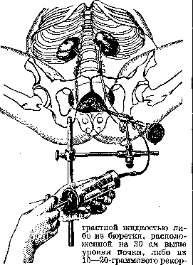
a syringe (see figure). The advantage of the first method is that it avoids excessive stretching of the pelvis. The use of a syringe is more convenient but requires great caution when introducing the contrast substance. With a slow movement of the piston, the liquid heated to body temperature is introduced through the ureteral catheter into the pelvis. The liquid is introduced until the first unpleasant sensations appear in the renal region or along the course of the ureter. At this moment the X-ray apparatus is turned on and an X-ray is taken. If it is necessary to obtain a picture not only of the pelvis but also of the ureter, especially its upper half, the ureteral catheter after filling the pelvis should be pulled out 10-15 cm. After the X-ray an attempt is made with the same syringe to aspirate back the contrast substance. In this way it is possible to reduce pain if it has occurred and to protect the mucous membrane of the pelvis from the irritating effect of a hypertonic solution. This is especially important in cases
expansion of the renal pelvis with reduced contractility of the latter. If, despite this, pain still occurs, one can resort to injecting morphine or pantopon. The administration of narcotics before pyelography is contraindicated, as it reduces sensitivity, depriving the examining physician of the criterion for when to stop the injection of contrast fluid. With moderate pain, a warm bath or hot water bottle can suffice. Some authors recommend after pyelography injecting into a vein for prophylactic purposes 5.0g of a 40% solution of urotropin. In pneumopyelography, oxygen is introduced into the renal pelvis from a Rekord syringe, or more commonly, air. The sterility of the air is achieved by drawing it through the flame of an alcohol lamp. The insufflation of air continues, as with ordinary pyelography, until an unpleasant sensation appears in the renal area. For greater contrast of the image, it is advisable to empty the pelvis of urine before filling it with air, which is achieved by aspirating it with a syringe. The advantage of pneumopyelography consists, first, that it makes a preliminary radiograph unnecessary, second, that the shadows from kidney stones become more contrast, and third, that air does not irritate the mucous membrane of the renal pelvis at all, which is especially valuable in hydro- and pyonephrosis. In intravenous pyelography, a simple preliminary radiograph is taken immediately before the injection of the contrast substance. This radiograph makes it possible to check whether the intestines are sufficiently free of gas. If such are present, pyelography should be postponed, since the contrast substance excreted by the kidneys is poorly visible on the radiograph. The preparation (40.0 Uroselectan, resp. 20.0 Abrodil, etc.) is dissolved in 100, resp. 50 cm3 of water, the solution is sterilized, and in a warm state (at body temperature) is slowly introduced into the vein. With normal kidney function, the injected substance is excreted by the kidneys after 15-20 minutes already in such a concentration that the outlines of the renal pelvis, ureter, and urinary bladder are visible on the radiograph. The shadow of the kidney itself appears more contrast on the radiograph than usual because, so to speak, the entire renal parenchyma is impregnated with the preparation. With reduced kidney function, the renal pelvis fills sufficiently with contrast urine later - after 1/2 hour, an hour or more. With poor kidney function, the kidney is unable to excrete the substance introduced into the blood in sufficient concentration, and on the radiograph, the contours of the renal pelvis on the corresponding side are not visible. The advantages of intravenous pyelography consist in that this method is applicable also in those cases where retrograde pyelography is impossible due to the inability to cystoscope or catheterize the ureters. The value of the method also lies in that a radiograph of both sides is obtained and that the latter makes it possible to judge the functional activity of the kidneys. The picture obtained in intravenous pyelography is usually less contrast than in retrograde pyelography, but it corresponds more to reality, since it eliminates the artificial stretching of the renal pelvis and ureter by the contrast solution injected through the catheter. The disadvantage of intravenous pyelography is that with poor kidney function it is not possible to obtain a sufficiently contrast radiograph. Pyelography is usually performed in the horizontal (lying) position of the subject, but if nephroptosis is suspected, it is necessary to take radiographs in the vertical position, since only in this way is it possible to determine the actual position of the kidney and to determine the presence of a kink in the ureter. If it is desired to determine the excursion of the kidney depending on the position of the patient, it is necessary to take two radiographs - one in the lying and the other in the standing position of the subject. In pyeloscopy, filling of the renal pelvis occurs in front of the screen. When introducing liquid contrast substance, it is visible how the upper part of the ureter gradually fills, then the renal pelvis and finally the calyces. There is no need to wait for the appearance of painful sensations, as is done in ordinary retrograde pyelography. When examining the renal pelvis, peristaltic contractions of the calyces, renal pelvis, and upper part of the ureter can be seen. In front of the screen, it is possible to determine in what time the renal pelvis empties of contrast substance. With normal function, it empties within 5-7 minutes. If it is desired to determine the motor ability of the renal pelvis, after filling it, the ureteral catheter should be removed. With proper technique (thin ureteral catheter, slow and careful introduction of contrast solution, observance of asepsis), pyelography is a safe method and can be performed on an outpatient basis. Indications and contraindications. Pyelography is indicated in all cases of surgical lesions of the kidneys and ureters when other methods (chromocystoscopy, catheterization of the ureters, etc.) fail to establish the diagnosis. But even in those cases where recognition is possible by simpler methods, pyelography clarifies a number of anatomical and functional details which may prove to be very valuable from the point of view of both indications for surgical intervention and the choice of the latter method. Considering the safety and diagnostic value of pyelography, it is used in all lesions of the upper urinary tract except 1) patients with high temperature, 2) very weakened patients, 3) patients with kidney bleeding in the presence of infected urine (infection can be introduced into the circulatory system). In the latter group of patients, pyelography should be postponed until the bleeding stops. In bilateral lesions, it is advisable to perform pyelography on each side on different days, although some authors prefer to image both sides simultaneously. The presence of one kidney is not a contraindication to pyelography, but such patients require special care. Correct interpretation of the pyelogram is possible only on the basis of evaluation of the patient's complaints, data from the history and clinical picture, as well as the results obtained by other methods of investigation. Normal renal pelvis and normal ureter. Two main types of renal pelvis are distinguished: ampullary and branching. In addition to the main types, there are many transitional forms. In the ampullary type, the renal pelvis itself (pelvis) is a cavity divided into 2 or 3 large calyces (calices majores), which in turn are divided into small calyces (calices minores). In the branching type, the renal pelvis itself is of very small size, and the ureter as it were directly divides into the calyces. The capacity of the ampullary renal pelvis is 7-8 cm3, of the branching type 2-3 cm3. The normal renal pelvis is located on the pyelogram at the level of L1 and L2; the 12th rib divides the renal pelvis on the left into two equal parts, while on the right, 2/3 of the renal pelvis is located above the 12th rib. If a straight line is drawn connecting the edges of the upper and lower calyces, the axis of the renal pelvis is obtained. In the normal position of the kidney, the axis of the renal pelvis (as well as the kidney) is directed from above and inward downward and outward. If the axes of both renal pelves are extended, they will intersect in the area of the thoracic vertebrae, forming an acute angle. The contours of the renal pelvis and calyces in the normal state should be clear and well-defined [see separate table (Vol. XIX, pp. 135-136), figure &]. The lumbar part of the ureter, not counting the bend in the very upper part, is located approximately parallel to the spine, and if mentally extended in the direction of the renal pelvis, it forms with the latter's axis two adjacent angles - a lateral one of 45° and a medial one of 135°. The upper curved part of the ureter with the lower contour of the renal pelvis and the medial contour of the lower calyx usually forms an arc equal to half a circle. The ureter forms several bends on the pyeloureterogram. In the upper part, the ureter forms a pelvic bend with convexity directed toward the spine, with maximum approach to the latter at the level of L4. The lower, more sharply expressed bend - the prevesical one - is located in the cavity of the small pelvis, and its convexity is directed, compared with the upper bend, in the opposite direction. Anomalies of the kidneys and ureters. Recognition of the significant majority of kidney anomalies became possible only with the application of pyelography. Before that, anomalies were accidentally discovered either at autopsies or on the operating table. Thanks to the widespread use of pyelography, it was possible to establish that anomalies of the kidneys and ureters occur much more frequently than was previously assumed. Pyelography gives in almost all anomalies an exhaustive anatomical picture of the upper urinary tract. In aplasia of the kidney, the ureteral orifice may be present in the bladder in the normal place. In individual cases, the ureter may even exist for some distance and fill with contrast substance during pyeloureterography. The contours of the renal pelvis are absent on the radiograph. The contours of the kidney are also not visible. In dystopia of the kidney, the kidney may be located either in the pelvis (pelvic dystopia) or higher (lumbar dystopia). The ureter is short, without kinks. The renal pelvis is of irregular shape. In some cases, it is located laterally, while the calyces are medially. In such cases, the ureter arises from the lateral side of the renal pelvis. In crossed dystopia, both kidneys are located on one side with the normal position of both orifices in the bladder.
On the pyelogram, it is visible how one of the ureters crosses the spine and how both renal pelves are located one above the other. In a horseshoe kidney, on the pyelogram both pelves are located lower than normal; the axes of both kidneys have a direction opposite to normal - from top inside downward and outward, and when extended, the axes intersect in the caudal direction, forming an angle open cranially. The calyces are located medially, as in dystopia, and the shortened ureters depart either from the middle or medially from the pelvis. The contours of the pelvis and calyces have a characteristic shape. Duplication of the renal pelvis and ureter is the most common anomaly. All variants of duplication can only be recognized by pyeloureterography. On the P., crossing of the ureters is visible in their duplication. In duplication of the pelves, the upper pelvis is located higher than normal, very often it is small, rudimentary, consisting of a small ampulla devoid of calyces, while the lower pelvis usually has normal contours. When there are two ureteral orifices in the bladder on one side, suspicion of duplication of the ureters and pelves always arises. For P., catheters are inserted into both orifices and both pelves are simultaneously filled with contrast fluid from two syringes. The presence of two pelves in a split ureter is usually discovered accidentally during II. When there is suspicion of a split ureter with two pelves, filling with contrast substance is performed through a catheter inserted only a few centimeters, or intravenous P. is performed. Accurate recognition of nephroptosis became possible only by the use of P., especially in cases where palpation does not give precise results for one reason or another. Since the kidney returns to its normal position when the patient is lying on their back, P. should invariably be performed with the patient in a standing position when nephroptosis is suspected. The pelvis may be unchanged in nephroptosis; but it is located lower than normal depending on the degree of kidney descent. Depending on the rotation of the kidney around one of its axes, the axis of the pelvis accordingly also changes its direction. In some cases, the calyces are located below the pelvis. The ureter descends along with the kidney, then it becomes more tortuous than in normal conditions. In individual cases, the ureter is fixed in its upper third and therefore forms a bend here, creating unfavorable conditions for urine outflow. In such cases, P. simultaneously indicates whether secondary changes in the pelvis in the form of pyelectasis or hydronephrosis have occurred. When hydronephrosis is suspected, gaseous contrast substances should be preferred to avoid complications from irritation of the mucosa by hypertonic solutions of haloid salts. In hydronephrosis, they are introduced in large quantities and are difficult to remove due to the loss of tone by the hydronephrotic sac. Pyonephrosis in far-advanced cases gives the same pyelographic picture as hydronephrosis. One large cavity filled with contrast substance, spherical or scalloped, is visible with complete absence of the outlines of the normal pelvis and calyces. In an earlier stage, the picture of pyonephrosis is characterized by the predominance of calyceal dilation over dilation of the pelvis itself. The newly formed cavity increases mainly due to the destruction of the renal parenchyma in the area of the pyramid apices, unlike hydronephrosis, in which first the pelvis dilates, then the calyces, and only later under the influence of pressure, the renal tissue atrophies. Israel distinguishes dilation of the pelvis itself, dilation of some calyces without change in the pelvis, and combined dilation of the pelvis and calyces. Tuberculosis of the kidney can be recognized in most cases without the use of P. (see Kidneys). In rarer cases, when dysuria is absent and cystoscopy does not indicate the presence of a tuberculous process in the bladder, diagnosis can be made on the basis of P. In early stages, when there are no cavities communicating with the pelvis, the pyelogram shows blurred and eroded contours of the calyces with slightly altered outlines of the pelvis itself. When cavities communicating with the pelvis exist, they also fill with contrast fluid and give a characteristic picture: on the X-ray, narrow septa are visible connecting the cavity of the calyx with the cavity of more distant cavities. In far-advanced cases, when most of the parenchyma has been destroyed, P. shows the picture of ordinary pyonephrosis, and only from the characteristic picture of the ureter can a diagnosis of tuberculosis be made. When the ureter is involved in the process, it shortens and appears on the pyeloureterogram as straight and devoid of physiological tortuosity. Kidney and ureteral stones can be diagnosed based on simple X-ray films, but to determine the secondary changes in the pelvis and ureter caused by the presence of a stone, as well as to establish exactly whether the stone is in the pelvis or in one of the calyces, is possible only by P. In kidney stone disease, one can either perform P. with liquid contrast substance and compare the plain film with the pyelogram to determine the location of the stone, or even better, use pneumopyelography. By using gas (air), on one hand, the contrast of the stone can be increased and an image of so-called 'invisible' stones (small urates that do not give a shadow on a plain film) can be obtained on the X-ray, and on the other hand, such an X-ray immediately determines the location of the calculus. Stones may be stationary in one of the calyces or in the pelvis and not impede urine outflow. In such cases, the pelvis remains unchanged. If the stone impairs urine outflow, then P. gives a picture of pyelectasis or hydronephrosis. The exact location of the stone is especially important to know in cases where surgical intervention is indicated. Clarification by pyelography of all anatomical features makes it possible to determine the method of surgical intervention - pyelotomy, nephrotomy, etc. In stones of the ureter, the pyeloureterogram shows the condition of the urinary system above the location of the stone - whether the ureter is dilated, whether the pelvis is changed. In case of a suspicious shadow in the area of the ureter - especially in the pelvic part - the ureterogram helps to determine whether the shadow is due to a stone in the ureter or is caused by another reason (phlebolith). Kidney tumors, in cases when the kidney is not palpable, can only be diagnosed by P. A tumor growing in the renal parenchyma begins to compress one or another calyx or the pelvis. In far-advanced cases, the tumor may grow into the pelvis cavity and even fill it entirely. The pyelogram shows either compression of the calyx or its complete absence; the latter as if amputated. When the pelvis is filled with tumor, the contrast substance fills only the ureter. In cystic degeneration of the kidneys, the pelvis and calyces are elongated, and the picture obtained on P. shows the pelvis and calyces as if enlarged in scale without significant disturbance of their mutual relationships. Since this disease usually affects both sides, in doubtful cases it is necessary to resort to bilateral P. With significant enlargement of the kidney in size, the ureter, as in large kidney tumors, may be displaced toward the spine. Echinococcus and solitary kidney cysts are somewhat more difficult to recognize on the pyelogram. The task is facilitated if the wall of the cyst or echinococcus is calcified. In diseases of the ureter, ureterography makes it possible to detect narrowings, compressions of the ureter by an accessory vessel, determine the presence of ureteral empyema, dilation and atony of the ureter, ureteral diverticulum, its neoplasms, etc. In retroperitoneal tumors, lateral displacement of the ureter is visible on the ureterogram. When the function of the sphincter apparatus of the ureteral orifices is impaired, contrast substance can penetrate into the ureters and pelves during filling of the bladder with contrast substance and give a one- or two-sided pyeloureterogram by so-called reflux. This picture is observed in congenital atony and in tuberculosis of the urinary system.
Related articles
Mentioned in
Cite this page
“Pyelography.” Soviet Medical Encyclopedia. English translation of Bolshaya Meditsinskaya Entsiklopediya, 1st ed. (Moscow, 1928–1936), ed. N. A. Semashko. https://sovietmedicalencyclopedia.pages.dev/article/pyelography/