Catheterization
Historical document, translated for reference. It reflects medical knowledge of the 1920s–30s and is not medical advice.
Summary
This historical encyclopedia article from the 1930s details the medical procedure of catheterization, focusing primarily on the urinary tract. It covers asepsis rules, instrument types (soft, semi-soft, and metal), techniques for insertion, potential complications, and clinical precautions.
Encyclopedia article (1928–1936)
CATHETERIZATION, the introduction of a catheter into channels of the human body opening outward; most commonly used in the clinic of the urinary tract. Catheterization must always be performed with the strictest observance of all rules of surgical asepsis both with regard to the operator (cleaning of hands), the patient (cleaning and disinfection of entry orifices), and the instruments (sterilization). Sterilization of instruments is performed by boiling, formalin vapors, and disinfecting solutions according to the same rules as bougies (see Bougies, Bougienage). Before introduction to the patient, instruments are lubricated so that they enter more easily and slide along the urethra. Either sterilized oil is used—which is more difficult to wash off, ruins rubber and soft instruments, and hinders their subsequent sterilization—or water-soluble substances that do not have these specified drawbacks, such as glycerin, thick sugar syrup, Kasper's catheter-purin (composition: Tragacanth 2.0, Glycerin 20.0, Mercury oxycyanate 0.246, Distilled water to 100.0). During catheterization, which frequently has to be performed repeatedly and often, the observance of these rules is especially important to avoid infectious complications, which easily occur and are especially grave in their consequences in patients suffering from urinary retention and residual urine. The very technique of catheterization for soft and semi-soft instruments is simple and boils down to introducing the tip of the instrument into the external orifice and carefully pushing the catheter further inward into the bladder, which is recognized by the appearance of a stream of urine from the catheter. Before introducing a Nelaton rubber catheter, it is necessary to test it by pulling for rupture and brittleness, since such catheters easily dry out when lying around, and then a part of the instrument can easily tear off and remain in the canal or bladder. Semi-hard instruments from prolonged use acquire cracks and fractures, along which they can also easily break off; but in general they are less dangerous in this regard than rubber ones. To avoid the introduction of infection, instruments should be held with the hand as far as possible from the tip so as not to touch and not to hold with the fingers that part which will enter the posterior urethra and bladder. In this regard, semi-hard instruments are better than soft ones, and metal ones surpass both of the first types. In case of an obstruction, the instrument should not be pushed with force, as this usually does not lead to the goal, but one needs either to try to pull the instrument out a little and re-introduce it, giving it slight rotatory movements and feeling out the course, or, slightly pressing it inward, hold it like that for some time under uniform and constant pressure. The latter technique is good when there is an obstruction in the depth from a spasm of the sphincter, which after a certain time yields to pressure, and the instrument itself easily slips into the bladder. With soft instruments it is almost impossible to damage the urethra to any significant extent, and therefore only they can be given into the hands of the patient if circumstances force him to catheterize himself. Allowing the patient to introduce the instrument himself can only be done in extreme cases, as this usually soon leads to chronic cystitis, which remains with these patients forever. Bleeding can also be caused by a soft instrument in case of looseness and hyperemia of the mucous membrane of the posterior urethra, especially in prostatics. The introduction of soft instruments is usually performed without any anesthesia. In exceptional cases of special sensitivity of the patient, one has to resort to anesthesia of the urethra by injecting a 2-3% novocaine solution with adrenaline using an ordinary Tarnovsky syringe in the amount of 5-8 cubic centimeters, slowly and at very low pressure; it is retained in the urethra for 3-5 minutes by pinching the external orifice. During the first catheterization, it is generally better not to resort to anesthesia until the structure of the patient's urethra is yet unknown: the obstructions, sensitivity, and pain of one or another section of the urethra will be better determined, and there will be less opportunity to cause trauma. This is especially important when introducing metal instruments, which for the first catheterization for the same considerations should never be taken thinner than caliber 18-20 Ch. The introduction of metal instruments is obligatory in the supine position of the patient, in the lithotomy position. The latter is convenient because it allows the operator to approach from the side of the patient's feet. In any case, the legs must be spread and, preferably, slightly bent at the knee and hip joints. A basin for the drainage of urine is placed between the legs. For the introduction of a metal catheter, 3 classical techniques have been developed and are described. 1. The turn over the abdomen (Tour über den Bauch). The physician stands on the left side of the patient. The right hand holds the catheter by the flange between the thumb, index, and middle fingers, like a writing pen, and rests the little finger on the middle of the abdomen along the linea alba. The beak of the instrument is directed downward. The left hand holds the penis by the glans and pushes it onto the instrument "like a worm onto a fishing hook" (Oberlander). Thus the beak of the catheter must
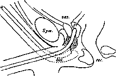
Figure 1.
be advanced to the bulbous part of the canal (Fig. 1 a). Further advancement of the instrument is produced by lifting its handle from the surface of the abdomen, adhering to the plane of the midline (sagittal) and carefully advancing it inward (Fig. 1 b); at the same time, the left hand placed on the perineum can feel the end of the catheter and by slight pressure help its passage through the urethra. Here the first obstruction may be felt if the instrument rests against the lower fold of the dilation of the bulbous part (Fig. 1 a). It is bypassed if the catheter is withdrawn slightly and trying to be re-introduced, keeping the end closer to the upper wall of the canal and pressing from the side of the perineum. When the catheter forms approximately a 45° angle with the horizontal line, its end is already behind the lower edge of the pubic symphysis. Further advancement is achieved by a greater abduction of the handle from the abdomen and advancement inward until the vertical position of the straight part of the catheter is reached. Here a 2nd obstruction is possible in the posterior part of the canal at the protruding seminal colliculus, into which the end of the instrument can sometimes abut. It is also avoided by keeping to the upper wall of the canal. The same technique overcomes the 3rd obstruction described by Dittel—a strongly developed internal sphincter of the urinary bladder. Here a technique in the form of slight and prolonged pressure is useful, overcoming the muscle spasm, and the instrument immediately slips easily into the bladder without increasing violence on the part of the operator. This obstruction is most common and is encountered in almost every catheterization. Finally, the 4th obstruction can be presented by the bulging apex of the trigone of Lieutaud (enlarged middle lobe of the prostate gland). This obstruction may prove insurmountable for an instrument of the chosen curvature and may require the use of an instrument of another shape or make the case generally impassable for a metal catheter. Here one has to act especially carefully, since it is easiest to make a false passage, and apply the techniques described above, without showing excessive persistence and by no means allowing any significant violence. It is always necessary to remember that pushing a metal instrument by force is dangerous. One must learn to hold the instrument in the hand firmly enough and at the same time elastically in order to feel its passage through the canal, let it "find its own way," but by no means push the instrument by force. 2. The second catheterization technique, the so-called grand tour de maître, consists in the fact that, standing on the right side of the patient, the instrument is introduced while holding it also with the right hand, but so that the handle is directed downward between the patient's legs, i.e., the long axis is .
, located at an angle of 180° /
\ to the described first !4a
angle is introduced until it easily pro
\l y/y gresses inward, i.e., 3*-----p\f-----' usually to the fossa of the bulb
\
I arrows, at the same time 1
/ \
I advancing it deeper *
)2|
f by the gradual approach
Fig. 2 lifting of the handle upwards, so that it describes a spiral which ends when the handle is on the midline and the instrument is already in a vertical position relative to the body of the lying patient (Figure 2, positions 2, 5, 3, 4, 1). With this maneuver, in experienced hands the beak easily bypasses the first, second, and sometimes all obstacles. This maneuver is very good when performed with a uniform, smooth, and sufficiently rapid movement, and therefore is appropriate only in the hands of an experienced operator, as indicated by its very name. 3. The third maneuver (petit tour de maître) often leads easily to the goal and is generally less risky; it differs from the previous one in that the introduction is begun by holding the instrument horizontally and perpendicularly to the length of the body of the lying patient, i.e., at an angle of 45° in relation to the beginning of the grand tour, and completing all other moments as in this maneuver, with the spiral being described by the handle along a ¼ circumference (Fig. 2, positions 3, 4, 1). Most commonly used is the French maneuver, in which the introduction of the instrument is begun by holding it horizontally, parallel to the inguinal fold, and proceeding further as in the previous cases (Fig. 2, positions 5a, 3a, and 4a). All these maneuvers are useful to know, because in a difficult case any one of them may individually lead to success. Dittel points out that this is especially necessary in the presence of a false passage already known to the operator and the need to catheterize the patient nonetheless. Knowing the position and location of the false passage, one must apply the catheterization maneuver in which the beak goes closer to the wall of the canal opposite the site of injury. This avoids the entry of the catheter into the false passage. Great difficulties for catheterization may be encountered in strictures. In extreme degrees of stricture, inflammatory swelling of the mucosa in the area of the stricture sometimes occurs, resulting in retention of urine, which will already require catheterization. It is here that special difficulties are observed. Only thin silk, elastic catheters (Nos. 5, 6, 7 Ch.), straight ones, with a Mercier curve, bicoudé, etc., can be applicable. Often one has to resort to the "bundle technique," as with a filiform bougie, introducing 3-4 catheters at once up to the obstacle and then acting with each one separately in turn, and one of them will find its way. Preliminary injection of novocaine with adrenaline into the urethra, as well as warm oil, sometimes facilitates the matter. But if the stricture is such that even the finest catheter, through which urine will flow only drop by drop, does not pass, one must turn to the finest, thread-like bougies and, having introduced one, leave it in the canal a demeure. This maneuver leads to the fact that after some time urine begins to flow in drops next to the bougie, the bladder is emptied somewhat, and in about a day it becomes possible to introduce a thicker instrument, and at the same time the proper treatment of the stricture itself. Only if all described attempts do not lead to the goal can puncture of the bladder or high section of it be considered indicated. Catheterization is a method of symptomatic treatment of prostatic hypertrophy. It is clear that catheterization must be used in complete urine retention, acute and chronic, but even in incomplete retentions (residual urine) it is the main therapeutic maneuver. In prostatic hypertrophy, catheterization is especially difficult and often unsafe, and its use requires special caution, strict indications, and the strictest asepsis. In the question of the technique of the operation itself, the selection of an instrument appropriate to the case plays a major role. Soft catheters are usually used. Instruments with a Mercier curve often turn out to be better than ordinary ones. Sometimes local anesthesia (cocaine-adrenaline) is necessary, and a preliminary injection of 5-10 cm3 of sterilized oil into the canal by a syringe may also prove useful. If (as often happens) the soft catheter does not pass, it is best to achieve the goal with elastic ones, which are generally the instruments of choice for prostatotics. Straight instruments in most cases do not pass and very often cause bleeding. Flat catheters and Thiemann catheters are especially good for prostatotics. They pass especially easily even in the most difficult cases. Only in case of extreme necessity (and even then with special caution during the first catheterization) can a metallic instrument be used in a prostatotic. It is introduced with extreme caution, and if it does not pass, an instrument with a different curve is taken and thus a shape most suitable for the given case is selected. Any violence during introduction must be carefully avoided, remembering the possibility of a false passage. If this complication happens and a false passage is made, catheterization is stopped until the injury heals, and only in extreme cases, when there is urine retention, is catheterization with a soft instrument permissible, or they try to introduce and leave a catheter a demeure, keeping it until the false passage heals. For a catheter a demeure, one must use a soft or elastic catheter to avoid bedsores of the mucosa. Metal instruments are not suitable for this; it is especially impermissible to leave them for the night, in order to avoid mechanical damage during sleep. Pezzer and Malecot catheters are especially convenient for this purpose, because they are introduced on a mandrin, and upon its removal they hold themselves without external fixation. When releasing urine in a prostatotic, either a diagnostic goal is pursued—determining the degree of bladder atonia and the amount of residual urine—or a therapeutic goal—emptying during retention—or systematic catheterization for the treatment of atonia and concomitant cystitis with washings. When embarking for the first time on catheterization in chronic urine retention and a severely distended bladder, one should not immediately empty the bladder to the end, because after this sometimes, especially in weakened old patients with an unstable heart and already degenerated kidneys, the so-called "emptying reaction" (Entlastungsreaktion) sets in. The resulting immediate change in the conditions of urine outflow from the kidney (elimination of backpressure) produces significant changes in the kidney tissue and its vascular system, leading to a drop in the excretory capacity of the kidney. This results in either acutely (1–2 days) or more slowly developing progressive weakness: the patient's diuresis drops, and death occurs with phenomena of deep intoxication. Emptying the bladder in such patients must be done only gradually, either releasing urine little by little with each catheterization or introducing a thin catheter for the ureter (No. 6–7) and allowing urine to continuously flow out of it in drops, with the bladder being emptied gradually and very slowly—12–24 hours. Among other complications of catheterization, infectious fever (Katheterfieber) must be mentioned, sometimes with shaking chills, which occurs sometimes very soon (an hour later) after catheterization, despite all aseptic precautions, and moreover after each introduction of the instrument. This is usually observed in individuals who are carriers of infection (cystitis, especially prostatitis and lesions of the posterior urethra). In stronger organisms, therapeutic measures (washings with therapeutic solutions, quinine, salol, urotropin internally, intravenous injections of a 40% urotropin solution) lead more or less quickly to the attenuation and cessation of these temperature jumps and an improvement in the quality of the urine and general condition. However, sometimes such a "catheterization fever" of the old authors can turn into a genuine urosepsis with a chronic course, as a result of which exhaustion and weakness develop, appetite disappears, persistent thirst appears, and the patient dies with the progressive development of these symptoms. Thirst, a dry coated tongue, dryness in the mouth, and loss of appetite are symptoms that are harbingers of danger in such cases. A fairly frequent and unpleasant complication of catheterization is also inflammation of the epididymis, developing in patients who have to be catheterized repeatedly and for a long time, especially if catheterization proceeds with difficulty and the catheter has to be left a demeure for a long time. Rough technique and the presence of infection processes in the urinary tract contribute to the occurrence of epididymitis. In contrast to gonorrheal ones, these septic epididymitis cases often end in suppuration and even more or less extensive necrosis of the cellular tissue and can be sources of other septic complications (parotitis, general sepsis) and the cause of death in more decrepit and weakened patients. Strict asepsis, careful and gentle technique, possible restriction of catheter introduction, wearing a suspensory, and urinary antiseptics should be recommended as prophylactic measures against this unpleasant complication, from which it is completely impossible to guarantee a patient undergoing long and systematic catheterization. Upon the onset and repeated outbreaks of epididymitis and the necessity to catheterize the patient, one has to resort to the operation of ligation of the vas deferens (see Vasoligation) to stop the repetition of these inflammations. The invention of the cystoscope (Albarran, Casper) and its adaptation for the introduction of catheters into the ureters and renal pelvis has given into the hands of clinicians a new and highly valuable method of diagnosis and therapy of surgical diseases of the kidney. The technique of the method is simple in principle, but requires dexterity and prolonged practice.
The catheterizing cystoscope is equipped with a channel for the catheter and a tongue controlled from the outside, which makes it possible to give the catheter entering the bladder a certain angle relative to the long axis of the instrument. This device makes it possible, upon seeing the ureteral orifice in the field of view of the cystoscope, to bring the tip of the catheter to it and, by advancing the latter into the channel of the cystoscope, force the tip, which was in the bladder, to enter the ureteral orifice and advance along it, if necessary, to the renal pelvis. The excess length of the catheter is pushed into the bladder, where it coils into rings, after which the cystoscope can be withdrawn without pulling back that part of the catheter which is located in the ureter. By repeating the entire operation immediately with the second catheter, one can also catheterize the second kidney and leave the catheters simultaneously in both ureters. There are cystoscopes with two channels (from all manufacturers) for the simultaneous catheterization of both ureters, but ceteris paribus they are thicker and usually do not pass catheters thicker than No. 6, which is a disadvantage that is hardly compensated by the convenience of the simultaneous catheterization of both kidneys. Urine soon begins to drip periodically from the inserted catheter: a few drops in a row and then a pause. This corresponds to the periodic peristaltic contractions of the renal pelvis and ureter, by which urine is normally driven into the bladder. If the drops go without pauses, continuously, this indicates that the catheter has fallen out of the ureter and lies in the bladder or that its end has passed to a greatly dilated renal pelvis (hydronephrosis), which is emptied through the catheter. In the latter case, pressure on the region of the corresponding kidney increases the outflow from the catheter. Catheterization of the ureter is used predominantly for diagnostic purposes, but in many cases it is also used with great success for the treatment of diseases of the renal pelvis and ureter (pyelitis, small hydronephrosis, kinks and strictures of the ureter, for the extraction of a ureteral stone, etc.). Proceeding from the premise (fully confirmed by clinical data) that in a healthy person both kidneys excrete the same amount of completely identical urine over a certain given period of time, urine is collected simultaneously and separately from each catheter for 1/2–1 hour. Over such a period, depending on the diuresis, 25–100 cm3 of urine is usually collected. It is more advantageous to collect urine not under conditions of very high diuresis. The study of both portions shows from which kidney pathological impurities are obtained, and quantitative analysis, i.e., the determination of specific gravity, freezing point, chlorides, urea, sugar (in the case of phlorhizin injection), etc., will show which of the kidneys ceteris paribus yields more concentrated urine and to what extent it thus possesses greater functional capacity. The introduction of dyes subcutaneously or intravenously will make it possible to judge the intensity of their excretion. All this, together with general clinical data, makes it possible to conclude which of the kidneys is diseased, as well as to judge the degree of its damage and destruction. Such diagnostic catheterization of the ureters has acquired enormous significance for determining kidney diseases and resolving the question of the indications and feasibility of surgery. Catheterization of the ureter is also used to perform pyelography, i.e., obtaining an X-ray image of the ureter, pelvis, and calyces (see Pyelography). In the treatment of diseases of the middle ear, catheterization of the Eustachian tube is used. The catheter is inserted into the nose along the inferior nasal meatus to the posterior wall of the pharynx. Then the catheter is turned with its concave side toward the nasal septum and extracted backward until the posterior edge of the nasal septum is felt by the curvature. If the catheter is now turned 180°, its head with the opening will be opposite the orifice of the Eustachian tube and can be brought to it. Now, through the catheter, inflation of the tympanic cavity of precisely the desired side can be performed using a Politzer bag, without involving the other, healthy side in the therapeutic procedure. A finer catheter, resembling a ureteral catheter, can be passed through a metal catheter into the Eustachian tube itself and the tympanic cavity for the purpose of its emptying and the introduction of drugs. These latter instruments and techniques are rarely used in practice, in contrast to the very common catheterization with inflation. Laryngeal intubators can be partly classified as catheters (see Intubation).
Related articles
Mentioned in
Cite this page
“Catheterization.” Soviet Medical Encyclopedia. English translation of Bolshaya Meditsinskaya Entsiklopediya, 1st ed. (Moscow, 1928–1936), ed. N. A. Semashko. https://sovietmedicalencyclopedia.pages.dev/article/catheterization/