Zkovskaya Fistula
Historical document, translated for reference. It reflects medical knowledge of the 1920s–30s and is not medical advice.
Summary
This article describes the Zkovskaya fistula, an experimental method for partially excluding liver function by creating a shunt between the inferior vena cava and the portal vein. It details the historical development of the technique, the specific surgical steps for its creation, necessary precautions, and its use in animal models.
Encyclopedia article (1928–1936)
Zkovskaya Fistula, an experimental method for the partial exclusion of liver function, first proposed by Eck (1877). The method consists of surgically creating a communication between the inferior vena cava and the portal vein, with the simultaneous ligation of the latter above the communication, as a result of which the blood flow, bypassing the liver, is directed directly into the inferior vena cava. In its original form, the creation of the E. f. was a very difficult operation, as it required specially manufactured instrumentation (e.g., a special device for miniature scissors) and great technical skill. Subsequently, due to the particular importance of the E. f. as a method for studying liver function, attempts were made to improve and simplify the technique of its creation, as a result of which Fischler, in collaboration with Schroder, published in 1909 a simplified method for the E. f. operation, which over time was only slightly improved. In the latest modification, the operation for creating the E. f. consists of the following. A skin incision is made along a semi-oval line, beginning at the upper end in the left hypochondrium region, 3-4 cm to the side of the median line. Then the incision crosses the latter and, describing a semi-oval, descends downwards for 6-7 cm, almost along a straight line. If access to the inferior vena cava and portal vein is difficult after opening the abdominal cavity (the abdominal wound cannot be widely spread), the incision is continued further for the required distance. When the abdominal cavity is opened, the abdominal organs are spread to the sides with gauze swabs so that a wide access to the aforementioned veins (v. cava inf. and v. portae) is opened. Then they are carefully dissected bluntly from the surrounding tissues on a space sufficient for their free approximation. When this is achieved, a long ligature is passed under the v. portae, at the place of its entry into the liver hilum, with a Dechamp needle, the ends of which are seized with a Kocher or Pean clamp and brought out of the abdominal cavity, leaving them free (Fig. 1, a). After this, a first suture is applied between the veins as high as possible (as close as possible to the liver hilum), tightly fixing their walls to each other (Fig. 1). Usually 7 such sutures are applied, occupying a space of about 2.5-3 cm. Then, on a round needle, a so-called cutting ligature (of relatively thin but strong silk) is passed from below upwards through the entire thickness of the wall of the v. portae between and at the levels of the 2nd upper and 6th lower sutures. After exiting the lumen of the v. portae at the level of the 2nd upper suture, it is thrown across to the side of the v. cavae inf. and, at an equal distance from the sutures connecting the two veins, is passed in the same order, but only from above downwards through the wall of the v. cavae inf. and the lower end is brought out outside (Fig. 1). The next stage of the operation is the closure of the cutting ligature so that it ends up between the closely apposed walls of the v. cavae inf. and v. portae. This is achieved by applying a second row of sutures to the inferior vena cava and portal vein, symmetric to the first, but located on the other side of the cutting ligature (Fig. 2). The last (7th) suture is not tied in a knot and remains temporarily free. Then, when this is achieved, the ends of the cutting ligature are firmly seized and pulled out by means of rapid sawing movements. At this moment, both walls of the sutured veins are cut through and a communication (anastomosis) is established between them, as a result of which blood from the portal vein begins to flow into the inferior vena cava. The formation of the anastomosis is indicated by bleeding from the lower angle of the unsecured connecting suture (7th), which immediately tightens and the bleeding stops. Then the ligature is tightly tied, previously freely applied to the v. portae (Fig. 1, a), as a result of which blood now flows completely from the v. portae into the v. cava inf. The operation for creating the E. f. ends at this point. The precautions that must be observed during the E. f. operation mainly consist in avoiding significant bleeding, which is achieved by careful selection of surgical instrumentation and, especially, needles. The latter must be of small size, round cross-section, and sharply curved. The number of silk is selected according to the available needles so that it is slightly thicker than the eye of the needle. Bleeding from the sutures is stopped by tamponade. The E. f. operation is usually performed on dogs weighing 15-16 kg. The choice of animal is of special importance; it must have a wide chest, a flat abdominal wall, and, if possible, a wide subcostal angle. All these body features ensure a low position of the liver, which significantly facilitates access to the inferior vena cava and portal vein. Among suitable breeds one can mention bulldogs, pugs, and especially a cross between a dachshund and a common mongrel. Females are more suitable for the E. f. than males. In the postoperative period, the animal must receive a carbohydrate-milk regimen. Meat and the general abundant introduction of animal protein are excluded due to the rapid development of symptoms of poisoning.
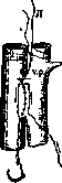
Figure 1. The first connecting suture; arrows show the direction of the cutting ligature (r. l.): V. c. inf. - inferior vena cava; V. p. - portal vein.
out of the abdominal cavity, leaving them free (Fig. 1, a). After this, a first suture is applied between the veins as high as possible (as close as possible to the liver hilum), tightly fixing their walls to each other (Fig. 1). Usually 7 such sutures are applied, occupying a space of about 2.5-3 cm. Then, on a round needle, a so-called cutting ligature (of relatively thin but strong silk) is passed from below upwards through the entire thickness of the wall of the v. portae between and at the levels of the 2nd upper and 6th lower sutures. After exiting the lumen of the v. portae at the level of the 2nd upper suture, it is thrown across to the side of the v. cavae inf. and, at an equal distance from the sutures connecting the two veins, is passed in the same order, but only from above downwards through the wall of the v. cavae inf. and the lower end is brought out outside (Fig. 1). The next stage of the operation is the closure of the cutting ligature so that it ends up between the closely apposed walls of the v. cavae inf. and v. portae. This is achieved by applying a second row of sutures to the inferior vena cava and portal vein, symmetric to the first, but located on the other side of the cutting ligature (Fig. 2). The last (7th) suture is not tied in a knot and remains temporarily free. Then, when this is achieved, the ends of the cutting ligature are firmly seized and pulled out by means of rapid sawing movements. At this moment, both walls of the sutured veins are cut through and a communication (anastomosis) is established between them, as a result of which blood from the portal vein begins to flow into the inferior vena cava. The formation of the anastomosis is indicated by bleeding from the lower angle of the unsecured connecting suture (7th), which immediately tightens and the bleeding stops. Then the ligature is tightly tied, previously freely applied to the v. portae (Fig. 1, a), as a result of which blood now flows completely from the v. portae into the v. cava inf. The operation for creating the E. f. ends at this point. The precautions that must be observed during the E. f. operation mainly consist in avoiding significant bleeding, which is achieved by careful selection of surgical instrumentation and, especially, needles. The latter must be of small size, round cross-section, and sharply curved. The number of silk is selected according to the available needles so that it is slightly thicker than the eye of the needle. Bleeding from the sutures is stopped by tamponade. The E. f. operation is usually performed on dogs weighing 15-16 kg. The choice of animal is of special importance; it must have a wide chest, a flat abdominal wall, and, if possible, a wide subcostal angle. All these body features ensure a low position of the liver, which significantly facilitates access to the inferior vena cava and portal vein. Among suitable breeds one can mention bulldogs, pugs, and especially a cross between a dachshund and a common mongrel. Females are more suitable for the E. f. than males. In the postoperative period, the animal must receive a carbohydrate-milk regimen. Meat and the general abundant introduction of animal protein are excluded due to the rapid development of symptoms of poisoning.
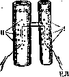
Figure 2. Method of sewing the cutting ligature (r. l.): I - first row of connecting sutures; II - second row of connecting sutures.
These features ensure a low position of the liver, which significantly facilitates access to the inferior vena cava and portal vein. Among suitable breeds one can mention bulldogs, pugs, and especially a cross between a dachshund and a common mongrel. Females are more suitable for the E. f. than males. In the postoperative period, the animal must receive a carbohydrate-milk regimen. Meat and the general abundant introduction of animal protein are excluded due to the rapid development of symptoms of poisoning. The operation of the so-called 'reverse' E. f., proposed by Fischler, is performed analogously to the 'direct' E. f., with the sole difference that, above the anastomosis, it is the inferior vena cava that is ligated, not the portal vein. The reverse E. f. is used to obtain pure hepatic blood by means of deep catheterization (through the v. jugularis) of the inferior vena cava at the level of the entry of the hepatic vein (Fischler). This last operation (reverse E. f.) was used by Mann and Magath as a preparatory stage for their famous operation of complete liver function exclusion E. f. Despite its deficiencies in physiological respects (incomplete exclusion of hepatic circulation, since the a hepatica remains intact), it still has great significance in experimental physiology and pathology of the liver.
P 0P physl0l°Sy ot liver, ibidem, v.LXIx" S. Chechulin.
Related articles
Cite this page
“Zkovskaya Fistula.” Soviet Medical Encyclopedia. English translation of Bolshaya Meditsinskaya Entsiklopediya, 1st ed. (Moscow, 1928–1936), ed. N. A. Semashko. https://sovietmedicalencyclopedia.pages.dev/article/zkovskaya-fistula/