Cavae Venae
Historical document, translated for reference. It reflects medical knowledge of the 1920s–30s and is not medical advice.
Summary
The article describes the anatomy of the superior and inferior vena cava, their formation, course, tributaries, and anastomoses with other venous systems, including the portal vein. It also discusses developmental anomalies of these major vessels.
Encyclopedia article (1928–1936)
CAVAE VENAE, hollow veins. There are superior and inferior hollow veins (Figure 1). V. cava superior is formed behind, under the cartilage of the right first rib at the right edge, and drains blood from the head, neck, thoracic wall, and upper extremities. From its place of formation to its entry into the right atrium, the length of v. cava superior varies within 4-5 cm. On the anterior thoracic wall, the vein corresponds (vertically) to the ends of the I and II intercostal spaces and the sternal end of the III rib. In front, at the place of entry into the atrium, it is covered by the pericardium; its dorsal and lateral surfaces lie outside the pericardium. To the right of v. cava superior lie the right mediastinal pleura and right lung, separating the vein from the anterior thoracic wall. Between v. cava superior and the mediastinal pleura on the right lies the n. phrenicus dexter. In its lower part, v. cava superior crosses the right bronchus and the root of the lung, lying in front of them. To the left of it lies the aorta. Into v. cava superior flow the vv. anonymae, v. azygos, vv. pericardicae, vv. mediastinales ant.-V. cava inferior is formed by the union of both common iliac veins (v. iliacae communes) and begins at the level of L5 behind and to the right of art. iliaca com. dextra. From here the vein goes upward, along the spine, lying to the right of the abdominal aorta [see separate table, Volume IV (articles 147-148), Figure 3], At the lower posterior edge of the liver, v. cava inferior enters the right longitudinal groove of the liver, deviating to the right from the aorta, and through the foramen venae cavae of the diaphragm enters the thoracic cavity, pierces the pericardium and opens into the right atrium. Behind and to the left of v. cava inferior lie the bodies of the lumbar vertebrae, from which it is separated by the trunk of the sympathetic nerve, aa. et vv. lumbales and the right crus of the diaphragm (above). The root of the mesentery of the small intestine at the level of the body of L5 crosses v. cava inferior obliquely in front. The pars horizontalis inf. duodeni, the head of the pancreas, v. portae (partially and at some distance) and the liver lie in front of v. cava inferior. Medially to it lies aorta abdominalis, laterally-pars libera of the right ureter and the inner edge of the right kidney. Anastomoses of v. cavae inferior can be divided into two groups: 1) connections with the system v. cavae superior and 2) connections with the portal vein. Anastomoses between the system v. cavae superior and v. cavae inferior. This collateral pathway is carried out by means of v. iliaca ext. (resp. v. femoralis), vv. epigastricae inf. superfic. et prof., v. epigastricae sup. et v. mammariae int., v. subclaviae et v. cava sup. In addition, there are also longitudinal anastomoses between individual lumbar veins and vv. azygos and hemiazygos. Anastomoses between the system v. cavae inferior and v. portae are divided into superficial and deep (Fig. 2). The superficial connection is carried out by means of v. paraumbilicalis, v. epigastrica inf. tegumentosa and v. femoralis.-The deep pathway of connection between v. portae and v. cava inferior can be formed in five different ways. Most often, collateral circulation develops by means of the following anastomoses: from the portal vein, blood through v. coronaria ventriculi sup. sin. flows into the veins of the esophagus. From there through the intercostal veins it first enters v. azygos, and then v. cava sup. Other collateral paths of this group are much less common: v. portae, v. paraumbilicalis xiphoidea (Braune), v. epigastrica sup. prof. sin., v. mammaria int., v. anonyma, v. cava sup.-Sometimes in the development of collateral circulation, the veins of the diaphragm participate. In such cases, blood from the portal vein flows into v. coron. ventric. sup. and through connecting branches enters the diaphragmatic veins and vv. musculo-phrenicae. The latter communicate with v. cava sup by means of v. mammaria int. Sometimes blood flows through the same anastomoses, bypassing the superior gastric vein. To this same group of collaterals should be attributed the anastomosis by which blood from the portal vein flows back into the splenic vein and through a connecting branch into v. azygos and v. cava sup.-There are many deep pathways of connection between the portal and inferior vena cava. In some cases, blood flows through vv. coronar. ventric. sup. or through the veins of the liver capsule (Kolliker's) into the diaphragmatic veins. Sometimes collateral circulation develops with the participation of the superior and inferior mesenteric, hemorrhoidal, and pudendal veins. In other cases, blood from the portal vein returns to the superior or inferior mesenteric veins, and from there through small branches of veins of the duodenum, ascending part of the colon and rectum or through branches of the veins of the descending part of the colon and rectum and the left kidney, it flows into v. cava inf. V. lienalis can also serve as a collateral conductor for blood: from the portal vein blood flows back into v. lienalis and through vv. gastr. breves and v. diaphragm. inf. sinistra flows into v. cava inf. By means of gastric veins, two collateral paths can develop: the first-by means of v. coron. ventr. inf. sin. or dext., small anastomoses with v. renalis and the v. renalis itself (Fig. 4 and 6); the second-by means of v. coron. ventr. inf. dext., which connects with the veins of the pylorus of the stomach. The latter are in communication with v. diaphragm. inf., through which they connect with the inferior vena cava. To this same group of anastomoses belongs the communication between the superior gastric vein, inferior esophageal veins through small connecting branches with the left suprarenal (Saxer; Figure 3). Communication between the system v. portae and v. cava inf is also possible by means of v. paraumbilicalis, the vein of Burow, v. epigastrica inf. prof. et v. iliaca. Anastomoses are also described that directly connect the trunk of the portal vein with the veins of the perirenal fat, and through them with the renal and inferior vena cava (Fig. 5) (Val'ker). In addition to these anastomoses that develop mainly in case of circulatory disturbance in the system v. portae or v. cavae inferior, there are
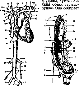
Figure 1.
Figure 2.
Figure 1. System of both hollow veins: 1 and 2-v. cava inf. et sup.; 3 and 4-v. anonyma dex. et sin.; 5-v. jugularis int.; 6-v. subclavia dex. et sin.; 7-trunk of vv. cervic. prof, vertebral.; 8-v. thyreoid. inf.; 9-v. mammar.; 10-v. intercostales supr. dex.; 11-v. azygos; 12-v. phrenicae; 13-vv. hepaticae; 14-v. suprarenalis dex. et sin.; 15-v. renalis dex. et sin.; 16-v. spermatica dex. et sin.; 17-v. iliaca com. dex. et sin.; 18-v. sacralis med.; 19-v. hemiazygos; 20-v. jugularis ext.; 21-arcus venosus juguli; 22-d. thoracicus; 23-d. lymphaticus dex.; 24-v. thyreoid. ima; 25-vv. lumbares (From Rauber.) Figure 2, Location of large veins and their anastomoses with the system v. portae: 1-v. subclavia dex.; 2-v. mammaria int.; 3-vv. hepaticae; 4-v. portae; 5-vv. paraumbilicales; 6-v. mesenterica sup.; 7-v. epigastrica inf.; 8-v. femoralis; 9-piles haemorrhoidales; 10-v. haemorrhoidalis sup.; 11-v. mesenterica inf.; 12-v. lienalis; 13-v. coronaria ventriculi; 14-vv. cardiacae; 15-vv. oesophageae; 16-v. azygos; 17-v. cava sup.; 18-v. anonyma sin.; 19-v. jugularis com. dex. (From Cornlng.) still anastomoses, which can be considered constant and probably functioning under normal conditions. To their number should be attributed the connection between the veins of the large intestine and the ureter, which were described by Toracheva (Fig. 7).

Development. V. cava sup. is formed from the right Cuvierian duct, which due to the downward displacement of the heart obtains an oblique direction, and partly from the caudal segment of v. cardinalis ant. dextra, while the left Cuvierian duct reduces, and V. cardinalis
Figure 3.

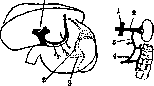
Figure 4. Figure 5. Figure 3. Anastomosis described by Saxer: 1-v. portae; 2-v. suprarenalis; 3-v. renalis sin. Figure 4. Anastomosis between v. renalis sin. and v. coli ascend.: 1-v. cava inf.; 2-v. renalis sin.; 3-anastomosis; 4-v. colli ascend. Figure 5. Anastomosis between v. portae and the veins of the perirenal fat: 1-anastomosis; 2-v. renalis dex.; 3-v. portae.
sinistra connects with v. card. dextra by an anastomosis giving rise to v. anonyma sin.-V. cava inf. is formed: 1) from a small vessel collecting blood from the walls of the abdominal cavity and after its union with vv. hepaticae flowing into the venous sinus; 2) from the caudal end of v. cardinalis post. dextrae, which first connects by anastomoses with the caudal part of v. cardinalis post. sin. (see Cardinales venae, Circulatory system, development).-Anomalies. The most common anomaly of the superior and inferior vena cava is their duplication. In the work of Odgers (1928) 131 cases of double superior vena cava (of which 47 in children) described in the literature are cited. To this number Odjer added one more of his own cases. Three types of double v. cavae sup. are distinguished. 1. V. cava sup. consists of two non-crossing trunks. 2. Between both v. cavae sup. there is an anastomosis, which
Figure 6. Anastomosis between v. lienalis and v. renalis sin.: 1-v. portae; 2-v. lienalis.; 3-v. cava inf.; 4-v. renalis sin.; 5-anastomosis.
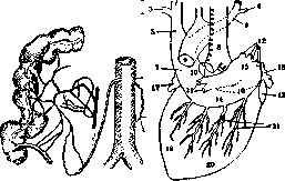
Figure 7.
Figure 8. Figure 7. Pre-existing anastomoses between the veins of the ascending colon, the appendix, and the internal spermatic vein. Figure 8. Left-sided position of the superior vena cava (posterior view): 1 and 2 - left and right jugular veins; 3 and 4 - left and right subclavian veins; 5 and 6 - left and right anonymous veins; 7 - superior vena cava; 8 - trachea; 9 - aorta; 10 - left pulmonary artery; 11 - left pulmonary veins; 12 - right pulmonary veins; 13 - inferior vena cava; 14 - coronary sinus; 15 and 16 - left and right atria; 17 and 18 - left and right auricles; 19 and 20 - left and right ventricles; 21 - left and right posterior coronary veins. The left superior vena cava is smaller than the right. 3. There is an oblique anastomosis; the left superior vena cava is very small. In duplication of the superior vena cava, the left superior vena cava crosses the aortic arch and the root of the left lung, extends to the great coronary sinus and opens into the right atrium. Duplication of the inferior vena cava should be considered as the preservation of both posterior cardinal veins up to the point of entry of the renal veins. Duplication of the proximal part of the inferior vena cava above the renal veins up to the point of entry into the heart is extremely rare. In addition to the anomalies of the superior and inferior vena cava in the form of their duplication, there are also abnormal relationships with neighboring organs [ureter, left-sided position (Fig. 8), perforation of the thymus gland by the superior vena cava, etc.]. Cases of absence of the inferior vena cava have also been described. Comparative anatomy. In reptiles, the anterior cardinal vein segments, located symmetrically together with the ducts of Cuvier, are called superior vena cavae. Into each of the superior vena cavae flow the jugular, subclavian, and vertebral veins of the corresponding side. In some mammals (egg-laying, marsupials, ungulates, insectivores, rodents, bats), the cardinal vein system remains double (symmetrical): there are two superior vena cavae, with the cardiac veins flowing into the right one. In other mammals (edentates, cetaceans, carnivores, prosimians, primates), instead of the ducts of Cuvier, only the coronary sinus of the heart remains, into which the cardiac veins flow. It is completely separated from the left cardinal (vena cava) vein, which flows into the right superior vena cava. In addition, the azygos vein and one anonymous vein on each side flow into the superior vena cava. The rudiments of the inferior vena cava in the form of an unpaired venous trunk flowing into the venous sinus or directly into the heart are already found in dipnoan fish and amphibians. The posterior part of the inferior vena cava develops from the posterior part of the right posterior cardinal vein. The anterior part of the inferior vena cava arises independently. In reptiles, the inferior vena cava predominates over the posterior cardinal vein system. In its anterior part, they atrophy and no longer communicate with the ducts of Cuvier. The main mass of blood from the posterior part of the body flows into the inferior vena cava or through the paired (sometimes unpaired) abdominal vein (epigastric superior) into the portal system of the liver. A small part of the blood flows into the kidneys (the renal-portal system undergoes retrograde development). In mammals, there is a well-developed system of inferior vena cava. The renal-portal system is preserved only in the embryo. Pathology. Diseases of the vena cava can be divided into the following groups: 1) wounds, 2) compressions, 3) thrombosis, 4) fibrous shrinkage, 5) diseases of the wall. In wounds of the superior vena cava, usually fatal, strong bleeding is observed, both external and internal; air embolism is possible. The causes of narrowing or closure of the lumen of the superior vena cava are: a) aneurysms of the aortic arch, b) tumors of the anterior mediastinum, c) retrosternal goiters, d) compression by scars, e) thrombosis. In aneurysms and tumors of the anterior mediastinum causing compression of the superior vena cava, cough irritation, a feeling of pressure in the chest, dizziness, headache, cyanotic coloration of the upper half of the body up to the costal arch, edema of the eyelids, a blue-red rim around the eyeball, blue sclera with a thin venous network, swelling of the neck veins, extensive phlebectasias on the anterior surface of the chest; the neck merges with the swollen face (sometimes a collar-like swelling of the neck). On auscultation, a stenotic noise and bronchial breathing are determined. On percussion - dullness in front up to the IV rib. The disease develops gradually. In thrombosis of the superior vena cava or its compression by scars, symptoms of increased intrathoracic pressure may be absent. Diseases of the wall of the superior vena cava are expressed by arteriosclerotic changes, ulcers as a result of malignant tumor growth, septic endophlebitis, etc. Injuries to the inferior vena cava occur relatively often during operations on the right kidney. In such cases, ligation of the inferior vena cava is indicated. Experimental data show that ligation of the inferior vena cava peripheral to the renal veins in animals does not lead to a fatal outcome. However, ligation of it central to the renal veins is always fatal. Collateral circulation after ligation of the inferior vena cava occurs through the azygos, hemiazygos, epidural veins, and spermatic veins (Leotta). The veins of the anterior abdominal wall do not have significant importance in these cases. Compression and complete obliteration of the inferior vena cava occur much less frequently than narrowing or closure of the lumen of the superior vena cava. The causes of compression can be: 1) pericardial exudates (in this case the upper part of the inferior vena cava is compressed); 2) left-sided pleuritic exudate; 3) tumors of the abdominal cavity; 4) echinococcus; 5) aneurysm of the abdominal aorta; 6) infectious thrombophlebitis (more often on the basis of puerperal sepsis, less often - influenza, typhus, tuberculosis, syphilis, cholera, etc.) and 7) thrombosis. Symptoms of difficulty in blood outflow through the inferior vena cava are edema of the legs (sometimes absent, sometimes even one-sided), the scrotum, penis, labia majora, the abdomen and lumbar region, dilation of collaterals, pain in the lower back, sacrum, abdomen, limbs, weakness, cyanosis of the lower half of the body, prominently appearing small venous networks, a sensation of cold in the limbs. In obstruction of the lumen of the inferior vena cava in its middle or upper third, above the point of entry of the renal veins, the edema is more pronounced, albuminuria, disorders of liver circulation, enlargement of the liver and spleen are observed. Ascites may be absent. In septic thrombosis of the inferior vena cava (or infectious phlebitis), fever, delirium, signs of general infection, pyemic abscesses, endocarditis, pain in the lower back accompanied by hematuria are observed. There is no syndrome different from cardiac edema and Bright's disease. Death usually occurs with signs of cachexia, uremia, weakening of cardiac activity; sometimes - from gastrointestinal bleeding, hematuria, pulmonary artery embolism, embolism of the right heart. Collaterals usually function worse than in thrombosis of the superior vena cava (Eppinger). Obliteration or narrowing of the lumen of the inferior vena cava may be limited to only its lower third. Then large networks of collaterals develop, significantly more developed than in cirrhosis. The wall of the inferior vena cava can be affected by arteriosclerotic processes, and ulcers can also occur (septic processes, tumor infiltration, etc.). Treatment for thrombosis and obliteration of the inferior vena cava is almost hopeless; cases have been described where after eliminating the cause of compression of the inferior vena cava (tumor), the signs of stenosis disappeared. More often, a thrombus forms at the site of compression, which subsequently organizes and turns the inferior vena cava into a fibrous cord. Ligation of the inferior vena cava is indicated in injuries and puerperal sepsis. According to observations by Warnekros, in puerperal sepsis, it is best to ligate the common iliac vein of the affected side, or the hypogastric vein, or the internal spermatic vein, and only with extensive thrombosis is the inferior vena cava ligated. Often on the operating table, a thrombus cannot be found despite the clinical indications of its presence. There are so many collaterals of the pelvic veins that it is impossible to completely exclude the entire venous bed of the pelvic region. Witt points out that after ligation of the main venous trunks, about 20 collaterals of the pelvic veins remain. Most authors consider the transperitoneal ligation of the inferior vena cava (median incision) to be easier. However, one can approach the vein through the extraperitoneal incision of Pirogov (lumbo-ilio-inguinalis). Mortality after ligation of the inferior vena cava for puerperal sepsis is about 60% (L. Koch). Among the complications sometimes observed after this operation, ecchymoses and ulcers of the gluteal region and gangrene of the lower limbs should be noted. The operation of anastomosis between the inferior vena cava and the portal vein is called the Eck fistula and was proposed for experimental purposes. Recently, such an anastomosis has been used (Krestovsky and others) for the treatment of ascites. For the same purpose, transplantation of the superior mesenteric vein into the inferior vena cava (operation of Bogoraz) is performed.
Related articles
Mentioned in
Cite this page
“Cavae Venae.” Soviet Medical Encyclopedia. English translation of Bolshaya Meditsinskaya Entsiklopediya, 1st ed. (Moscow, 1928–1936), ed. N. A. Semashko. https://sovietmedicalencyclopedia.pages.dev/article/cavae-venae/