Pleura
Historical document, translated for reference. It reflects medical knowledge of the 1920s–30s and is not medical advice.
Summary
This article describes the anatomy of the pleura, the serous membrane covering the lungs and thoracic cavity. It details the parietal and visceral layers, the pleural cavity, the various sinuses, and the topographic boundaries of the pleura.
Encyclopedia article (1928–1936)
PLEURA (pleura), a serous membrane lining the inner surfaces of both halves of the thoracic cavity and covering the lungs, which appear as if they have grown into closed pleural sacs. Just as in the peritoneum, two layers are distinguished in the pleura: the parietal, or parietal (pleura parietalis), lining the walls of the thoracic cavity, and the visceral, or pulmonary (pleura pulmonalis, s. visceralis), covering the outer surface of the lungs with the exception of the hilum and the site of attachment of the pulmonary ligaments (Fig. 1). The pleural ligaments (lig. pulmonalia) are stretched in the frontal direction between the visceral pleura in the region of the medial surface of the lung and the mediastinal pleura, originating from the latter. Like the ligaments of the peritoneum, they represent a duplication of the pleura, reach the base of the lung with their lower end, and are easily visible when attempting to remove the lower lobes of the lungs during an autopsy of the thoracic cavity. Both layers of the pleura delimit the pleural slit or cavity (cavum pleurae) located between them and transition into one another at the place where vessels and nerves approach the lung from the side of the lung hilum (porta pulmonis). The layer of visceral pleura, covering the lung from the surface and fusing tightly with it, descends into the depth of the pulmonary fissures separating the lobes from each other, where it passes from the surface of one lobe to another. The layer of parietal pleura lies directly against the thin fibrous plate of the endothoracic fascia (fascia endothoracica), which is well
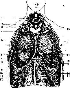
Figure 1. Pleural cavity, opened from the front: 1-m. sterno-cleido-mast.; 2-m. scalenus ant.; 3-clavicle; 4-I rib; 5-lobus sup.; 6-lobus med.; 7-processus xiphoideus; 8-lobus inf.; 9-VII rib; 10-lower border of the costal pleura; 11-m. transversus abdominis; 12-linea alba; 13-pericardium; 14-pleura mediastinalis; 15-cupula pleurae.
expressed in the lateral and anterior sections of the chest, and is separated from the latter by a layer of loose subpleural tissue. According to the degree of expression and the density of the connection in the subpleural tissue, three zones or belts are distinguished (Rudnev). The first zone is located in the posterior section, along the sides of the spine, from which it extends 5-6 cm in both directions. Here the tissue is abundant, loose, and contains a number of the most important vessels and nerves (nn. sympathici, aa., vv. et nn. intercostales, v. azygos et hemiazygos) and (even with severe emaciation) is easily detached from the underlying fascia, exposing the aforementioned neurovascular trunks. The second, middle zone, extending to the axillary line, contains less abundant but still quite loose tissue, allowing the separation of the pleura without its damage. In the anterior zone, the subpleural tissue thins out to such an extent that it is almost impossible to separate the pleura from the endothoracic fascia. In topographic terms, several sections or regions are distinguished in the parietal pleura. The part adjacent to the spine and covering the ribs, intercostal spaces, and the surface of the costal cartilages with part of the sternum is called the costal pleura (pleura costalis). In adults, the costal pleura is so closely fused with the costal cartilages by means of the endothoracic fascia that it is impossible to separate it from the ribs and intercostal muscles without damaging the pleural layer. In newborns and children, the costal pleura is connected to the ribs much more loosely. The part of the parietal pleura adjacent to the mediastinum (mediastinum) and located anteriorly is called the mediastinal pleura (pleura mediastinalis); at the hilum of the lung, it seamlessly transitions into the visceral pleura. In the region of the hilum, the mediastinal pleura forms a triangular fold in the direction of the diaphragm, in the form of a ligament (lig. pulmonale), connecting the mediastinal surface of the lungs with the mediastinal and diaphragmatic pleura (Figs. 2 and 3). The mediastinal pleura ventrally lies against the pericardium, with which it is connected by means of delicate and weakly expressed connective tissue, and throughout the entire indicated segment is called the pericardial pleura (pleura pericardialis). In the space between the outer layer of the pericardium and the pericardial pleura runs the phrenic nerve (n. diaphragmaticus, s. phrenicus), embedded in loose connective, and sometimes adipose, tissue. On the left, the mediastinal pleura covers part of the aorta and the left subclavian artery, the trunk of the left sympathetic nerve and the hemiazygos vein, and on the right—the right side of the superior vena cava and the innominate artery, the azygos vein and the trunk of the right sympathetic nerve, as well as the right side of the trachea. By diaphragmatic pleura (pleura diaphragmatica) is meant that part of the lower layer of the parietal pleura which covers the upper surface of the diaphragm to a varying extent on the left and right, remaining free from the attachment of the diaphragmatic layer of the pericardium. The described sections of the pleura are more or less delimited from each other and form closed cavities of pleural sacs around the lung. The dimensions of the pleural sacs are somewhat larger than the lungs contained within them, as a result of which, during exhalation, the protrusions of the pleural sacs clearly stand out. These protrusions, at the bottom of which different sections of the pleura transition into one another, are called sinuses (sinus pleurae); the lower
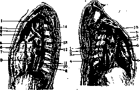
Figure 2.
Figure 2. Mediastinal spaces and their relation to the pleural sacs (left): 1-a. subclavia sin.; 2-a. carot. comm.; 3-n. vagus; 4-aortic arch; 5-v. pulmonalis sup.; 6-bronchus; 7-v. pulmonalis inf.; 8-n. phrenicus; 9-lig. triangulare sin.; 10-n. splanchnicus major; 11-n. vagus; 12-n. sympathicus; 13-a. pulmonalis; 14-v. azygos. Figure 3. Mediastinal spaces and their relation to the pleural sacs (right): 1-v. azygos; 2-n. sympathicus; 3-v. pulmonalis; 4-n. splanchnicus; 5-lig. triangulare dex.; 6-a. pulmonalis et bronchus; 7-right bronchus; 8-n. phrenicus; 9-v. cava sup.; 10-n. vagus. The pleural line lies below the lower border of the lungs. Only in a state of deep inspiration, as well as in certain pathological conditions (e.g., pulmonary emphysema), do the slit-like sinuses smooth out and their cavity is filled by the lungs. Usually, three main protrusions of the pleural sacs are distinguished: 1) the costomediastinal sinus (sinus pleurae costo-mediastinalis)—between the anterior end of the costal pleura and the pericardium, clearly visible on a horizontal section of the thoracic cavity (Figure 4); 2) the costodiaphragmatic sinus of Gerhardt—between the lower edges of the costal and diaphragmatic pleura, representing the largest depressions of the sacs and also clearly visible on frontal sections through the thoracic cavity (Fig. 5); 3) the phrenicomediastinal sinus (sinus pleurae phrenico-mediastinalis)—the least sharply expressed and located at the site of connection
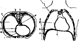
Figure 4.
Figure 5.
Figure 4. Horizontal section through the chest: 1-aorta; 2-v. azygos; 3-pleura costalis; 4-cavum pleurale dext.; 5-n. phrenicus; 6-pleura mediastinalis; 7-pericardium parietale; 8-sinus pleurae costo-mediastinalis; 9-oesophagus. Figure 5. Frontal section through the chest: 1-pleura costalis; 2-pleura mediastinalis; 3-pleura diaphragmatica; 4-sinus phrenico-costalis; 5-pericardium parietale. of the diaphragmatic pleura with its layer covering the heart sac. The first two sinuses—the costomediastinal and the costodiaphragmatic—are reserve cavities into which the lungs primarily move during inspiration and from which they depart during exhalation, as a result
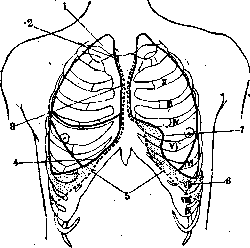
Figure 6. Pulmonary and pleural borders from the front: 1-apex pulmonis; 2-linea ant. pleurae dext.; 3-margo ant. pulmonis dext.; 4-margo inf.; 5-linea inf.; 6-sinus diaphragmaticus; 7-incisura cardiaca.
of which both layers forming the sinuses lie closely against each other. Extremely important in practical terms is the study of the boundaries of the extreme displacement of the pleura on the anterior chest wall. Starting on both sides in the region of the sternoclavicular joint, the boundary line descends downwards, and for some distance on the posterior surface of the manubrium and body of the sternum, a triangular area remains, free from the pleural covering and bordering directly on the loose connective tissue of the mediastinal space. At the level of the sternal ends of the IV costal cartilages, the lines of the anterior pleural borders diverge: on the left, the line is directed in a slightly curved arc outwards to the VI costal cartilage, where at the level of its sternal end it passes into the line of the lower fold of the pleura. As can be seen in Fig. 6, the ventral ends of the IV and V intercostal spaces are devoid of pleural covering. On the right, the line goes parallel to the costal arch to the VII costal cartilage, where on both sides the lower border of the pleural cavity begins, going in accordance with the protrusion of the diaphragmatic-costal sinus in an arc downwards and outwards. Along the mammary line, the border crosses the VII rib, along the axillary—the X, and approaches the spine at the level of the middle of D XII. Thus, the XII ribs are crossed by the lower border of the pleura on the line of the
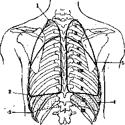
Figure 7. Lungs from behind. Lower border of the lungs and pleura: 1-apex pulmonis; 2-margo inf. pulmonis sin.; 3-linea inf.; 4-sinus phrenico-costalis; 5-incisura interlobaris.
union of the upper and middle third of the rib (Figure 7), which has practical significance for surgical access to the kidneys and renal pelves. Along the indicated boundaries on the anterior wall, there remain two places free from the pleura, corresponding to the manubrium and the body of the sternum. The pericardial sac lies against the body of the sternum, which determines the method of surgical access to it during puncture or incision without damaging the pleural sacs in cases of their typical location (in the region of the IV or V intercostal space). In newborns and children, the thymus gland lies against the upper section corresponding to the manubrium of the sternum, and in adults, adipose tissue representing its remnants. Thus, the pleural sacs, diverging along the midline, leave behind the sternum two free fields not covered by the pleura: above—the thymic field (area thymica), and below—the pericardial field (area pericardiaca). Deviations from the typical course of the pleural boundaries are often observed, and the variations in their course are quite significant and can be reduced to two main extreme forms of displacement of the boundaries to the right or left, which is clearly visible in the attached diagrams of these displacements on the anterior chest wall (Fig. 8). The above-described cases of atypical course of the pleural sacs must be taken into account during punctures or incisions of the pleural cavities. The upper section of the pleural sacs, called the cupula of the pleura (cupula pleurae), in the form of a cone-shaped outgrowth, extends through the superior thoracic aperture into the region of the lower part of the neck, reaching the level of the middle of Cvii, and encloses the apex of the corresponding lung.
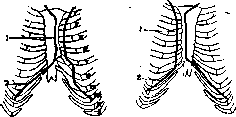
Figure 8. Diagram of extreme displacements of the pleural borders to the right and left on the anterior chest wall: 1—limes anterior pleurae dext.; 2—limes inferior pleurae dext.
The degree of protrusion of the apices of the lung, and thereby the cupula of the pleura, is subject to individual fluctuations and is in the closest relationship with the constitutional features of the chest: in the paralytic form of the chest, the apex of the pleura protrudes 3-5 cm above the upper edge of the I rib, and in the cone-shaped form it barely extends beyond the ribs. Usually, the right cupula of the pleura is slightly higher than the left, which is apparently
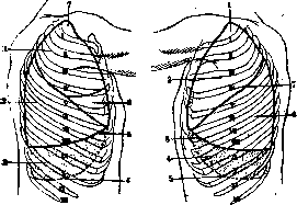
Figure 9.
Figure 9. Boundaries of the right lung and lower pleural boundary: 1—lobus sup.; 2—lobus inf.; 3—sinus phrenico-costalis; 4—limes inf. pleurae; 5—margo inf. pulmonis; 6—lobus med.; 7—apex pulmonis. Figure 10. Boundaries of the left lung and lower pleural boundary: 1—apex pulmonis; 2—lobus sup.; 3—margo inf. pulmonis; 4—sinus diaphragmaticus; 5—limes inf.; 6—lobus inf.; 7—incisura interlobaris. in connection with the right-sided position of the liver, due to which the right pleural sac is shorter than the left, but slightly exceeds it in width. The pleural cupula superiorly and laterally lies against the scalene muscles, posteriorly and medially—against the trachea and esophagus, anteriorly—against the subclavian artery and vein, and finally superiorly—against the lower trunk of the brachial plexus. The lower boundary of the pleural sacs does not reach the diaphragm, passes onto the chest wall, and lies on the left side lower than on the right (Figs. 9 and 10). In a state of maximum expiration (and on a cadaver), the lower boundary of the pleural sacs forms a straight line, starting on the right at the place of attachment of the VI rib to the sternum, and on the left—at the middle of the same rib, and ends posteriorly at the place of attachment of the XI rib, which corresponds to the level of the spinous process of Dx. In a living person, based on the results of percussion, the lower boundary of the lungs is taken on the right as: VI rib along the parasternal line, upper edge of the VII rib along the mammary line, lower edge of the same rib along the axillary line, IX rib along the scapular line, and finally X rib along the paravertebral line. On the left, the boundary is located a finger's breadth or a rib's width lower. During inhalation, the boundary moves several centimeters lower. Structure of the pleura. The pleura, like the peritoneum and pericardium, consists of a connective tissue elastic base, a thin vitreous membrane, and an outer covering of single-layered polygonal squamous epithelium. The epithelium of the parietal pleura contains special slit-like openings (stomata) that are in connection with lymphatic vessels. The thickness of the pleura varies in its different sections; it is thinnest on the lungs, where it is firmly fused with them; the costal pleura is thickest and easily detached from the ribs. On the sharp edges of the lungs, there are a large number of pleural villi (villi pleurales) similar to synovial villi, often containing vessels and nerve fibers (Luschka).
Blood vessels of the pleura originate from various sources and form wide capillary networks. The arteries of the parietal pleura originate from neighboring arteries, namely: from branches of the internal thoracic artery (a. mammaria int.), the mediastinal (a. mediastinalis), and the diaphragmatic artery (a. diaphragmatica); the costal part of the parietal pleura also receives branches coming from the intercostal arteries (aa. intercostales). The visceral pleura, tightly fused with the lung tissue, is vascularized like them by the bronchial arteries (aa. bronchiales). The venous drainage trunks of the parietal and visceral pleura fully correspond to the branching of the arteries (see above). Lymphatic vessels form dense and rich plexuses in the intercostal spaces and from the visceral pleura run together with the superficial lymphatic vessels of the lungs (see Lungs). The lymphatic vessels of the costal section of the parietal pleura are directed to the anterior intercostal nodes (lgl. intercostales ant.); the lymphatic vessels of the intercostal spaces located immediately under the pleura also go to them. From the indicated anterior intercostal nodes, the lymphatic vessels ascend upwards together with the internal thoracic arteries and veins and drain on the left into the thoracic duct (ductus thoracicus), and on the right—into the right broncho-mediastinal trunk (truncus broncho-mediastinalis dexter). The lymphatic vessels of other sections of the parietal pleura drain into the lymphatic vessels of the walls and are directed with them to the vessels accompanying the intercostal, internal thoracic, and diaphragmatic arteries. Nerves of the visceral pleura come from the pulmonary sympathetic plexus, which explains its insensitivity; the parietal pleura, however, is innervated not only by the sympathetic nerve, but also by the phrenic and intercostal nerves, which causes its pain sensitivity. In addition, the anterior, ventral branches (rami ventrales) of the intercostal nerves run for some distance directly under the parietal pleura, passing further into the costal grooves (sulci costales). This circumstance explains the ease of involvement of the intercostal nerves in the pathological process during pleurisy in the form of severe, sometimes intercostal neuralgias.
N. Melik-Pashayev. Pathology of the pleura. Wounds, pathological processes, anomalies, etc., only very rarely affect the pleura primarily. For the most part, the pleura is involved in processes developing in organs and tissues located in immediate proximity to it. These include the skin and soft tissues covering the chest cage, ribs, lungs, mediastinum, heart, spine, diaphragm, and abdominal cavity. The close proximity to the listed organs makes secondary involvement of the pleura and the pleural cavity during processes in them almost mandatory. Traumatic injuries of the chest cage and lung, especially penetrating wounds of them, almost always accompanied by the development of pneumothorax, are a vivid example of such involvement of the pleura and pleural cavity (see Lungs-surgery). Analogous to the peritoneum, the pleura responds to the introduction of infection into it with hyperemia and exudates (see Pleurisy), and to chemical and thermal irritations—more often with dry inflammation (pleuritis sicca). Hemorrhagic diatheses, hemophilia, etc., can lead to the accumulation of blood in the pleural cavity (haemothorax); diseases accompanied by general dropsy (nephrosis, heart diseases, hunger edema, severe anemias, cachexias, etc.) lead to non-inflammatory accumulation of fluid in the pleural cavity (see Hydrothorax). Tuberculosis of the pleura is observed in 2 forms: a) a pure form of tuberculosis of the pleura, proceeding with infiltration of the pleura and the development of tubercles, and b) tuberculous pleurisy. Often, however, these 2 forms are encountered in combination. The first form is for the most part the result of the transition to the pleura of a tuberculous process from caseously degenerated bronchial glands, from ribs, or the spine affected by a tuberculous process, and is also observed in general acute miliary tuberculosis (see). Actinomycosis of the pleura is also almost always the result of the transition of the process to it from the lung, esophagus, or skin. The inflammatory process, as it develops, usually makes a path for itself outward through the thickness of the chest wall, forming fistulas that discharge specific pus. Since the clinical picture is often extremely similar to the picture of tuberculosis, a correct diagnosis is sometimes made only on the basis of an examination of the pus. Syphilis of the pleura is rarely observed in the form of a secondary form (roseola, papules), giving phenomena of bronchitis and pleurisy; more often, a gummatous form is encountered, also complicated by pleurisy. Echinococcus of the pleura is for the most part the result of the transition of the process to it from the liver, spleen, or ribs, but mainly from the lung. The primary form is observed very rarely. The process is almost always accompanied by secondary pleuritis. In the presence of daughter cysts, dissemination throughout the entire pleural cavity occurs rapidly. Clinical symptoms vary depending on the location and size of the echinococcal cysts. Treatment is surgical: thoracotomy, often with resection of a part of the chest wall. With simultaneous involvement of the liver or spleen—transthoracic laparotomy. Alveolar echinococcus affects the pleura by direct transition from the liver or spleen; it has not been observed primarily. In transdiaphragmatic wounds of the pleura, ascarids and tapeworms that have emerged from a damaged intestinal tube have been found in its cavity. In China, Korea, and especially in Japan, Distomum pulmonale has also been found in the pleural cavity, having entered it as a result of the decay of the lung tissue affected by it.
Foreign bodies are observed in the pleural cavity mainly in the form of minute particles of soot and dust that have entered it from the lung in workers in the corresponding types of industry (see Anthracosis, Pneumoconioses). As free foreign bodies (pieces of wood, fragments of stones, shells, bullet casings), they were rarely encountered as a consequence of penetrating wounds. Rubber drainage tubes are often encountered in chronic purulent pleurisy (see Pleurisy-purulent pleurisy). To remove foreign bodies from the pleural cavity that cause extensive adhesions, it is sometimes necessary to perform very extensive thoracotomies, which result in high mortality. Among tumors, lipomas, fibromas, chondromas, osteomas, angiomas, sarcomas, endotheliomas, and cancer are observed. Malignant tumors are for the most part metastases or spread to the pleura from neighboring organs and tissues. Among tumors developing in the pleura primarily, Sauerbruch observed endotheliomas, sometimes reaching large sizes. Tumors of the pleura present a diverse clinical picture depending on their size, location, and character. With tumors of significant size, the phenomena of dyspnea, occurring even with slight exertion, and pain radiating to the corresponding shoulder, usually come to the fore. With malignant tumors, an increase in temperature and serous effusion in the pleural cavity are observed. Klemperer notes a significant development of veins on the skin over the tumor. In making a correct diagnosis, fluoroscopy and the character of the effusion (hemorrhagic) play a large role. Treatment of tumors of the pleura is surgical: thoracotomy, extensive resection of a part of the chest wall; it is successful but technically difficult in benign or primary malignant tumors of the pleura, and does not yield success in far-advanced cases and secondary tumors.
Related articles
Mentioned in
Cite this page
“Pleura.” Soviet Medical Encyclopedia. English translation of Bolshaya Meditsinskaya Entsiklopediya, 1st ed. (Moscow, 1928–1936), ed. N. A. Semashko. https://sovietmedicalencyclopedia.pages.dev/article/pleura/