Supraclavicular Fossa
Historical document, translated for reference. It reflects medical knowledge of the 1920s–30s and is not medical advice.
Summary
The supraclavicular fossa is a triangular depression on the lateral surface of the neck, bounded by the sternocleidomastoid muscle, omohyoid muscle, and clavicle. This article describes its anatomical boundaries, layers, contents, and clinical significance in the 1930s Soviet medical understanding.
Encyclopedia article (1928–1936)
SUPRACLAVICULAR FOSSA, supraclavicular region (regio, s. fossa supraclavicularis, s. trigonum omo-claviculare), occupies the lower part of the lateral surface of the neck and externally has the appearance of a triangle. Its boundaries: in front - the posterior edge of the sternocleidomastoid muscle, above - the omohyoid muscle, below (base) - the middle part of the clavicle. Normally the supraclavicular region is slightly depressed; in thin people and with difficult breathing it is more or markedly sunken, in fat people it disappears. When the head is turned to the opposite side, the shoulder is lowered, and the shoulder blade is thrown back, the depth of the fossa decreases, the organs are located more superficially and are more accessible during operations. Under opposite conditions the fossa becomes deeper, the muscles relax, and examination of the organs is facilitated.-Upon layered examination of the supraclavicular region we have 1) thin, movable, easily displaced skin; 2) subcutaneous tissue with a superficial fascia; 3) the subcutaneous muscle (platysma) of the anterior-inferior part of the region (fig.1); 4) still deeper - the first cervical fascia, which covers the sternocleidomastoid muscle on both sides, and unites along its posterior edge into one sheet lining the entire lateral region of the neck, attached below to the upper edge of the sternum and clavicle and passing below it into the superficial thoracic fascia; posteriorly, at the medial edge of the trapezius muscle, the first fascial sheet divides, covering this muscle on both sides (figure 2). Between the platysma and the first cervical fascia pass a) cutaneous nerves of the cervical plexus (nn. supraclaviculares et acromiales), which emerge from behind the middle of the posterior edge of the sternocleidomastoid muscle and descend downward and outward; b) small arteries, branches of the transverse scapular and cervical arteries (a. transv. scapulae et colli) and the superficial cervical artery (a. colli sup.); c) corresponding veins, emptying into the external jugular vein; the latter is mostly in the inner angle of the supraclavicular region, descending from the anterior surface of the sternocleidomastoid muscle downward and outward, it pierces the first and second cervical fasciae and empties into the subclavian vein. Along the course of the veins, mainly the external jugular, go superficial lymphatic vessels and glands. After removal of the first cervical fascia, the omohyoid muscle is visible, which originates from the upper edge and transverse ligament above the scapular notch, goes upward and forward, passes under the sternocleidomastoid muscle and above the main vascular-nerve bundle attaches to the hyoid bone (figure 3). The supraclavicular region is divided by it into two triangles: 1) upper (trig. omo-trapezoides), filled with adipose tissue, in which pass insignificant blood vessels and branches of the cervical plexus; above them from behind the posterior edge of the middle of the sternocleidomastoid muscle to the trapezius goes the accessory nerve of Willisii; 2) the lower, smaller triangle (trig. omo-claviculare), bounded above by the omohyoid muscle, below by the clavicle, in front by the sternocleidomastoid muscle. The omohyoid muscle is covered on all sides by the second cervical fascia, which in the form of a triangular plate goes from the upper edge of this muscle downward and outward; below it attaches to the scapular notch, to the posterior edge of the clavicle and passes into a fibrous plate covering the subclavian muscle and vein. The continuation of the second cervical fascia medially surrounds and accompanies the other anterior presternal muscles of the neck (mm. ster-no-thyreo-hyoidei). Between the first and second cervical fasciae there is adipose tissue; in it passes along the upper-posterior edge of the clavicle the transverse scapular vein. After removal of the omohyoid muscle and the second fascia, an excavation, or bed of the supraclavicular region, is opened, having the form of a triangular pyramid; its anterior and posterior edges correspond to the external boundaries of the supraclavicular fossa, and the median one to the transverse processes of the cervical vertebrae. Of the three walls of the pyramid, the lateral one separates the bed from the external coverings and was just described layer by layer; the posterior one is formed by muscles going from the cervical vertebrae to the I and II ribs - mm. scaleni medius et posticus. The median wall is formed by the sternocleidomastoid muscle and deeper - by the anterior scalene muscle (t. scalenus anticus), originating on the transverse processes of C6-C7 and attaching to the Lisfranc tubercle on the I rib (fig. 2). The median wall has two slits: the anterior scalene - between the anterior scalene and sternocleidomastoid muscles, through which pass the subclavian vein, thoracoabdominal nerve, and transverse scapular artery; the posterior scalene slit - between the anterior and middle scalene muscles; through the posterior scalene slit pass the subclavian artery and brachial nerve plexus (figures 3 and 4). The scalene muscles are covered by an aponeurotic process from the third cervical aponeurosis (fascia praevertebralis). The base of the supraclavicular bed is triangular in shape; its lateral edge is formed by the clavicle, posterior by the edge of the scapula, median by a line drawn from the posterior edge of the sternocleidomastoid muscle to the transverse process of C7; it is divided by the I rib into an outer part, occupied by the clavicle, subclavian muscle, and I rib, and into a median part, corresponding to the superior thoracic aperture (apertura thoracis superior), in which is located the dome of the pleura and the apex of the lung. Here to the dome of the pleura in the angle formed by the anterior scalene muscle and the long muscle of the neck with its vertex on the transverse process of C7 (angulus scaleno-vertebralis), are adjacent: the first part of the subclavian artery with its branches - a. vertebralis, a. mammaria int. and the third cervical sympathetic ganglion (fig. 5). The bed of the supraclavicular fossa is supplied with more or less developed adipose tissue, surrounding the organs of this region and filling the spaces between them and the walls. Medially it passes into the tissue of the sternocleidomastoid region, below into the tissue
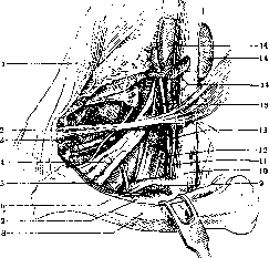
Figure 3. Right supraclavicular fossa. Sternocleidomastoid muscle removed. Clavicle pulled downward: 1-m. trapezius; 2-m. omo-hyoideus; 3-a. and v. transversa colli; 4-brachial nerve plexus; 5-a. subclavia; 6-v. subclavia; 7-v. transversa scapulae; 8-clavicle; 9-truncus lymphaticus axillaris; 10-v. jugularis interna; 11-truncus lymphaticus jugularis; 12-m. scalenus anticus; 13-n. phrenicus; 14-upper deep cervical lymph glands (lower ones removed). (By K. Yesinov.)
omohyoid muscle is covered on all sides by the second cervical fascia, which in the form of a triangular plate goes from the upper edge of this muscle downward and outward; below it attaches to the scapular notch, to the posterior edge of the clavicle and passes into a fibrous plate covering the subclavian muscle and vein. The continuation of the second cervical fascia medially surrounds and accompanies the other anterior presternal muscles of the neck (mm. ster-no-thyreo-hyoidei). Between the first and second cervical fasciae there is adipose tissue; in it passes along the upper-posterior edge of the clavicle the transverse scapular vein. After removal of the omohyoid muscle and the second fascia, an excavation, or bed of the supraclavicular region, is opened, having the form of a triangular pyramid; its anterior and posterior edges correspond to the external boundaries of the supraclavicular fossa, and the median one to the transverse processes of the cervical vertebrae. Of the three walls of the pyramid, the lateral one separates the bed from the external coverings and was just described layer by layer; the posterior one is formed by muscles going from the cervical vertebrae to the I and II ribs - mm. scaleni medius et posticus. The median wall is formed by the sternocleidomastoid muscle and deeper - by the anterior scalene muscle (t. scalenus anticus), originating on the transverse processes of C6-C7 and attaching to the Lisfranc tubercle on the I rib (fig. 2). The median wall has two slits: the anterior scalene - between the anterior scalene and sternocleidomastoid muscles, through which pass the subclavian vein, thoracoabdominal nerve, and transverse scapular artery; the posterior scalene slit - between the anterior and middle scalene muscles; through the posterior scalene slit pass the subclavian artery and brachial nerve plexus (figures 3 and 4). The scalene muscles are covered by an aponeurotic process from the third cervical aponeurosis (fascia praevertebralis). The base of the supraclavicular bed is triangular in shape; its lateral edge is formed by the clavicle, posterior by the edge of the scapula, median by a line drawn from the posterior edge of the sternocleidomastoid muscle to the transverse process of C7; it is divided by the I rib into an outer part, occupied by the clavicle, subclavian muscle, and I rib, and into a median part, corresponding to the superior thoracic aperture (apertura thoracis superior), in which is located the dome of the pleura and the apex of the lung. Here to the dome of the pleura in the angle formed by the anterior scalene muscle and the long muscle of the neck with its vertex on the transverse process of C7 (angulus scaleno-vertebralis), are adjacent: the first part of the subclavian artery with its branches - a. vertebralis, a. mammaria int. and the third cervical sympathetic ganglion (fig. 5). The bed of the supraclavicular fossa is supplied with more or less developed adipose tissue, surrounding the organs of this region and filling the spaces between them and the walls. Medially it passes into the tissue of the sternocleidomastoid region, below into the tissue
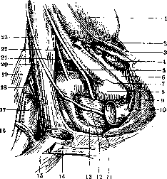
Figure 4. Right supraclavicular fossa: 1-thyroid gland; 2-lgl. praetrachealis; 3-v. thyreoidea inferior; 4-lgl. supraclaviculares med.; 5-n. vagus; 6-fascia of the vascular bundle; 7-a. carotis; 8 and 17-a. subclavia; 9-articular cavity for the removed clavicle; 10-v. anonyma; 11-pleura (dome); 12 and 23-n. phrenicus; 13-a. mammaria interna; 14-tendon of the sternocleidomastoid muscle; 15-v. subclavia; 16-v. transversa colli; 17-a. transversa colli (place of origin); 19-nerve plexus; 20-m. scalenus anticus; 21-a. cervicalis ascendens (a. thyreoidea inf. atypically originated deeper); 22-v. vertebralis. (By K. Yesipov.)
of the sternocleidomastoid region, below into the tissue
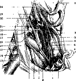
Figure 5. Right supraclavicular fossa. Deep department (angulus scaleno-vertebralis): 1-displaced thyroid gland; 2-lymph nodes paratracheales ('recurrens Kette'); 3-inferior thyroid vein; 4-recurrent nerve; 5-sympathetic nerve; 6-lymph nodes paratracheales; 7-brachiocephalic artery (cut); 8-subclavian vein; 9-pleura (dome); 10-vagus nerve; 11-internal mammary artery; 12-pleura; 13-subclavian vein (outer segment); 14-subclavian artery; 15-transverse vein of the neck; 16-anterior scalene muscle; 17-transverse artery of the neck; 18-Vieussens' loop (posterior knee); 19-phrenic nerve; 20-deep sympathetic ganglion; 21-inferior thyroid artery; 22-middle sympathetic ganglion (ganglion thyreoideum); 23-internal jugular vein; 24-internal carotid artery (According to K. Yesin.) <mediastinum and axillary fossa. In it are located the deep supraclavicular or lower deep cervical lymph glands (lymphoglandulae cervicales prof, inf.), which collect lymph from the lateral and median areas of the neck. Through anastomoses with the axillary glands, these glands can also be connected with the mammary gland. The subclavian vein (v. subclavia) enters the supraclavicular area, being on the 1st rib medially from the subclavian artery, and then-below and in front of it; it goes in a transverse direction hidden behind the clavicle, but in front of the anterior scalene muscle, which separates the vein from the subclavian artery. The fibrous fascia, going mainly from the second cervical fascia, fixes, encompassing, the subclavian vein and prevents collapse of its lumen, which is why when it is injured, as with other veins of the neck, the sucking action of the chest easily manifests, and air embolism occurs. This same fibrous plate protects the vein from damage by fragments in fractures of the clavicle. The subclavian artery, lying directly on the dome of the pleura, enters the supraclavicular area through the posterior scalene fissure behind the anterior scalene muscle; it then lies on the upper surface of the 1st rib, approaches under the m. subclavius in the area of the middle of the clavicle, and below already bears the name a. axillaris (axillary artery). From the branches of the subclavian artery in the supraclavicular area pass: the transverse artery of the scapula-goes in front of the anterior scalene muscle along the posterior surface of the clavicle to the scapular notch; the ascending cervical artery (a. cervicalis ascendens)-goes upward along the anterior scalene muscle; the costocervical trunk (truncus costo-cervicalis) with branches-the deep cervical artery to the muscles of the neck and the superior intercostal-to the I and II intercostal spaces; the transverse artery of the neck passes mostly between the bundles of the brachial plexus outward and backward along the outer surface of the middle scalene muscle under the t. levat. anguli scapulae (fig. 5 and 6). Corresponding to these arteries are veins, flowing into either the angulus venosus or the v. jugularis ext.-Into the angulus venosus or the v. subclavian flow the main lymph vessels of the neck, upper limb, and thoracic cavity, separately or by a common trunk: on the left-thoracic duct, on the right-right lymphatic duct. Along the anterior surface of the anterior scalene muscle descends the phrenic nerve (n. phrenicus) from C3-C5, lies below between the subclavian artery and vein and passes into the mediastinum. The brachial plexus (plexus brachialis) appears in the supraclavicular area through the lateral scalene fissure, above and behind the subclavian artery; its lower bundles lie adjacent to the dome of the pleura; from it depart in the supraclavicular fossa the subclavian nerve (nervus subclavius), the long thoracic nerve (n. thoracicus longus) and the dorsal nerve of the scapula (nervus dorsalis scapulae). The different shape of the supraclavicular area, as well as the degree of depression of the supraclavicular fossa, depend to a large extent on the configuration of the chest, resp. on the constitutional type of the subject. For example, in asthenics the supraclavicular fossa is sharply expressed even in normal conditions, and conversely, it is almost completely smoothed out in persons of hypersthenic constitution. A change in the shape of the supraclavicular area (sometimes significant) also occurs in pathological conditions. A sharp depression of the supraclavicular fossa, unilateral or bilateral, is observed in contraction of the apex of the lung and can be one of the important symptoms of fibrous tbc of the lungs (see Tuberculosis of the lungs). The same is observed also in cases of pleural adhesions after suffered pleuritis.
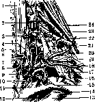
Figure 6. Deep department of the supraclavicular fossa in cross section: 1-longus colli muscle; 2-vertebral artery; 3-deep cervical artery; 4-inferior cervical sympathetic ganglion; 5-recurrent nerve; 6-esophagus; 7-trachea; 8-thyrocervical trunk; 9-carotid artery; 10-vagus nerve; 11-site of entry of the jugular vein into the angulus venosus; 12-internal mammary artery; 13-1st rib; 14-subclavian vein; 15-thoracic duct; 16-subclavian artery; 17-phrenic nerve; 18-transverse scapular artery; 19-anterior scalene muscle; 20-transverse artery of the neck; 21-costo-pleural ligament; 22-brachial plexus; 23-pleuro-transverse ligament; 24-middle scalene muscle. III ig also in cases of pleural adhesions after suffered pleuritis. Bulging of the supraclavicular area occurs in emphysema, phlegmons and abscesses and due to purulent inflammation of the supraclavicular lymph glands; in tumors originating from lymph glands (lymphogranulomatosis, lymphosarcomatosis, etc.); sometimes with low-descending bronchogenic cysts. The supraclavicular area is deformed in fractures of the clavicle, and rarely in hernial protrusions of the lung due to emphysematous expansion. Injury to the subclavian artery more often occurs in the third part of it after exiting the scalene fissure. Arrest of bleeding is done by applying a ligature from an incision parallel to the clavicle in the trigono claviculare or temporarily by pressing the artery against the 1st rib. Aneurysms of the subclavian artery occur more often than in other neck vessels; in men more often than in women, on the right more often than on the left.-In the supraclavicular fossa a number of operative interventions are performed on its organs: approach to the phrenic nerve, approach to the 1st rib by Coffey's method for its resection in tbc of the lungs, anesthesia of the brachial plexus by Kulenkampfi's method, etc.
g. Prikhodko.
Related articles
Mentioned in
Cite this page
“Supraclavicular Fossa.” Soviet Medical Encyclopedia. English translation of Bolshaya Meditsinskaya Entsiklopediya, 1st ed. (Moscow, 1928–1936), ed. N. A. Semashko. https://sovietmedicalencyclopedia.pages.dev/article/supraclavicular-fossa/