Stupor
Historical document, translated for reference. It reflects medical knowledge of the 1920s–30s and is not medical advice.
Summary
This article from the 1928-1936 Great Medical Encyclopedia discusses the clinical definition and various forms of stupor, including catatonic, depressive, and psychogenic types. It also begins coverage of the subclavian artery and vein, detailing their anatomy and anatomical variations.
Encyclopedia article (1928–1936)
STUPOR, complete immobility. According to Jaspers' definition, stupor is a state in which a patient, with complete motor rest, without saying a word and without giving any understandable signs of the mental processes occurring within them, does not react to any attempts to enter into a relationship of mental interaction with them. Stuporous patients often lie in bed for many weeks and months without changing position, without answering questions, and sometimes completely refusing to take food. Other patients stand or sit, sometimes huddled in strange, bizarre poses, turning their faces to the wall or into a corner or covering their heads with a blanket, remaining completely motionless in one and the same pose until they are forcibly moved to another position. A distinction is made between flaccid stupor and stupor with muscular tension. The most frequent form of stupor is catatonic stupor (see Catatonia). This is predominantly a stupor with tension. In it, according to Kraepelin, the possibility of producing movements is not impaired at all by itself, but any impulse immediately elicits an oppositely directed impulse, so that the movement is suppressed at its very inception. Pathophysiologically, catatonic stupor, like other catatonic phenomena, according to V. P. Osipov, is explained by a disturbance of innervation relationships between the cortex and subcortical areas (see Catatonia). Depressive stupor, observed in severe circular depressions, differs from catatonic in that it is based on a general psychomotor inhibition, making in extreme cases the production of movements impossible due to the extreme difficulty of any effort for the patient. Jaspers also singles out stupor in bewilderment, caused by the fact that the general state of mental stupor does not allow impulses to manifest themselves. A particular type of stupor in bewilderment is the so-called reactive or psychogenic stupor described by Raccke, which is understood by a number of authors as a peculiar expression of the " reflex of imaginary death," but in reality is a consequence of the fact that the shock reaction, under the influence of the acuteness and tension of the situation that caused it, is delayed in its manifestation. A special position is occupied by states of immobility in severe forms of postencephalitic parkinsonism, which are explained by a sharp increase in muscle tone and the inability to translate impulses into movements.
P. Zinovyev. SUBCLAVIA ARTERIA, VENA. The subclavian artery (see Carotid artery, figures 1 and 2) delivers blood mainly to the upper extremity, and then to the head, neck, posterior section of the brain, walls, and certain organs of the thoracic and abdominal cavities. (For structure, see Artery; for caliber, see Blood vessels, arterial system.) There are two subclavian arteries, right and left. The right arises from the brachiocephalic artery, its origin being projected onto the right sternoclavicular articulation; the left subclavian artery arises from the aortic arch and runs vertically upward among the organs of the mediastinum, contacting externally with the pleura up to its dome, internally with the lateral wall of the esophagus, alongside the inferior laryngeal nerve (recurrent laryngeal nerve); the trachea remains anterior to the artery; posteriorly, the left subclavian artery adjoins the spine and the thoracic duct running along it; anteriorly, the brachiocephalic vein runs almost transversely across it. From the region of the sternoclavicular joints, the subclavian arteries run obliquely backward and inward toward the front and outward in a gentle arch encompassing the dome of the pleura, and arch over the first rib. The initial part of the subclavian artery is located internal to the anterior scalene muscle (1st part) in the lower part of the sternocleidomastoid region at a depth of 3 cm for the right and 5 cm for the left subclavian artery; the next part of it passes behind the anterior scalene muscle (2nd part of the subclavian artery) and its 3rd part—outward from this muscle to the middle of the clavicle, where the arteries change their name to axillary arteries (axillary arteries). The right subclavian artery is shorter than the left by 20-25 mm, by the length of the brachiocephalic artery. Variations. The right subclavian artery arises to the left of the aortic arch; passes behind the esophagus; further passes in front of the anterior scalene muscle, through the posterior scalene muscle; 2-3 cases (Quain) of inversion of the mutual arrangement of the subclavian artery and vein have been noted: the artery in front of the anterior scalene muscle, and the vein behind it; the subclavian artery divides into two trunks...
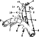
Figure 1. Scheme of the subclavian artery (A, B, C—first, second, and third parts of the subclavian artery): 1—common carotid artery; 2—inferior thyroid artery; 3—vertebral artery; 4—brachiocephalic artery; 5—internal mammary artery; 6—supreme intercostal artery; 7—first rib; 8—subclavian artery; 9—deep cervical artery; 10—transverse scapular artery; 11—transverse cervical artery; 12—ascending cervical artery.
passes anterior scalene muscle and again merges into a single trunk, forming a ring around the muscle; in the presence of cervical ribs, the subclavian artery loops over one of them—the lower. Each of the subclavian arteries gives off the following branches (see Blood vessels): medial to the anterior scalene muscle arises the vertebral artery (a. vertebralis) (Fig. 1), the internal mammary artery (a. mammaria interna), the thyrocervical trunk (truncus thyreo-cervicalis). Posterior to the anterior scalene muscle arises the costocervical trunk (truncus costo-cervicalis). In the space between the scalene muscles emerges the transverse cervical artery (a. transversa colli). The vertebral artery originates from the upper circumference of the subclavian artery and ascends behind the common carotid artery, enters the aperture of the transverse process of the sixth cervical vertebra and goes upward through similar apertures of the fifth to first cervical vertebrae. Between the atlas and the occipital bone, the vertebral artery pierces the dura mater; in the cranial cavity, the vertebral artery merges with its fellow to form the unpaired basilar artery (a. basilaris) (see Brain). Along its course, the vertebral artery gives off numerous branches. Variations of the vertebral artery: it arises from the common carotid artery, from the inferior thyroid artery, from the brachiocephalic artery, and even from the aorta; the vertebral artery may begin with two or even three trunks, subsequently merging into a single main trunk, while it may arise either from a single artery (subclavian) or as a single trunk from the subclavian artery, and the second from the aorta or from the inferior thyroid artery; its three trunks may originate from the subclavian artery, the inferior thyroid artery, and the aorta (Quain, Dubreil). With a difference in caliber, the left vertebral artery usually prevails over the right. Instead of penetrating into the transverse process of the sixth cervical vertebra, the vertebral artery enters the transverse process of the seventh or fifth, fourth, third, and even second cervical vertebra; having originated from the aorta behind the left subclavian artery, it may run behind the esophagus and enter the right transverse process of the sixth cervical vertebra. The thyrocervical trunk (truncus thyreo-cervicalis) begins on the anterior circumference of the subclavian artery, divides into: 1) the inferior thyroid artery (a. thyreoid. inf.), running to the posterior surface of the thyroid gland; along the way, the inferior thyroid artery gives off branches to the pharynx, esophagus, trachea, and larynx; 2) the ascending cervical artery (a. cervicalis ascendens); 3) the superficial cervical artery (a. cervicalis superf.) and 4) the transverse scapular artery (a. transversa scapulae); the latter, anastomosing with the transverse cervical artery (a. transversa colli), forms a collateral pathway on the posterior surface of the scapula from the subclavian artery to the axillary artery (see Shoulder, Fig. 7 and 8). The internal mammary artery (a. mammaria interna) emerges from the lower circumference of the subclavian artery, goes downwards and inwards SUBCLAVIA ARTERIA, VENA along the pleura, behind the subclavian vein, runs behind the cartilages of the first to eighth ribs at a distance of about 1 cm from the edge of the sternum (see Carotis arteria, Fig. 1 and 2). Initially, the internal mammary artery obliquely crosses the phrenic nerve, passing behind it and the brachiocephalic vein; on its path in the thoracic cavity, the artery is covered by the pleura and the transversus thoracis muscle; at the level of the sixth intercostal space, the artery divides into two terminal branches: the musculophrenic artery (a. musculo-phrenica) and the superior epigastric artery (a. epigastrica sup.); the latter anastomoses with the inferior epigastric artery (a. epigastrica inf.) (Fig. 2) and forms a paired pathway parallel to the aorta. Branches of the internal mammary artery: anterior mediastinal arteries (aa. mediastinales ant.), thymic arteries (aa. thymicae), bronchial arteries (rami bronchiales), the pericardiacophrenic artery (a. pericardiaco-phrenica); sternal branches (rami sternales), perforating branches (rami perforantes); one of these branches, emerging usually through the second intercostal space, is developed more strongly than the others, goes downward and branches in the mammary gland (rami mammarii); intercostal branches (rami intercostales), two for each intercostal space, anastomose with the intercostal arteries. Variations of the internal mammary artery: sometimes it arises from the subclavian artery by a common trunk with one of its branches—with the inferior thyroid artery or with the transverse scapular artery; very rarely from the aorta, the brachiocephalic artery, and even from the axillary artery; sometimes on one or both sides it is accompanied by an accessory mammary artery (a. mammaria accessoria); cases of transverse anastomosis between the right and left mammary arteries behind the xiphoid process are encountered. A case of lateral mammary artery (a. mammaria lat.) has been described—an additional branch of the subclavian artery, separating from the latter between the scalene muscles and running along the inner surface of the lateral wall of the chest cavity (Testut). The costocervical trunk (tr. thyreo-cervicalis) emerges from the posterior circumference of the subclavian artery, divides into the deep cervical artery (a. cervicalis profunda) and the supreme intercostal artery (a. intercostalis suprema). Variations of the latter: may be absent, may give off the internal mammary artery.
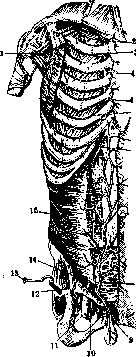
Figure 2. Internal mammary artery and epigastric artery: 1—axillary artery and vein; 2—internal mammary artery; 3—intercostal artery; 4, 5 and 6—branches of the internal mammary artery; 7—navel; 8—inferior epigastric artery, anastomosing above with the superior epigastric artery (branch continuing the internal mammary artery downward); 9—spermatic cord and testicle; 10—femoral vein; 11—femoral artery; 12—reflected outward aponeurosis of the external oblique muscle; 13—sartorius muscle; 14—external iliac artery; 15—transversus abdominis muscle.
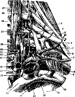
Figure 3. Topography of the left subclavian artery medial to the anterior scalene muscle (m. scalenus anticus): 1—posterior scalene muscle; 2 and 4—pleurovertebral ligament; 3—brachial plexus; 5—transverse cervical artery; 6—posterior scalene muscle; 7—transverse scapular artery; 8—phrenic nerve; 9—subclavian artery; 10—subclavian vein; 11—first rib; 12—thoracic duct; 13—cupula of the pleura; 14—internal mammary artery; 15—left brachiocephalic vein; 16—left internal jugular (common) vein; 17—vagus nerve; 18—left common carotid artery; 19—inferior thyroid artery; 20—trachea; 21—recurrent nerve (inferior laryngeal nerve); 22—esophagus; 23—inferior cervical sympathetic ganglion; 24—deep cervical artery; 25—vertebral artery; 26—carotid tubercle; 27—longus colli muscle.
neal artery of the neck begins from the upper circumference of the subclavian artery, passes through the brachial plexus; its descending branch runs along the vertebral border of the scapula between the rhomboid muscles and the serratus posterior superior. Variations of the transverse cervical artery: sometimes the artery passes not through the brachial plexus, but anterior to it; the transverse cervical artery may pierce the posterior scalene muscle. Topographically, the subclavian artery is divided, as stated, into three parts: the first part, located in the sternocleidomastoid region on the cupula of the pleura, is covered by the skin, sternocleidomastoid muscle, sternohyoid and sternothyroid muscles, and the first and second cervical fasciae. The relations of the subclavian artery to other vessels and nerves of this region differ on the right and left (Fig. 3 and 4). On the right, anterior to the subclavian artery is the junction of the internal jugular vein and the subclavian vein, the vagus and phrenic nerves; the latter cross the subclavian artery perpendicular to its direction; posteriorly—the inferior laryngeal nerve (n. laryngeus inf.); medially—the common carotid artery. On the left (Fig. 5), anterior to the origin lies the beginning of the left brachiocephalic vein and the peripheral segment of the thoracic duct. The left subclavian artery is not crossed by nerves, but runs at some distance from them by virtue of its almost vertical path. The subclavian artery lies in this part at a depth of 4–6 cm from the skin surface, gives off many branches, is surrounded by bulky venous trunks, therefore SUBCLAVIA ARTERIA, VENA ligation of this part of the subclavian artery is difficult and dangerous, and the surgeon has to resort to resection of the clavicle in order to facilitate the intervention. The second part of the subclavian artery on the right and left passes behind the anterior scalene muscle, which separates the artery from the subclavian vein anteriorly; by its lower circumference, the subclavian artery touches the first rib; for exposure
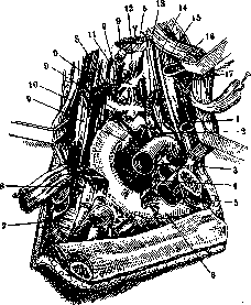
Figure 4. Right supraclavicular fossa—deep layers: 1—inferior thyroid artery; 2—vertebral artery; 3—internal mammary artery; 4—apex of the lung covered by pleura (cupula of the pleura); 5—anterior scalene muscle; 6—first rib; 7—first digit of the anterior serratus muscle; 8—posterior scalene muscle; 9—five roots of the brachial plexus, severed at its exit from the spine; 10—deep cervical artery; 11—carotid tubercle; 12—intertransverse muscle; 13—sympathetic trunk and its middle ganglion; 14—common carotid artery; 15—vagus nerve; 16—internal jugular (common) vein; 17—thyroid gland.
To reach it here, the anterior scalene muscle must be retracted inward or its lateral border incised, remembering that the phrenic nerve extends along the anterior surface of this muscle. Posterior and superior to the artery lies the brachial plexus (plexus brachialis). The third part of the subclavian artery occupies a more superficial position and is more accessible for intervention than the first two parts; the subclavian artery lies on the first rib, immediately posterior and lateral to the attachment site of the anterior scalene muscle to the first rib, i.e., from the scalene tubercle of Lisfranc (see Ribs); at this place the subclavian artery can be compressed against the first rib. Anterior to the subclavian artery pass the subclavian vein, covered by the clavicle, the transverse scapular artery, which also hides behind the upper border of the clavicle, and the peripheral part of the external jugular vein (Fig. 5). Slightly superior to the subclavian artery runs the omohyoid muscle. Here, upon approaching the artery, one must pass through the skin, the platysma, the first (superficial) and middle aponeuroses, and in the cellular tissue of the supraclavicular fossa proper (see Supraclavicular fossa) lies the subclavian artery. Delicate accompanying veins (venae comitantes) run with the subclavian artery. The subclavian vein (v. subclavia) continues upward from the axillary vein, from the subclavius muscle to the sternoclavicular articulation, where it joins the internal jugular vein. The subclavian veins run identically on both sides, both of the same length. In front of them is the clavicle, behind them is the subclavian artery, separated in one place by the anterior scalene muscle (see above), below is the first rib, in the lateral and medial parts is the pleural dome, above is the layer of the supraclavicular fossa and the sternocleidomastoid region. The subclavian vein is embraced by the middle aponeurosis of the neck, which is firmly fused with its walls; by virtue of this, the vein does not collapse when incised, but gapes, which promotes the occurrence of air embolism; the aponeurosis fixes the vein to the clavicle. At the beginning and end of the vein are valves that prevent the backflow of blood from the brachiocephalic vein to the subclavian vein. The subclavian vein has no constant branches; most frequently the highest intercostal vein (Testut) empties into it, more rarely the transverse cervical and scapular veins, which more often open into the external jugular vein (Spalteholz). The junction of the subclavian vein with the internal jugular vein is named Pirogov's venous angle (angulus venosus). Opening into this angle (frequently) are: the external jugular vein, the thoracic duct on the left, and the right lymphatic duct (Fig. 6). From Fig. 6 a, b, c it is seen that only in "a" is the lymphatic vessel developed according to the trunk type (correctly), in "b" this is expressed only partially, since the bronchomediastinal trunk opens separately, and a common lymphatic trunk, or, as Testut calls it, a lymphatic vein, does not exist, but there are three trunks opening independently into the vein. Usually the right lymphatic duct opens into the right venous angle. Variations of the subclavian vein: sometimes the vein runs above the subclavian artery; the vein may run behind the anterior scalene muscle together with the subclavian artery or alone, while the subclavian artery occupies the position of the vein
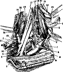
Figure 5. Vessels and nerves of the supraclavicular fossa (right side): 1 - m. omo-hyoideus (transected); 2 - a. thyreoidea int.; 3 - a. vertebralis; 4 - n. supraclavicularis; 5 - v. jugularis ext.; 6 - first rib; 7 - m. pectoralis major with platysma; 8 - cellular tissue of the axilla; 9 - a. subclavia; 10 - m. subclavius; 11 - clavicle (sawed through); 12 - a. transversa scapulae; 13 - a. transversa colli; 14 - m. trapezoideus; 15 - m. scalenus post.; 16 - plexus brachialis; 17 - m. scalenus ant.; 18 - n. phrenicus; 19 - a. carotis com.; 20 - n. vagus; 21 - v. jugularis int. (communis); 22 - sternocleidomastoid muscle; 23 - ansa hypoglossi.
in front of this muscle; in one case Luschka observed the vein divided into two branches - one in front, the other behind the anterior scalene muscle. Cases of the cephalic vein emptying into the subclavian vein are known. The internal mammary veins initially accompany each like-named artery in pairs, then at the level of the II-III intercostal space each merges into a single trunk and empties: the right anteriorly into the junction point of both brachiocephalic veins, the left into the left brachiocephalic vein; along the path, the sternal, anterior intercostal, and mediastinal veins empty into them. The internal mammary veins anastomose: among themselves - by means of sternal veins; with the posterior
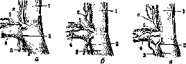
Figure 6. Variants of lymphatic vessels opening into the vein: 1 - v. jugularis int.; 2 - truncus lymphaticus broncho-mediastinalis; 3 - v. anonyma; 4 - v. subclavia; 5 - truncus lymphaticus subclavius; 6 - truncus (lymphaticus) jugularis.
intercostals - through the anterior intercostals, and with the subcutaneous veins of the abdomen; by these latter anastomoses, a continuous collateral venous pathway of the abdomen and chest is established, which provides a bypass for the venous blood flow in case of obstruction of the portal system and the inferior vena cava system. Clinic. The subclavian artery and subclavian vein may be wounded in non-firearm fractures of the clavicle. Out of 11 cases collected by Gallois and Pillet, the subclavian vein was damaged in 6 and the subclavian artery in 5; more frequently, wounds of the named vessels are inflicted by firearms or piercing weapons. At the same time, the pleura and the brachial plexus are often wounded. Wounds of the subclavian artery constitute 9.6% of all vascular wounds and 0.05% of the total number of wounds. Wounds of the subclavian vein are very rare - 14% of all vein wounds; the rarity of wounds of the subclavian vein observed in clinics is explained by the extreme severity of wounds of this vessel; the wounded die from air embolism and hemorrhage before they fall into the hands of the surgeon. Injuries to the subclavian artery are treated by ligation and vascular suture. Ligation of the artery in the first part yields 83% mortality, in the second - 44%, and in the third - 31.5% (Mikhailov). A wounded subclavian vein should preferably be ligated, since the operation is simpler and there is less risk of air embolism during the operation itself. Five cases of resection and circular suture of the vein with a good outcome have been described (Moskalenko). To expose the subclavian artery for surgical purposes, the most easily accessible section of the artery is its third part. The patient lies on their back with a bolster under the shoulders. The incision line runs parallel to and 1 fingerbreadth above the clavicle from the anterior border of the trapezius muscle to the sternocleidomastoid muscle. The skin with subcutaneous tissue, superficial fascia, platysma, and beneath it the supraclavicular nerves are incised; in the anterior angle, the posterior jugular vein is visible; the first aponeurosis is incised along a grooved probe; deeper lies the middle aponeurosis of the neck and within it the omohyoid muscle; the aponeurosis is opened between this muscle and the clavicle; the brachial plexus appears; inwardly, the lateral border of the anterior scalene muscle is felt with a finger; below on the first rib, the pulsating subclavian artery is palpated; the subclavian vein is retracted anteriorly with a finger; the carefully isolated subclavian artery (beneath it is the pleural dome) is encircled with a ligature (silk) thread from the side of the nerves, i.e., from top to back and down to front; layered sutures are applied to the wound. Suture of the subclavian artery has been applied in only a few cases so far, and therefore, due to greater simplicity, ligation remains the operation of choice (Moskalenko). To expose the subclavian artery, Krymov proposed temporarily resecting a piece of the clavicle, and Morozova proposed disconnecting the sternoclavicular articulation. Aneurysms of the subclavian artery are observed more often than aneurysms of other arteries of this region, occurring in men 16 times more frequently than in women. The frequency of aneurysms of the right subclavian artery is determined at 63%, of the left at 35.3%, and bilateral at 1.7% of the total number of subclavian artery aneurysms. The best method for the right side is Antyllus' method, and for the left - Brasdor-Wardrop. Mortality in the surgical treatment of subclavian artery aneurysms, according to Haberer, is 14%; the cause of death is most often hemorrhage from the ligation site, pyemia, or septicemia. Arteriovenous aneurysms of the subclavian vessels are very rare (7 cases, Moskalenko).
Related articles
Mentioned in
Cite this page
“Stupor.” Soviet Medical Encyclopedia. English translation of Bolshaya Meditsinskaya Entsiklopediya, 1st ed. (Moscow, 1928–1936), ed. N. A. Semashko. https://sovietmedicalencyclopedia.pages.dev/article/stupor/