Apoplexy
Historical document, translated for reference. It reflects medical knowledge of the 1920s–30s and is not medical advice.
Summary
Apoplexy refers to sudden paralysis of the central nervous system, primarily caused by acute circulatory disorders such as hemorrhage, thrombosis, or embolism. The article describes symptoms, etiology, pathological anatomy, and histological findings of cerebral apoplexy as understood in the 1930s.
Encyclopedia article (1928–1936)
APoplexy, apoplexia (from Greek apo-plesso-I stun, overwhelm), stupor (paralysis of the body or its parts), otherwise apoplectic stroke or attack; in common usage, this word denotes a sudden paralysis of the central nervous system, depending mainly on acute circulatory disorders in it (hemorrhages, thrombosis or embolism of vessels, etc.). The concept of A. is applied not only to the nervous system; one speaks, e.g., of A. of the uterus, ovaries, lungs, when one wants to indicate acute hemorrhages into their parenchyma or mucous membrane. Apoplexy of the brain (apoplexia cerebri) manifests as sudden loss of consciousness with falling to the ground, loss of voluntary movements and sensitivity, in the absence, at least, of sharply expressed phenomena of cardiac failure (distinction from syncope). The phenomena of stroke may also be expressed in an abortive form. Instead of complete loss of consciousness, only one or another degree of stupor is often observed, with the preservation of the ability to perceive and carry out, though with difficulty and not immediately, elementary commands, and to react weakly to external irritations. The disorders of voluntary movements and sensitivity may also be incomplete. The concept of cerebral apoplexy should be extended in the sense that the suddenness of onset is not its necessary sign: there are cases of so-called progressive A., in which a comatose state develops gradually. The etiology of A. includes all those factors that lead to diseases of the vascular apparatus: age, chronic intoxications, infections and among them especially syphilis, dyscrasias (mainly gouty diathesis), diseases of the heart valves (embolism). Specifically for hemorrhage into the brain, haemorrhagia cerebri, a special apoplastic habitus, habitus apoplecticus, is characteristic: elderly, well-fed subjects with a short neck, with frequent rushes of blood to the head, usually sanguine temperament, immoderate in food and drink. In younger age, the case usually concerns either syphilitic vascular lesions (mainly cerebral thrombosis) or a heart defect (which causes cerebral embolism). Heredity plays an undeniable role in cerebral hemorrhage. In terms of sex, no significant differences are noted; a certain predominance in the male sex is explained mainly by the greater frequency of specific infection in men. Also important are factors leading to damage to already pathologically altered vessels and capable of causing an increase in blood pressure: strong emotional excitement, especially anger; physical strain-lifting heavy weights, strain during defecation (constipation), during coitus, during vomiting, etc.; high ambient temperature (hot bath); intoxication; spontaneous rushes of blood to the head, appearing without visible cause. Pathological anatomy. At autopsy, either hemorrhage or softening of the brain is found. Often such foci are located symmetrically on both sides. The hemorrhagic focus can reach enormous sizes (see illustration), the hemorrhage can rupture into the ventricles and fill their cavity, and even rupture onto the surface of the hemisphere. Histological research reveals, as the most frequent cause of cerebral apoplexy, miliary aneurysms (Charcot), i.e., expansions of small intracerebral arteries in the form of ampullae (0.1-1 mm in diameter), arising as a result of disease of the muscular coat of the vessel and periarteritis. Subsequently, the focus of hemorrhage undergoes typical transformations, and as a result, a cyst with serous fluid usually forms, surrounded by a capsule from reactively proliferated glia. In rarer cases, the process of resorption of the clot and destroyed brain tissue leads to the formation of a scar. The path-anatomical picture of softening of the brain (endarteritis obliterans, thrombosis, embolism) also varies greatly depending on the duration. In the initial stage, during the first day, softening is generally found with some difficulty. Impregnation with serous fluid, however, even during this period gives the focus of softening
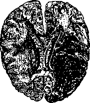
a certain consistency. In the initial stage, during the first day, softening is generally found with some difficulty. Impregnation with serous fluid, however, even during this period gives the focus of softening
a different color and consistency compared to the surrounding tissue. Subsequently, the focus softens more and more due to fatty degeneration of the necrotized tissue; here, infiltration with blood also often occurs—red softening. The breakdown of pigment gradually leads to a change in color and the formation of the so-called yellow softening. The end result is either a scar or a cyst. Clinical picture. Stroke may be preceded by warning signs: flushes of blood to the head, headache, noise in the ears, dizziness. In other cases, it occurs without any warning signs. Often, the absence of any visible external causes is noted; stroke strikes a person during complete rest, often at night, during sleep. In a fully developed cerebral apoplexy, consciousness is completely lost, the patient is in a state of deep coma, does not react to external irritations, and is completely immobile. Breathing is stertorous, the pulse is often tense and slowed, there is incontinence or, less commonly, retention of urine. Temperature is slightly lowered at the beginning, then quickly returns to normal. In severe cases, hyperthermia may occur, up to 40-41°, with significant tachycardia and frequent respiration (fatal cases). Pupils are somewhat dilated, often not reacting to light. Corneal and conjunctival reflexes and skin reflexes are absent. Often, at the very beginning of the apoplectic coma, Babinski's reflex can be detected on the side of the paralysis. Tendon reflexes are usually absent, with general hypotonia. The latter is uneven on both sides and more pronounced on the side opposite the focus of hemorrhage. Determining the side of hemiplegia is also helped by the inflation of the affected cheek during respiration ('the patient is smoking a pipe'), as well as the state of the reflexes, which are particularly diminished on the paralyzed side, and the appearance of Babinski's reflex on the same side. In rarer cases, instead of hypotonia, contractures are noted from the very beginning—so-called early contractures (the 'golmentonic' symptom complex of Davidenkov). They occur in cases with particularly extensive damage and with rupture of the spilled blood into the ventricles. Among trophic disorders, acute bedsore, decubitus acutus, should be mentioned, which has a very unfavorable course, appearing soon after the stroke and only in severe cases. Among vasomotor disorders, increased blood pressure on the affected side and increased skin temperature here are noted. In the initial period, protein often appears in the urine, sometimes sugar. Besides complete apoplectic strokes, abortive ones also occur, in which consciousness is only somewhat clouded or the condition is limited to simple dizziness. The form of stroke depends on the size of the focus, the nature of the process, and its localization. Thrombotic processes often do not cause loss of consciousness, and paralytic phenomena develop only gradually. With localization of the process outside the cerebral hemispheres (cerebellum, brainstem), changes in consciousness are less pronounced than with localization in the hemispheres. The diagnosis of apoplectic stroke can meet with considerable difficulties in the absence of an accurate history. Differentiation must be made from simple fainting, from intoxication comas (uremia, diabetes, intoxication, morphine poisoning, etc.), from apoplecticoid strokes in progressive paralysis, in disseminated sclerosis, from certain conditions in hysteria, in epilepsy. Syncope is characterized by a sharp decline in cardiac activity, a thread-like pulse. Intoxicative coma states differ from cerebral apoplexy by specific signs: uremic and diabetic comas—by corresponding changes in the urine, uremia, in addition, by the presence of edema and retinitis albuminurica, and diabetic coma—by the characteristic smell of acetone; severe alcohol poisoning—by a specific odor from the mouth and vomit; morphine poisoning—by sharp narrowing of the pupils, etc. Apoplecticoid strokes in tabes and disseminated sclerosis are essentially completely similar to cerebral apoplexy; differentiation is possible only on the basis of history and further course. The same should be said about apoplectic strokes in brain tumors (hemorrhage into the tumor substance). Pseudocomatose states in hysteria are characterized by the absence of organic symptoms (preservation of pupillary, tendon, and skin reflexes, absence of Babinski's reflex, etc.). For epilepsy, the following are characteristic: sharp pallor of the face, absence of hemi-symptoms, rapid complete return to normal, and history. The next task of diagnosis is to recognize the nature of the process that caused the stroke: it is important for prognosis and treatment to know whether it is a matter of hemorrhage, thrombosis, or embolism; further—whether there is an arteriosclerotic process or syphilitic vascular lesion. The age of the patient, the course of the stroke, the degree of change in consciousness, the presence of specific infection, heart defect, signs of general arteriosclerosis help in solving these questions. In cerebral hemorrhage, the face is usually intensely red, the pulse is tense and slowed, often the carotid arteries pulsate. Redness of the face is usually not present in thrombotic stroke. Syphilis much more often leads to thrombosis than to hemorrhage. With cortical localizations, the matter is more often about thrombosis, whereas hemorrhage usually gives a capsular localization. Prognosis depends on the localization and size of the focus. With localization of the process in the brainstem part of cerebral apoplexy, it almost always leads to a fatal outcome. The spread of paralysis to both sides is always a bad sign. A poor prognosis is given by cases with rupture of hemorrhage into the ventricles and early contractures. Large hyperthermia, significant decrease in temperature, presence of early bedsore, and duration of the comatose state (if it lasts more than a day) should also be considered as bad signs. If this comatose stage is passed, the immediate danger to life decreases, and the question of the prognosis of the remaining phenomena of deficit comes to the fore. The prognosis is determined by the course of the disease in the first months after stroke; if improvement in the first 1½-2 months is absent or minimal, one cannot count on the smoothing of paralytic phenomena. On the contrary, in cases where improvement occurs already in the first days, complete recovery is possible. Prevention and therapy. As preventive measures, all means that prevent the development of arteriosclerosis or are intended to cure already developed vascular sclerosis should be mentioned first. In this respect, elderly people and, mainly, people with habitus apoplecticus should be kept in mind. The strictest regime in regard to diet and lifestyle is necessary here: strict prohibition of alcohol in all forms; prohibition of smoking; moderate food, diet mainly without meat, without any spices, mainly a milk-vegetable regime; sufficient time outdoors, reasonable exercise (but in no way carried to fatigue); generally, elimination of causes of fatigue, both physical and mental. Great attention should be paid to the elimination of all kinds of emotional shocks, as they can serve as a direct cause of rupture of intracerebral vessels and lead to apoplexy; other factors leading to flushes of blood to the head should also be carefully eliminated—staying in high temperature (especially hot bath), etc.; proper sleep, only at night, prohibition of daytime sleep, especially sleep after meals; elimination of constipation. Of medicinal preparations, iodides, Trunbechev's serum, etc., are used, whose effect, unfortunately, gives only a very modest effect. During the stroke itself, rest and absence of irritations are primarily necessary. With redness of the face—counter-irritants: venesection, leeches, laxatives, ice on the head. In case of certain signs of softening of the brain, on the contrary—use of cardiac agents (camphor, caffeine). Treatment of the consequences of stroke is mainly reduced to symptomatic therapy, i.e., to treatment of paralysis and contractures by massage and gymnastics, electro- and hydrotherapy, and sometimes by surgical measures. In the presence of syphilitic endoarteritis, of course, energetic specific therapy should be carried out. (Apoplexy of other organs—see the respective organs.)
Related articles
Mentioned in
Cite this page
“Apoplexy.” Soviet Medical Encyclopedia. English translation of Bolshaya Meditsinskaya Entsiklopediya, 1st ed. (Moscow, 1928–1936), ed. N. A. Semashko. https://sovietmedicalencyclopedia.pages.dev/article/apoplexy/