Cerebellopontine Angle
Historical document, translated for reference. It reflects medical knowledge of the 1920s–30s and is not medical advice.
Summary
The cerebellopontine angle is a critical anatomical region between the cerebellum, pons, and medulla oblongata. This article discusses its clinical significance, common pathologies including acoustic neuromas, and surgical approaches for treating tumors in this region.
Encyclopedia article (1928–1936)
CEREBELLOPONTINE ANGLE (Klein-hirnbrückenwinkel, angle ponto-cerebelleuse, according to some angle ponto-bulbo-cerebelleuse) occupies a unique place in neuropathology, neurohistopathology, and neurosurgery. This term designates the angle between the cerebellum, medulla oblongata, and pons. On the cerebellar side, the angle corresponds to the extreme anterior portion of the lower surface of the organ, near the exit of the peduncle of the pons. Through this space in the frontal direction and almost horizontally enters a bundle consisting of nn. VII, VIII, and intermedius. Immediately behind and medially in the lateral diverticulum of the IV ventricle is located plex. chorioideus ventr. IV; * Cushing in his cases found lesions of nn. VI and VII in 2/3 of the total number. Geymanovich did not see such a frequency of lesion of n. VI. Brain symptoms are an early sign of tumors of the cerebellopontine angle, so processes in the pyramidal bone under certain circumstances must be differentiated from
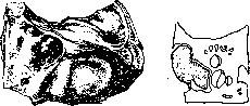
Figure 1.
Figure 2.
Figure 1. Tumor of the auditory nerve, growing into the canal. Figure 2. Relationship of the auditory nerve tumor to the canal. early stages of tumors of the cerebellopontine angle. The specific tumor of the angle is fibroma, resp. neurofibroma of n. VIII (fibrosarcoma, etc.). The history of the question of the symptomatology and pathology of the angle is connected with the history of tumors of this nerve (the first description was apparently made by Sandifort in 1777). Tumor of the auditory nerve (fig. 1 - 3), Acusticustumor, may be a manifestation of general neurofibromatosis. On this basis, both bilateral tumors of the angle and a combination of tumor of the angle with neurofibromatosis of other areas can develop. As for tumors of the area of the cerebellopontine angle, not originating from the auditory nerve, they may belong either to diffuse or limited tumors of the base (meninges, bone, in particular cholesteatomas of the base), or to tumors originating from the brain substance itself [see separate table (art. 71 - 72), fig. 3]. Other formations that can give similar symptom complexes may be aneurysms and hematomas, parasitic formations (cysticercus, echinococcus), abscesses (mainly ear), syphilis, rarely tbc. Syphilis
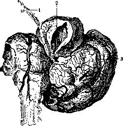
Figure 3. Tumor of the auditory nerve (1) with cystic (2) degeneration; 3-cerebellum.
can manifest here in the gummatous or serous-cystic form (Akopodzhanyants, Robustov), sometimes on both sides (bilateral syphilitic cyst, simulating a bilateral tumor of the cerebellopontine angle - Geymanovich). Changes in the cerebrospinal fluid in a typical tumor of the auditory nerve (at least in the early stage) amount to hyperalbuminosis (extracerebral compression), usually without cellular elements, and with great pressure - to xanthochromia. Hyperalbuminosis, phenomena from the side of n. acustici and hypesthesia of the cornea (especially if this is accompanied by cerebellar phenomena) serve as early symptoms of tumors of the cerebellopontine angle. Radiologically, expansion of the internal auditory canal is detected, but it
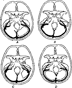
Figure 4. Dotted line indicates the position of the bone openings in relation to the sinuses: A - with unilateral intervention; B - through the labyrinth; C - combined and D - with bilateral intervention.
can also occur narrowing of it due to reactive hyperostosis.
A. Geymanovich. Surgery of the cerebellopontine angle. When establishing the diagnosis of a pontocerebellar tumor, the operation is performed as in a cerebellar tumor or through the labyrinth (fig. 4). The latter is recommended by otologists. Before going deep into the posterior cranial fossa (see Cerebellum, surgery), it is necessary to carefully examine the condition of the cerebellar hemispheres - compare their size, position, consistency, blood supply and pay attention to the relationship of the medulla oblongata to the foramen occipit. magnum and the degree of protrusion of the hemispheres from the opening of the dura mater. Too significant protrusion of the hemispheres rather indicates an intracerebellar tumor than a ponto-cerebellar tumor. In the absence of sharp changes in the hemispheres, examination of the cerebellopontine angle is performed. The hemisphere is usually deviated inward and upward with a large spatula (fig. 5). Often in the cerebellopontine angle serous cysts are found, in front of which are located true tumors. Finding tumors sometimes presents considerable difficulties, and it is necessary to significantly shift the cerebellar hemisphere, which is very undesirable. When the tumor is found, its removal is performed either by enucleating it with the finger
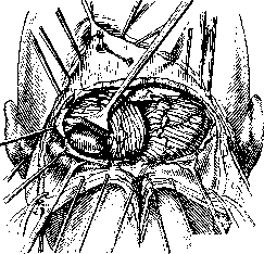
Ш\
Figure 5. (a method that no longer finds supporters) or by careful removal in parts. First, the capsule of the tumor is opened (fig. 6), then with a spoon the tumor is removed in parts (fig. 7 and 8), which in most cases is easily accomplished due to its fatty degeneration. During and after removal, there is significant bleeding from the walls of the capsule - it is stopped either by tamponade or by some fixing fluids (formalin solution, Zenger's fluid). The second method, which once attracted the attention of surgeons, - "the method of suctioning out the tumor (Krause; 1903). In this method, a glass tube is brought to the surface of the tumor, it is sucked out and pulled out; at the same time the capsule and neighboring healthy tissues and vascular bundles, which are recommended to be ligated, are pulled out. The latter is very difficult due to the narrowness, and usually during suctioning the matter is more about tearing, which is accompanied by significant bleeding. The third method of access to tumors of the cerebellopontine angle - through incision of the cerebellar hemisphere or by its removal (Borchardt, Frazier). Although these operations are tolerated by patients, and functional disorders are not so severe, they have found few followers, since in most cases such trauma is unnecessary. In operations in the area of the cerebellopontine angle, especially when removing tumors of n. acustici, the preservation of nn. acusticus, facialis is extremely important, and in neoplasms of the area of the cerebellopontine angle in the true
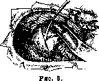
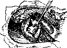
Figure 7.
sense of the word, one has to think about n. trigeminus and n. abducens. The preservation of these nerves is not always possible, moreover, it should be noted that the listed nerves are sometimes themselves affected by the pathological process (see above). Along with neoplasms of n. acustici and neoplasm of tissues in the area of the cerebellopontine angle, during surgical intervention, one most often finds serous limited meningitis, syphilitic and tuberculous meningitis. These diseases in terms of symptomatology in some cases are difficult to distinguish from true neoplasms, only during the operation the correct diagnosis is established. Recently in the literature one increasingly encounters reports by various authors indicating progress in topographical and functional diagnosis in relation to tumors of the cerebellopontine angle, but errors are still inevitable: a number of processes have been described that led to erroneous diagnosis: Meniere's disease, Bell's palsy, neuralgia of the facial nerve, disseminated sclerosis, multiple neuritis of cranial nerves, neuralgia of occipital nerves, torticollis, bulbar progressive paralysis, bulbar syndrome, tabes, tumors of the quadrigeminal bodies, psammoma of the third ventricle, tumors of the temporal lobe, tumor of Gasser's node and finally tumor of the frontal lobe. The surgeon must know about the possibility of such processes, so that with negative results of the examination, he can change the course of the operation and use the operation for diagnostic purposes. - Results of surgical intervention. Mortality in surgical interventions is quite high. Until the beginning of the last decade, it fluctuated between 68 - 75 - 86.6% (statistics of Tooth, Henschen, Krause, Eiselsberg). During the last 15 years Cushing published a series of reports, where it is indicated that the mortality rate successively fell from 40 to 33.3, 30, 24, 20 and to 11.1%. As for the functional results, the figures of recovery and significant improvements are still small.
N. Burdenko.
535 VI. Surgery of the M..................562 Cerebellum, 4 small brain, cerebellum, cervelet, Kleinhirn, one of the constituent parts of the brain, in charge of coordination and regulation of all movements, both voluntary and reflex. M. - an unpaired, symmetrical organ; it lies in the posterior cranial fossa (see Craniocerebral topography); it forms the posterior wall, or roof of the IV ventricle; it is located behind the medulla oblongata and Varolius' bridge, below the corpora quadrigemina, under the posterior lobes of the cerebral hemispheres, from which it is separated by a process of the dura mater (see), the so-called tentorium, or tent (tentorium cerebelli) (fig. 1 and 18). I. Phylogenetic development of the cerebellum. The cerebellum in various classes of animals develops in parallel with the entire central nervous system: rudimentary in lower animals, it reaches its maximum
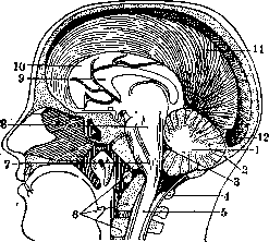
Figure 1. Sagittal section of the human head: 1-cerebellum; 2-protuberantia occipitalis interna; 3-IV ventricle; 4 and 6-upper cervical vertebrae; 5-spinal cord; 7-medulla oblongata; 8-Varolius' bridge; 9-corpus callosum; 10-frontal lobe of the brain; 11-falx cerebri major durae matris; 12-confluens sinuum.
in mammals. Its histological structure in animals of all classes, with the exception of the most primitive, is essentially the same; it consists of gray and white matter; the gray matter, forming its cortex, is divided into three layers (molecular, granular, and between them one row of large Purkinje cells) and covers the entire surface of the cerebellum. In addition to the cortex, accumulations of gray matter are also found in the deep parts of the cerebellum, forming subcortical nuclei. Under the cortex lies the white matter, formed by various functionally related fibers-associative, commissural, and projectional.-In amphioxus, in myxines, which live as parasites in the body of other fish or on stones, attached to them, the cerebellum is absent.-The most primitive cerebellum is found in
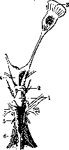
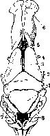
Figure 2.
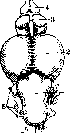
Figure 3.
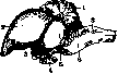
Figure 2. Brain of a shark: 1-cerebellum; 2-corpus quadrigeminum; 3-lobus olfactorius; 4-interbrain; 5-medulla oblongata; 6-n. vagus. Figure 3. Brain of a salamander: 1-cerebellum; 2-n. trochlearis; 3-corpus quadrigeminum; 4-n. opticus; 5-cerebral hemispheres; 6-lobus olfactorius; 7-medulla oblongata; 8-IV ventricle. m i n y (Petromyzon): it has the appearance of a plate, located posterior to the tectum opticum, and is as it were a continuation of the nuclei of the medulla oblongata (nuclei of nn. lateralis et vestibularis). This plate has characteristic cerebellar cells-granules; as for the Purkinje cells, there is no true layer of them, but there are individual large cells, scattered irregularly among the cells of the granular layer. This primitive cerebellum nevertheless has its afferent paths-nn. lateralis et vestibularis; the existence of a connection with the spinal cord is not proven, but there are fibers connecting it transversely with the hypothalamus, as well as with the midbrain. Efferent fibers originate from the large cells of the cerebellum and end in the motor area of the medulla oblongata and midbrain, partly crossing. In addition to the cells of the cortex, there is also an accumulation of gray matter at the border with the medulla oblongata - nucleus subcerebellaris; fibers from it after crossing go to the nuclei of nn. III. Figure 4. Brain of a salmon: 1-cerebellum; 2-corpus quadrigeminum; 3-cerebral hemispheres; 4-lobus olfactorius; 5-cranial nerves; 6-medulla oblongata. Thus the cerebellum here is not a simple plate, a simple fusion of the nuclei of the medulla oblongata, but an organ connected by afferent and efferent paths with surrounding formations. Cerebellum of f ish. Cerebellum of cartilaginous fish (Plagiostomata), such as shark, skate, is also located posterior to
M03J: tectum opticum; more complex in its structure, it has the appearance of an elongated process, attached by its base to the brain stem and free behind (figs. 2 and 4). In bony fish, its surface is smooth, in cartilaginous fish it is divided by grooves into convolutions, similar to the convolutions of higher vertebrates. In the cerebellum of fish, the middle part can be distinguished-corpus cerebelli and two lateral formations-auriculae cerebelli (cerebellar ears), which are homologous to the flocculi in mammals. In most fish, a transverse groove divides the corpus cerebelli into anterior and posterior parts, which in turn in large fish are subdivided into a number of convolutions: the larger the animal, the more convolutions and the more complex the pattern of the section; sometimes the division of corp. cerebelli into parts is not uniform-the anterior part is smaller than the posterior. The histological structure of the cerebellar cortex in higher fish is more differentiated, its connections are more complex. Inside the cerebellum is a ventricle, the place of which in higher animals is occupied by white matter, while in fish the white matter is poorly developed and has the appearance of a narrow strip located directly under the layer of Purkinje cells; it consists of centrifugal and centripetal fibers. There are no true subcortical nuclei in the cerebellum of fish, with the exception of a small accumulation of cells in the outer parts of the cerebellum (nucl. lateralis cerebellis, connecting with the nuclei of nn. vestibularis and lateralis). The connections of the cerebellum with surrounding formations are very rich-in the cerebellum end, in addition to part of the fibers of n. vestibularis, fibers from the spinal cord, partly crossing, then fibers from the medulla oblongata and from the midbrain. Of the efferent fibers, the following are described: 1) fibers going from Purkinje cells to motor centers of the medulla oblongata and spinal cord; a very small part of them ends near the nuclei of n. oculomotorii; 2) brachia conjunctiva, originating in nucl. lateralis cerebelli and partly in Purkinje cells; their fibers cross and end mostly in nucl. reticularis of the medulla oblongata, near the nuclei of n. III, the rest in the primitive nucl. ruber and in the hypothalamus; 3) tractus cerebello-vestibularis et cerebello-bulbaris, the fibers of which are coordinating between the cerebellum and the medulla oblongata. In amphibians (Amphibia) there is a difference in the structure of the cerebellum between tailed and tailless-in the latter it is better developed. Generally the cerebellum of amphibians is more primitive than that of fish; on the one hand it resembles the cerebellum of the lamprey-it has the appearance of a plate with thickenings on the sides, on the other hand-the cerebellum of fish due to the presence of auriculae cerebelli; in tailed amphibians (salamander) the latter are of very large size and adhere to the lateral walls of the midbrain, fusing with it (fig. 3); inside they contain a very wide cavity-recessus lateralis. Corpus cerebelli is located above the IV ventricle and consists symmetrically of two thickenings, connected by commissural fibers, among which small clusters of nerve cells can be seen. The molecular layer covers the entire surface of the cerebellum; in corpus cerebelli there are also large nerve cells-a rudiment of Purkinje cells; there are no granule cells. Of the afferent fibers, some end in corp. cerebelli, and some in auriculum cerebelli, therefore there is the same division as in fish. To the first fibers belong the spinocerebellar system (tractus spino-cerebellaris ventralis et dorsalis), fibers going from the hypothalamus (the connection is transverse) and tractus tecto-cerebellaris. In auricul. cerebelli end the fibers of nn. lateralis et vestibularis. The axons of large cells form the efferent fibers of the cerebellum (tr. cerebelli motorius), partly crossing and ending in the nuclei of nn. oculomotorii; another efferent system-brachium conjunctivum-after partial crossing ends in the tectum of the midbrain, where in higher animals the red nucleus is located. Finally in the cerebellum of amphibians there are own fibers connecting both halves of corp. cerebelli and forming a commissural system. - In tailless amphibians (frog) the difference in the structure of the cerebellum consists in the fact that corpus cerebelli is much better developed, while auriculae cerebelli remain poorly developed and at the same time the vestibular system is poorly developed. The cortex, covering
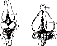
Figure 5.
Figure 6.
Figure 5. Brain of a snake: 1-cerebellum; 2-corpus quadrigeminum; 3-interbrain; 4-cerebral hemisphere; 5-medulla oblongata; 6-IV ventricle; 7-spinal cord. Figure 6. Brain of a rabbit: 2-cerebellar hemispheres; 2-vermis of the cerebellum; 3-corpus quadrigeminum; 4-cerebral hemisphere; 5-lobus olfactorius; 6-medulla oblongata. corp. cerebelli, consists of two layers-molecular and granular, between which Purkinje cells are regularly distributed. The connections of the cerebellum with surrounding formations are the same as in tailed amphibians. Cerebellum of reptiles (Reptilia) shows great diversity in size and shape. In snakes, due to the fact that they have only a trunk and no limbs, the cerebellum is very small and has the appearance of a thin transverse plate thrown across the IV ventricle (fig. 5); there are many Purkinje cells and they lie close to each other. The cerebellum of a lizard has a very peculiar structure in the form of a curved plate covering the posterior part of tecti optici; the molecular and granular layers are very well developed. In turtles and crocodiles the cerebellum has a well-developed shape with a cavity inside, partially filled with plexus choroideus; true cerebellar furrows
appear only in the crocodile. There are two transverse grooves that divide corp. cerebelli into three parts-lob. anterior, medius et posterior; the latter is connected with auricula cerebelli, which is very poorly developed, weaker than in fish. The cortex of the cerebellum is built according to the general principle. One can note an accumulation of cells that are homologous to the central nuclei of the cerebellum, namely the nuclei of the tectum and nuclei dentati. Of the afferent paths, the fasc. spino-cerebellaris is very well developed; of the efferent there are bundles originating in the nuclei (brachium conjunctivum), the hook-shaped bundle and one bundle from the cells of the cortex (tractus cerebelli motorius).
In birds (Aves), the cerebellum reaches very large development even in relation to the cerebral hemispheres (figure 7); it is formed almost exclusively by the middle lobe and only in some birds do lateral lobes, or hemispheres, first appear: they are barely indicated in the goose, chicken, sparrow, clearly visible in the pigeon, ostrich, duck, and stork. Generally, in those birds that rise into the air and can stay there, the lateral parts are better developed. The middle lobe is rich in furrows dividing it into transverse laminae, the number of which varies from 10 to 20 (according to Leuret); the larger the bird, the more developed the furrows and laminae. When comparing the weight of the cerebellum in various bird species, it was noted that its development is connected with a certain functional ability—in sea birds, which have to stay in the air longer, the cerebellum is better developed. The cerebellum of birds consists of cortex, white subcortical substance, and four central nuclei (two median and two lateral); in addition, in the peduncles connecting the cerebellum with the medulla oblongata, there is a nucleus belonging to the VIII pair of cranial nerves. In the cerebellum of birds, the presence of several systems of afferent fibers going to the cerebellum from the base of the posterior horns of the spinal cord, from the nuclei of the posterior columns, from corpus bigeminum and ending in the cortex of the cerebellum was established. Fibers taking their origin in the cortex of the cerebellum belong to commissural, association, as well as projection fibers; of these, the latter end in the central nuclei. The greater part of efferent fibers take their origin in the central nuclei and go to the nuclei of the vestibular nerve, to the motor nuclei of the trigeminal nerve, facial nerve and spinal cord, to the olives of the medulla oblongata and in the fasciculus longitudinalis posterior, with which they reach the nuclei of the oculomotor nerves and the spinal cord. The cerebellum of mammals (Mammalia) is characterized by an increase in the size of the lateral lobes forming the hemispheres, as well as the flocculi (figs. 6 and 8). The hemispheres, as newly appearing formations, are assigned the name neocerebellum, while the middle part (vermis and flocculus) as old formations is called palaeocerebellum. The development of the neocerebellum is connected with the development of the main olive and the cortex of the cerebral hemispheres (corticopontine fibers). The development of furrows occurs in a strict order. First, the main furrows appear, dividing the cerebellum into three lobes—lobus anterior, medius et post.—(as in birds), and thus the middle lobe is isolated, which in lower animals is the basis of the cerebellum. The posterior part plays the main role, designated by Bolk as lobus complicatus. The ratio between the central part (vermis) and the lateral parts (hemispheres) changes depending on the species of animal: the central part is very well developed in rodents, much less in ruminants and herbivores. The hemispheres are already very strongly developed in monkeys and reach their maximum in humans; their development is subordinate to the development of the cerebral hemispheres, and in the same direction goes the development of the pons Varolii and the middle peduncles of the cerebellum; in monkeys, for example, the size of the pons Varolii, middle peduncles and hemispheres of the cerebellum is very considerable, as well as the cerebral hemispheres. The number of furrows and convolutions increases with the size and weight of the cerebellum. In mammals, there is a very close relationship between the central nuclei of the cerebellum and the nuclei of the vestibular nerve; the nucleus of Bechterev stretches almost to the nucleus fastigii in the form of separate small islands of gray matter; the inferior olive reaches very large development and takes on a scalloped appearance, its phylogenetically old part (accessory olives, frontomedial part of the main olive) is connected with the vermis, which is also a phylogenetically older formation; the remaining part of the olive is connected with the cortex of the hemispheres.—In mammals, in addition to the cerebellar paths existing in lower animals, new ones appear, namely the cerebropontocerebellar paths, connecting the cortex of the cerebral hemispheres with the cortex of the cerebellar hemispheres through the nuclei of the pons Varolii.—The nuclei of the cerebellum in the lowest mammals (Insectivora) are found in the number of one; in other lower ones—two (nucleus fastigii et nucleus dentatus) or three (also nucleus interpositus) and finally in higher ones—four central nuclei; each nucleus corresponds to a certain part of the cortex, with which it is connected by projection fibers.—The principle of dividing the cerebellum into palaeocerebellum, or vermis, and neocerebellum, or hemispheres, is generally accepted. The correctness of such a division is confirmed by phylogenetic, ontogenetic, anatomical and pathological-anatomical data. When studying phylogenetic development, one can note the presence of the vermis in all classes of animals, while hemispheres appear in higher ones. Ontogenesis teaches that the development and differentiation of the cerebellar cortex occurs earlier in the vermis and in the flocculus; furrows also develop earlier in the palaeocerebellum; the presence of myelinated fibers is already clearly expressed in the 7th month of intrauterine life, whereas in the hemispheres there is still no myelin at birth; their connections with surrounding formations are different—the vermis is connected with the lower parts of the central nervous system (spinal cord and medulla oblongata), while the hemispheres are connected with the upper parts (cerebral, midbrain), whereas there is no difference between them in histological relation. In some pathological-anatomical processes, for example in atrophies, the neocerebellum more often changes as a younger, and therefore less stable formation. But despite all these data, the principle of dividing the cerebellum longitudinally into vermis and hemispheres is not recognized by all, and it is proposed to divide the cerebellum transversely; according to the opinion of these authors, the vermis cannot be separated from the hemispheres; by experiments it was possible to prove that in the vermis partially end the same systems as in the hemispheres; in pathological-anatomical investigation of cases of atrophy, changes were also found in the vermis. According to the opinion of these authors, the oldest parts of the cerebellum should be sought not along the median line, but in its basal and peripheral parts, which form the foundation on which younger parts can develop; the term 'vermis', 'hemispheres' is used by them to define only the central or lateral parts of individual lobes. In opposition to the division into vermis and hemispheres, Bolk distinguishes in the cerebellum only two parts—lobus anterior, very insignificant, covered with transverse furrows, and lobus posterior, containing almost the entire cerebellum; between them is the primary furrow; lobus posterior in turn is divided by Bolk into two parts—anterior and posterior: the anterior (lobus simplex) is comparatively little developed and also covered with transverse furrows, while the posterior part consists of the common middle lobe and two lateral ones; the middle one is small in size, is limited on the sides by the paramedian fissure and is divided into three parts—anterior, middle and posterior. In the lateral lobes, three parts are also distinguished—lobus ansiformis, lobus paramedianus and formatio vermicularis; this division is especially clear in the brain of the dog. II. Ontogenetic development of the cerebellum. The neural tube, from which the entire central nervous system develops, has the appearance of thick curved lateral plates, connected by anterior and posterior commissures; each of these plates divides into two—ventral (basic) and dorsal (lateral, alar) (figure 9); these plates have different morphological significance. The neural tube is divided into the cerebral and spinal sections, passing into each other; the cerebral section is first divided into three primary brain vesicles arranged one after another—anterior, middle and posterior. These vesicles undergo a series of changes; the posterior vesicle divides into the metencephalon and myelencephalon vesicles. The cerebellum together with the pons Varolii develops from the metencephalon vesicle; the cavity inside the vesicle turns into the fourth ventricle. In connection with the enhanced growth in length, the almost straight neural tube begins to bend in the sagittal plane; three bends are obtained—two convex in the dorsal direction and one in the ventral. The posterior dorsal bend corresponds to the metencephalon or cerebellum. The cerebellum develops from the lateral (alar) plates of the neural tube in the region of the metencephalon. These plates at the end of the second month of intrauterine life connect with each other by a curved leaflet, the convexity of which protrudes into the cavity of the fourth ventricle; it is a rudiment of the central part of the cerebellum—the vermis (figure 10). Further development proceeds faster in the vermis (figs. 11-14), which thickens and
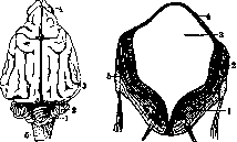
Figure 8.
Figure
9.
Figure 8. Brain of a dog: 1—cerebellar hemisphere; 2-vermis; 3-cerebral hemisphere; 4-lobus olfactorius; 5-medulla oblongata. Figure 9. Cross-section of the posterior cerebral vesicle of a human embryo 41/2 weeks: 1-basic plate; 2-lateral plate; 3-IV ventricle; 4-roof of IV ventricle; 6-vagus nerve.
Figure 10.
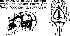
Figure 11.
^
Figure 10. Sagittal section of a human embryo brain, 2nd/nd month: 1-cerebellum; 2-IV ventricle; 3-spinal cord; 4-medulla oblongata; 5-Varolius bridge; 6-III ventricle; 7-thalamus opticus; 8-cerebral hemispheres; 9-corpora quadrigemina; 10-aqueduct of Sylvius. Figure 11. Human embryo brain, 3 months: 1-cerebellar hemisphere; 2-vermis; 3-corpora quadrigemina; 4-longitudinal sulcus of midbrain; 5-cerebral hemispheres; 6-IV ventricle; 7-medulla oblongata; 8-spinal cord. On the cerebral hemispheres, cerebellar convolutions begin to appear only in the middle of the fourth month; at the end of the second month, a small prominence appears on the lower surface of the cerebellum, which corresponds to the flocculus. In the fifth month, the cerebellum acquires its final form; four deep sulci divide it into five large parts; the first corresponds to the upper surface of the hemispheres and the vermis, the second to their posterior edge, the third, fourth, and fifth to the lower surface. Together with the development of the cerebral hemispheres, the cerebellar peduncles to the Varolius bridge appear; the fibers are directed forward and inward, and surrounding the pyramidal bundles, they form the basis of the Varolius bridge. At the end of the third month, within the cerebellar hemispheres, the dentate and fastigial nuclei appear; from the first, the cerebellar peduncles to the corpora quadrigemina originate, which also develop from the lateral plate at the level of the isthmus; fibers are directed to the fastigial nucleus from the vestibular nerve. In the last months of intrauterine life, the cerebellum increases in size, and also the number of secondary

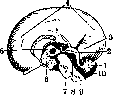
Figure 12.
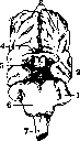
Figure 13. Figure 12. Outer surface of the human embryo brain, 3rd/rd month: 1-cerebellum; 2-corpora quadrigemina; 3-cerebral hemispheres; 4-temporal lobe. Fig. 13. Inner surface of the human embryo brain, 4th month: 1-cerebellum; 2-corpora quadrigemina; 3-occipital lobe; 4-cerebral hemispheres; 5-thalamus opticus; 6-III ventricle; 7-Varolius bridge; 8-aqueduct of Sylvius; 9-medulla oblongata; 10-IV ventricle. and tertiary sulci and sulci, which divide the lobes of the cerebellum into smaller lobules, convolutions, and lamellae and thereby give it a very complex structure, especially characteristic in sections of the cerebellum. Myelination of the cerebellum [see separate table (pp. 63-64), figures 1, 2 and 3]. In a fetus 42 cm in length, the cerebellar hemispheres have no myelinated fibers, whereas in the vermis there are already quite a lot; the fibers of the anterior commissure (see below) are stained especially intensely by Weigert (specific staining for myelin), while in other parts of the vermis there are fewer myelinated fibers and their staining is very weak. In the white matter of the hemispheres, the myelinated fibers belong mainly to the corpus restiforme; around the nucleus dentatus, fibers covered with myelin are visible and apparently belong to the cerebellar peduncles to the corpora quadrigemina. In a newborn, myelinated fibers enter all the small branches of the lobules, but they are not yet intensely stained, just like the white matter of the hemispheres; myelination of the cerebellar fibers continues after birth and ends at 9-10 years. III. Anatomy and histology of the cerebellum. The shape of the cerebellum is very irregular, it is compared to a sponge growing on trees [see separate table (pp. 515-516), figures 1 and 2]. The cerebellum consists of a middle part-the vermis and two lateral parts-the cerebellar hemispheres.
Dimensions of the cerebellum: transverse diameter from 10 to 11 cm, sagittal-for the vermis 3-4 cm, for the hemispheres 5-6 cm. Weight of the cerebellum about 150 g. The cerebellum has two surfaces-upper and lower, according to some authors (Thomas) three-also the anterior, facing the IV ventricle; two edges-anterior and posterior. The upper surface is divided into three parts-the vermis and 2 hemispheres, the division between them is not especially clear; on each side of the vermis there is an indentation-the superior longitudinal fissure. The cerebellar hemispheres are longer in the anteroposterior direction than the vermis; they are separated from each other by a notch-the posterior cerebellar incision, or incisura marsupialis; the notch on the anterior edge of the cerebellum, surrounding the trunk, is called the anterior cerebellar incision, or semilunar incision. The lower surface of the cerebellum is less even, the hemispheres are more developed and protrude downward more than the vermis; between them and the vermis lie large depressions-the vallecula, or Reil's groove. The third, anterior surface, turned toward the IV ventricle, can be seen by removing the cerebellar peduncles and separating the cerebellum from the Varolius bridge and the medulla oblongata. Along the posterior edge of the cerebellum, between the upper and lower surfaces, runs a deep groove-the great horizontal sulcus of Vicq d'Azyr; it extends from the posterior cerebellar incision to the middle cerebellar peduncles. On a sagittal section, the vermis has the appearance of a leaf [see separate table (pp. 515-516), figure 3], the veins of which are formed by white matter; in general, a complex picture is obtained, which older authors have named the 'arbor vitae'. From the front, at its base, there is a depression corresponding to the tent of the IV ventricle. The apex (fastigium) of this tent is formed by the white matter of the vermis itself, the anterior upper wall by the cerebellar sail (velum medullare superior, s. anticum), the posterior wall is partially formed by velum medullare infer., s. posticum, which passes downward into the epithelial obturator membrane. The vermis and hemispheres are divided by sulci into lobes (lobi cerebelli), which are divided by less deep sulci into lobules (lobuli), convolutions (gyri) and lamellae (lamellae). Between the lobules and convolutions of the hemispheres and vermis there is no complete correspondence [see separate table (pp. 515-516), figs. 1 and 2], and the relationships between them are established only on the basis of embryological data. Lobi superiores haemisphaerii Lingula Lobus centralis (Malacarna) Culmen
L°abnUtSerlor atUS) Lobus Monticulus{
Lobus hinatiK >4ua-dran- \ Declive
^obusJj£atus/ gularis Folium cacuminis
Lobus semilunaris supe- rior Vermis superior Frenulum lingulae Ala lobuli centralis Vermis inferior Tuber valvulae или Lobi inferiores haemisphaerii Lobus semilunaris inferior Tuber cerebelli (бугор) Lobus gracilis Pyramis (Malacarna)
Lobus biventer, s. cunei- formis Uvula
Amygdala (see), s. tonsil Nodulus Flocculus (клочок) I The anterior surface of the cerebellum corresponds to the anterior convolutions of the superior vermis-lingula, lobus centralis, diverticulum of the IV ventricle and anterior convolutions of the inferior vermis-nodulus, uvula. ; Of the sulci, on the upper surface should be noted the deep sulcus between lobus semilunaris superior and lobus quadrangularis-superior fissure of Vicq d'Azyr, on the bottom of which is a deep convolution; lobus quadrangularis is divided into two parts by the anterior superior sulcus. On the lower surface, the deep sulcus (inferior fissure of Vicq d'Azyr) passes between lobus semilunaris inferior and lobus gracilis, and in front of it-the inferior anterior sulcus. On the vermis, the sulci bear the name of those lobules near which they pass, with the addition of the particle prae or post [see separate table (pp. 515-516), figs. 1 and 2]. The relationship between the division of the cerebellum into lobes Bolna: Scheme of Bolna
Vermis Lobus anterior Lob. centralis (
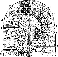
Culmen Lob. simplex Declive -P.{ Lob. medianus Lob. ansiformis Tuber valvulae Pyramis Uvula Nodulus are accepted by and the scheme of the Hemisphere Al lob. centralis Lob. lunatus anterior Lob. quadratus posterior 2,\ Lob. paramedianus Lob. vermicularis Lob. semilunaris superior Lob. semilunaris inferior Lob. gracilis digastricus ; Amygdala Flocculus Microscope, structure of the C. On a cross-section of the cerebellum, it can be seen that it consists of gray and white substance [see separate table (art. 515-516), figure 4]; the gray substance forms the cortex and central nuclei, but the main mass of the cerebellum is formed by white substance, within which are located the nuclei. The cortex covers the entire surface of the C., its thickness is uneven not only on various convolutions, but even on different slopes of the same convolution, and varies from 1 to 21/2 mm. On cross-sections, two layers are distinguished in it - the superficial, or molecular (A), stratum cinereum, and the deep, or granular layer (B), stratum granulosum. At the boundary between these two layers is located a very narrow layer, stratum gangliosum, formed by a single row of large Purkinje cells (1) (figure 15). Purkinje cells are more or less uniformly distributed along the convolutions; there are somewhat more of them at the top of the convolution than in its depths; they are of very large size - 60 μ in length, 30 μ in width and 25-30 μ in thickness; they have a pear-shaped form, are arranged with their long diameter perpendicular to the granular layer, into which their thick end extends; from this end arises the axon-cylinder, which penetrates through the granular layer into the white substance; almost at its very beginning it gives off collaterals, which end partly in the granular layer, and partly near neighboring Purkinje cells. From the thin or upper end, powerful dendrites extend into the molecular layer, which branch into numerous processes and give the cell the characteristic appearance of deer antlers. Such is the appearance of the Purkinje cell on sagittal sections, since all its protoplasmic processes branch in one plane, but on frontal sections the appearance of the cell is quite different (figure 16). The branching of the processes always occurs in one plane, perpendicular to the length of the convolutions, i.e., in the anterior-posterior direction; they end freely and are covered with spines arranged perpendicularly.- The molecular layer, in addition to these
and
WITH AB
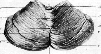
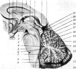
Figure 15/Microscopic structure of the cerebellar cortex: A-molecular layer; B-granular layer; C-medullary layer; 1-Purkinje cells; 2-small superficial cells of the molecular layer; 3-basket cells; 4-'baskets' around Purkinje cells; 5-granule cells; 6-large stellate cells; 7-mossy fibers; 8-'climbing' fibers; 9-cellules empanachees; 10-Bergmann fibers; 11-glial cells-astrocytes of the granular layer. branches contains stellate nerve cells of two kinds: small multipolar (2) are located superficially and have a short branching axon (Golgi cells, second type), large cells are located in the depths of the molecular layer (3), their dendrites extend in all directions, but the axon runs parallel to the surface of the convolution and after a fairly long path ends, branching near a Purkinje cell and forming around it a plexus in the form of baskets (Kölliker), which is why the cells themselves are called 'basket cells'; on its path the axon gives off collaterals, which also branch near Purkinje cells, thus one basket cell has a relationship with many Purkinje cells. In the molecular layer also branch the axis cylinders from the granular layer and from the white substance.- The granular layer consists of round cells, the smallest in size in the central nervous system (from 5 to 10 μ); their granule cells (5) with their numerous protoplasmic processes surround neighboring cells, and the axon-cylinder extends into the Figure 1. Upper, or posterior surface of the cerebellum: 1-incisura cerebelli anterior; 3-lobulus centralis; 3 and 4-monticulus (3-culmen, 4-declive); 5-folium vermis; 6-gyri cerebelli; 7-incisura cerebelli posterior; 8-lobulus semilunaris superior; 9-sulci cerebelli; 10 and 11-lobuli quadrangulares (10-pars anterior, 11-pars posterior); 12-ala lobuli centralis. Figure 2. Lower, or anterior surface of the cerebellum: 1-vermis; 2-lobulus centralis; 3-lingula cerebelli; 4-vinculum lingulae cerebelli; 5-velum medullare posterius; 6-nodulus; 7-uvula (vermis); 8-vallecula cerebelli; 9-pyramis (vermis); 10-tuber vermis; 11-lobulus semilunaris superior; 12-sulcus horizontalis cerebelli; 13-lobulus semilunaris inferior; 14-lobulus biventer; 15-tonsilla cerebelli; 16-pedunculus flocculi; 17-flocculus; 18-brachium pontis; 19-brachium conjunctivum; 20-velum medullare anterius; 21-hemisphaerium cerebelli (facies superior). Figure 3. Sagittal section through vermis of the cerebellum: 1-corpus mamillare; 2-fossa interpeduncularis; 3-nervus oculomotorius; 4-fastigium; 5-pons Varoli; 6-tela chorioidea ventriculi IV; 7-nodulus; 8-medulla spinalis; 9-uvula; 10-pyramis; 11-tuber vermis; 12-folium vermis; 13-laminae medullares; 14-monticulus (declive); 15-corpus medullare; 16-ventriculus IV; 17-lingula cerebelli; 18-lobulus centralis; 19-velum medullare anterius; 20-aquaeductus Sylvii; 21-lamina quadrigemina; 22-splenium corporis callosi; 23-corpus pineale; 24-ventriculus III; 25-thalamus; 26-truncus corporis callosi; 27-columna fornicis; 28-septum pellucidum; 29-chiasma opticum; 30-hypophysis; 31-monticulus (culmen). Figure 4. Cross-section of the cerebellum passing through the subcortical nuclei of vermis and hemispheres: 1 and 10-cortex of the cerebellar hemispheres; 2-nucleus dentatus; 3-embolus; 4-nucleus fastigii; 5-commissural fibers of the cerebellum; 6-vermis superior; 7-vermis inferior; 8-pedunculus cerebelli ad corpora quadrigemina; 9-white substance of the cerebellum, (From Spalteholz.) (For illustration of art. Cerebellum), 30 29 28 27 26 25 24 23 22
4 3 2
Ш 20 19
molecular layer and there T-shapedly divides into 2 branches, which run parallel to the surface of the convolutions and end in a free button. In the deep parts of the granular layer are also located large stellate multipolar cells with a short axon (type II Golgi) (6), which branches around its cell and around part of the granules; the protoplasmic processes end in the granular or in the molecular layer; from the white substance
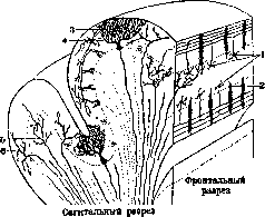
Figure 16. Schematic drawing of the cerebellar cortex: 1-granule cells and their axons, T-shapedly dividing; 2-Purkinje cells on a frontal section; 3-Purkinje cell on a sagittal section; 4-basket cells; 5-mossy fibers; 6-'climbing' fibers.
into this layer penetrate mossy fibers (7) (Kahal), ending near the grains; others so-called 'creeping' fibers (8) only penetrate this layer and end in the molecular layer. Myelinated fibers do not form in the cortex of M. laminae medullares, so characteristic of the cerebral cortex; parallel fibers and axons are not covered with myelin; the layer of grains is somewhat richer in medullated fibers and besides radial fibers also has transverse fibers, especially abundant under the Purkinje cells. Stimulation that reaches the cortex of M. is transmitted simultaneously to several Purkinje cells either directly or through the grains. Each Purkinje cell is connected with collaterals to neighboring ones, in addition association fibers form a close connection between individual laminae. Since these cells are the only elements of the cortex of M., the axons of which reach the subcortical nuclei, their dominant functional significance is obvious. The arrangement of neuroglia in the cortex of M. presents some peculiarities: absence of a glial network in the superficial layers and presence of special cells-cellules empanachees de Cajal (9), the body of which is located in the outer parts of the layer of grains, and the processes, known under the name of fibers of Bergmann (10), penetrate the molecular layer and end under the capsule in the form of a button-like thickening. Neuroglial fibers are few in the cortex (11), but the astrocytes are very numerous in the white matter. The central nuclei in M. are four in number on each side: nucl. dentatus, nucl. fastigii, nucl. emboliformis and nucleus globosus (see separate table, figure 4). Nucleus dentatus (s. olivace-rebelli, dentate nucleus, olive of the cerebellum) is located in the white matter of the hemispheres in its inner-lower part, formed from a plate of gray matter, bent in a zigzag and open inward, where are the gates of this nucleus or hilus nuclei dentati; this plate is surrounded by a large number of myelinated fibers which form capsula nuclei dentati cerebelli. Nucleus emboliformis (s. embolus, bushing, plug) is located in the white matter of the hemisphere, next and inward from the upper edge of nucl. dentatus, with which it is connected by a thin stalk. Nucl. globosus (globulus, spherical nucleus) is located medially from the preceding one. Both these nuclei are of much smaller size and do not have a scalloped shape; they are considered as nucl. dentati accessorii. All these three nuclei have the same structure: they contain a very large number of cells, myelinated fibers and nerve branch endings; the cells are of medium size, multipolar; in nucleus dentatus their axons are directed toward hilus; the nerve endings belong mainly to fibers coming from the cortex of M. (from Purkinje cells), but there are also extracerebellar ones. Nucleus fastigii (s. nucl. tecti, s. noyau du toit, tectal nucleus, tentorial nucleus, Kölliker's nucleus) belongs entirely to vermis, is located in its white matter directly at the median line, above the IV ventricle; its cells are of larger size (40-70μ) and strongly pigmented; this nucleus has connections with the nuclei of Bechterev and Deiters, its protoplasmic processes are few, finely branching, but very long and thick. The white matter occupies the central part of each lamina of the cerebellum, and from there fibers enter the cortex; more inward the white matter of all laminae and lobules collects together and in aggregate forms a very large mass in the hemispheres, in vermis there is much less white matter. Among the white matter are located the subcortical nuclei. According to their function the fibers composing the white matter are divided into association, commissural and projection fibers. Association fibers are divided into short and long; short ones occupy the periphery of each gyrus and connect neighboring gyri (Stilling's garland fibers), while the long ones occupy the center of the white matter of each lobule and connect distant lobules of the same hemisphere. Commissural fibers connect the opposite hemispheres. The commissures of M. (commissura cerebelli) are especially well seen on a sagittal section; a small posterior commissure is located near declive, tuber valvulae. The large anterior commissure is located forward and above nucl. fastigii. Projection fibers are divided into centripetal and centrifugal. Centripetal fibers are the terminal fibers of pedunculi cerebelli inferior et medius (middle and lower peduncles of M.); centrifugal fibers, going from the cortex (from Purkinje cells), end in the subcortical nuclei, fibers from the cortex of vermis-in nucl. fastigii, and from the cortex of hemispheres-in nucl. dentatus; centrifugal fibers of the subcortical nuclei leave M. and go to other parts. M. is connected with other parts of the central nervous system by three pairs of peduncles (see separate table) pedunculi cerebelli infer., medius, super. At this two peduncles consist mainly of afferent fibers-pedunc. cerebelli infer, and medius. and the third-ped. cerebelli super.-belongs to the efferent system of fibers. The lower peduncle of M. (pedunculus cerebelli infer., s. ped. cerebelli ad medullam oblongatam, s. corpus restiforme, restiform body) connects M. with the spinal cord and medulla oblongata and consists of two systems-spinal and bulbar. The center of corp. restiformis is the external nucleus of Burdach or nucleus of Monakow, which gives fibers to corp. restiforme; then the central part is occupied by the bundle of Flechsig, taking origin in the spinal cord, a small number of fibers from the bundle of Govers, which, deviating backward, enter corp. restiforme, fibers from the nuclei of funiculi gracilis and cuneati, forming fibrae arcuatae externae poster., fibers from the nuclei of the lateral column of the medulla oblongata and from nucleus arciformis of the opposite side, entering into the composition of fibr. arcuat. ext. anter. The peripheral part of corp. restiformis is formed mainly by fibers going from oliva infer of the opposite side (fibr. olivo-cerebellar.) and in very small quantity from the olive of its own side. Having penetrated into M., the fibers of corp. restiformis go around nucleus dentatus and end in the cortex of hemispheres and vermis, and a very small part-in the central nuclei. There is a constant relationship between the individual parts of the olive and M.; the lateral parts of the olive are in connection with the cortex of the opposite hemisphere, while the inner parts of the olive-with the cortex of vermis and the adjacent part of the hemisphere; the ventral folds of the olive connect with the lower surface, the dorsal-with the upper surface of the cerebellum. The olives do not receive fibers from the cerebral cortex, but receive into themselves fibers of the so-called central tegmental bundle, which takes origin in the upper parts of the brain stem; thus the olives connect mesencephalon and rhombencephalon with the cortex and with nucleus dentatus of the opposite side of the cerebellum. The second, or middle peduncle of M. (pedunculus cerebelli ad pontem Varolii, s. pedunculus cerebelli medius, s. brachium pontis) goes into M. from Varol's bridge; reaches maximum development in man, since its development is proportional to the development of corticopontine and pyramidal fibers; it is formed by the own fibers of Varol's bridge (fibrae propriae pontis Varolii), which take origin in the gray matter, resp. in the nuclei of the base of Varol's bridge; on leaving the nuclei these fibers are divided by pyramidal fibers into 3 layers-stratum superficiale, complexum, profundum, pass to the opposite side, forming a decussation, and at the outer edge of Varol's bridge collect together to form the middle peduncle of the cerebellum, which constitutes the main amount of white matter of M. The middle peduncles end in the cortex of the hemispheres of M. A small number of fibers in these peduncles goes in the reverse direction-begins in the cortex and ends in the nuclei of the bridge. In the gray matter of the bridge, where the cerebellar peduncles take origin, end fibers coming from the cortex of the same side, namely collaterals from pyramidal fibers, frontopontine, temporopontine and occipitopontine systems; thus the nuclei of the bridge serve as a link between the cerebral cortex of the same side and the cortex of M. of the opposite side and together with pedunculus cerebelli ad pontem Varolii enter into the systems: occipito-, temporo- and fronto-ponto-cerebellaris, as well as pyramido-ponto-
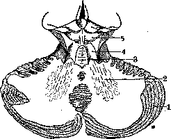
Figure 17. Section of the cerebellum (diagram): 1-cerebellum; 2-nucleus dentatus; 3-velum medullare anticum; 4-pedunculus cerebelli ad corpora quadrigemina; 5-corpora quadrigemina.
cerebellar. It is assumed that each area of the cerebral cortex is connected with a specific part of the pontine nuclei and with a specific area of the cerebellar cortex. Among the fibers related to the cerebellar cortex and not passing through the aforementioned pathways, it is necessary to note the striae medullares, which run along the floor of the fourth ventricle and do not belong to the auditory fibers. They originate in the nuclei of the substantia reticularis and terminate in the cortex of the flocculus. The third, superior cerebellar peduncle (pedunculus cerebelli super., s. pedunculus cerebelli ad corpus quadrigeminum, s. brachium conjunctivum) consists of efferent fibers originating in the cerebellum in the nucleus dentatus (fig. 17); directed upward and forward, the superior cerebellar peduncles form the walls of the fourth ventricle and penetrate into the pons Varoli, and then into the cerebral peduncle, in the lower parts of which their complete crossing-decussatio Wernekinki occurs; after the crossing, the brachium conjunctivum divides into two branches-a descending and an ascending one. The descending branch, which is very thin, terminates very quickly in the reticular nuclei of the pons. The ascending branch, larger in volume and significance, pierces the red nucleus, to which it gives off a considerable number of fibers, and terminates in the thalamus opticus, in
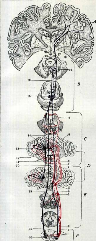
CEREBELLAR CONDUCTING PATHWAYS: A-cerebral hemispheres; B-cerebral peduncles; C-pontine Varoli; D-cerebellum: E-medulla oblongata; F-spinal cord; G-bundle of Flechsig; H-bundle of Gowers; I-corpus restiforme; J-pedunculus cerebelli ad medullam oblongatam; K-nucleus fastigii; L-fibrae vestibulo-cerebellares; 7-fibrae cerebello-vestibulares; 8-nucleus vestibularis; 9-fasciculus vestibulo-spinalis; 10-fibrae vestibulo-oculomotorae; 11-fibrae propriae pontis Varoli; 12-pedunculus cerebelli ad pontem Varoli; 13-fibrae cerebello-dentatae; 14-pedunculus cerebelli ad corpora quadrigemina; 15-decussatio pedunculi cerebelli; 16-nucleus ruber; 17-thalamus opticus; 18-fibrae thalamo-corticales; 19-fasciculus rubro-spinalis; 20-anterior horns of the spinal cord. B. M. E. Section Cerebellum. The cerebellum in the ventral part of its outer nucleus. From the large cells of the red nucleus originates the fasciculus rubrospinalis, the fibers of which after crossing terminate in the anterior horns of the spinal cord. Pedunc. cerebelli ad corpus quadrigeminum represents the efferent pathway for the hemispheres of the cerebellum. The efferent fibers of the vermis pass through the inner part of the inferior cerebellar peduncle; they originate in the nucleus fastigii, in the nucleus globosus and possibly in the embolus and terminate in the nuclei of the vestibular system-nuclei of Deiters, Bechterev and nucleus triangularis dorsalis; part of the fibers going to these nuclei from the nucleus fastigii of the opposite side, on their way, curves around the outside of the superior cerebellar peduncle at its exit from the nucleus dentatus and is isolated under the name of the hook-shaped bundle of Russell (faisceau en crochet Russel). The vestibular nuclei with the cerebellum and with all fibers connecting them with the nucleus vestibularis form the vestibulo-cerebellar system. From the nuclei of the vestibular system originate fibers which partly through the fasciculus vestibulo-spinalis go to the cells of the anterior horns of the spinal cord and partly through the fasciculus longitudinalis posterior are directed to the nuclei of the III. Projection fibers originate in the cerebellar cortex and terminate in the central nuclei. The nucleus dentatus and embolus receive fibers from the cortex of the hemispheres, the nucleus fastigii and globulus-from the cortex of the vermis and flocculus. According to some authors, the nucleus fastigii receives fibers from the cortex of the hemispheres and vermis; the relationships are direct, i.e., the cortex sends fibers to the nuclei of its own side. Association fibers are few and short; longer ones are found in the vermis, but have an anterior-posterior direction, so they do not extend beyond the cerebellum. The vermis and hemispheres are almost independent, as a small number of fibers goes from the vermis only to the nearest lobules of the hemispheres. The existence of commissural fibers between the two hemispheres is not proven. Thus, on the basis of the connections that exist between the cerebellum and other formations, two systems can be distinguished in it: the vermis is connected with the spinal cord and medulla oblongata through the pedunculus cerebelli inferior and forms the spinocerebellar system, while the hemispheres are connected with the brain through the fibers of the pedunculus cerebelli medii and super., forming the cerebrocerebellar system. The meninges of the cerebellum. The cerebellum, like the brain, is surrounded by three meninges-dura mater, arachnoidea and pia mater: the dura mater at the level of the cerebellum gives off two processes-the tentorium cerebelli, separating the cerebellum from the inferior surface of the brain (fig. 18), and the falx cerebri minor, extending between the two hemispheres of the cerebellum. The pia mater enters all the small subdivisions of the cerebellum, connecting them; between the pia mater and arachnoidea at the level of the cerebellum, cisterns are formed, in which in patho-logical cases a large amount of cerebrospinal fluid accumulates (see Brain, Meninges). - Blood supply of the cerebellum. The cerebellum receives blood from three pairs of cerebellar arteries: a. cerebel. super., a. cerebelli inferior ant. and a. cerebelli inferior post., which are branches of the a. basilaris. This artery begins with two trunks and from the art. ver-
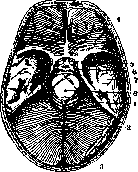
and arteria vertebralis. According to some data (Jakob) there is also a fourth artery-the a. cerebelli inferior media, but this artery is not constant. In general, there are many variations regarding the number of arteries, their place of origin and their branching, which indicates large individual deviations in the blood supply of the cerebellum. A. cerebelli inf.post. originates from the a. vertebralis (in 68.5%), more rarely from the a. basilaris, or on one side from the a. basilaris, and on the other from a verte- Ris-18-OСновадиe of the skull. one of <a. venous Sinus. Posterior cranial fossa bralis, or finally covered by the tentorium cerebelli: 1-cerebellum; 2-tentorium cerebelli; 3-confluens sinuum; 4-anterior cranial fossa; 5-n. opti-tebralis and from art. ba- ™s; 6-n. oculomotorius; 7-middle cranial fossa; 8-cerebral peduncle with strong connections; 9-superior orbital fissure; 10-venous sinus, and then they я-мозговая нонша c Силь-СОедиНЯЮТСЯ B ОДИН виевым водопроводом. общий ствол, which supplies blood to the medulla oblongata, and then, curving around the tonsil, goes to the inferior surface, where it divides into two secondary branches (more rarely into three, four)-medial and lateral, which are subdivided into a number of smaller arteries supplying the inferior surface of the vermis and hemispheres, the anterior surface of the flocculus (fig. 19*).- A. cerebelli inf. anter. originates from the a. basilaris, sometimes with a common trunk with the previous artery, then divides into two, more rarely into three secondary branches; on its way it gives branches to the cerebellopontine angle, to the nerves VII and VIII, plexus chorioideus. In the cerebellum it supplies blood to the nodulus, flocculus and the external parts of the inferior surface of the cerebellum (fig. 20).-A. cerebel. super, the most constant of the cerebellar arteries; it originates from the art. basilaris, behind its division into terminal branches, and behind the nucleus oculomotorius divides into two (more rarely into three) large branches-lateral and medial; each of these branches in turn divides in various combinations and in different numbers into tertiary branches. The lateral branch supplies the upper surface Fig. 19. Inferior surface of the cerebellum. Area of vascularization of a. cerebelli inferior posterior: 1-tonsil; 2-nodulus; 3-uvula; 4-pyramis; 5 and 6-lobus semilunaris inferior et superior; 7-tonsilla; 8-lobus gracilis; 9 and 10-lobus biventer; 11-pars anterior lobi quadrangularis. of the hemispheres (fig. 21), at the outer edge it anastomoses with the a. cerebelli inf. anter. and in case of weak development or absence of the latter, it takes over the vascularization of the external parts of the inferior surface of the cerebellum. The medial branch supplies the upper surface of the vermis and the internal parts of the hemispheres. Anastomoses exist between the arteries, both between the main trunks and between the secondary and ter
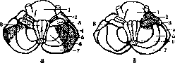
Fig. 20. Inferior surface of the cerebellum: a-area of vascularization of a. cerebelli inf. ant. (on one and the other side various variants of artery distribution are indicated); b-area of vascularization of a. cerebelli inf. media; 1-pontis Varoli; 2-lobus quadrangularis (pars anterior); 3-flocculus; 4-lobus quadrangularis (pars posterior); 5-lobus biventer; 6-tonsilla; 7-lobus semilunaris inferior; 8-medulla oblongata.
typical; between arteries, on the other hand, anastomosis occurs in the hemispheres, and between arteries of different sides - in the vermis. Nucl. dentatus is very rich in vessels, which it receives mainly from the secondary medial trunk; according to some data, a. cerebelli inf. post. and a. cerebelli inf. anter. participate in the supply of this nucleus. The arteries of nucl. dentati are a frequent "cause of hemorrhage into the cerebellum. Nucl. fastigii receives blood from almost all the arteries supplying the vermis. The chemical structure of the cerebellum - see the main HEAD. Injection of vessels. For the study of angio-architecture of the brain (for determining the terminal vessels in the brain and the relationship of vessel distribution to myelo-architectonics and cytoarchitectonics, especially in connection with the doctrine of patoclyses), in particular of the cerebellum, the vessels of the brain are injected with colored solutions, which must necessarily be "setting". Several such solutions have been proposed: 1) a gelatin solution to which vermilion or ink was added (Tandler's method), 2) plaster or chalk and talc in equal doses, to which is added
the surface of the cerebellum.
The area of vascularization of the superior cerebellar artery: 1-segment superior; 2 and 3-lobus quadrangularis (pars anterior et posterior); 4 and 5-lobus semilunaris superior et inferior; 6-decussatio; 7-culmen. (According to Chernyshev and Grigorovich). Glycerin is added until a soft paste consistency is obtained, vermilion or ink is added to the desired color, and water is added until the consistency of cream is achieved (Reitlinger's method), 3) a solution of glycerin, distilled water and ink (Bone's method) and others. Injection of the brain can be done either in situ or on the removed brain. On the corpse, injection is performed through the a. vertebralis at its origin from the a. subclavia: cannulas are inserted into these arteries on both sides, all other neck vessels are ligated, the prepared solution is injected through the cannulas with a syringe until the retinal vessels are colored (approximately 1 liter of solution is required). Then the skull is partially opened, the head is severed and transferred for 2-3 days to 10% formalin, after which the skull is completely opened, the brain is removed, freed from its membranes and studied macroscopically, and then after embedding and microscopically. There are modifications: injection is performed not only through art. vertebralis but also through art. carotis interna; this method more reliably ensures the penetration of the colored liquid into the smallest arteries. The injection solution must be prepared ex tempore; the brain must be very fresh. The liquid must be injected under very slowly increasing pressure. The brain for injection outside the corpse must be removed with great care, preserving the integrity of the dura mater; it is then immersed for 2 days in a 10% formalin solution, and after that the injection of vessels is performed; with the integrity of the circle of Willis, injection is performed into art. vertebralis and carotis, and with its violation-into individual cerebral arteries; during injection, it is easy to observe how the coloring substance penetrates all the smallest vessels. When injecting the Cerebellum separately, all vessels that are not related to the Cerebellum are ligated (a. cerebri posterior, a. spinalis); the Cerebellum is injected through a. vertebralis. Injections can also be made into individual arteries of the Cerebellum, ligating the other vessels for this purpose. IV. Physiology of the cerebellum. Questions about the functions of the Cerebellum and about the localization of these functions in its various parts have been studied for a long time by scientists of all countries, but even to this day they have not been finally resolved. This is explained by: 1) the special position of the Cerebellum in the vicinity of a large number of vital structures, the diseases of which reflected the conditions of the Cerebellum and which, with their symptoms, obscured the picture of cerebellar phenomena, 2) the impossibility of fully transferring to humans the phenomena observed in animals, and finally 3) insufficient familiarity for a long time with the anatomy of the Cerebellum due to the complexity of its structure. In the history of the study of the Cerebellum, two periods can be distinguished: the first from the end of the 17th century to the 1880s of the 19th century, and the second from the 1880s to our days. In the first period, the experiments of physiologists on the Cerebellum were very crude and inaccurate due to insufficient knowledge of anatomy and the imperfection of neurosurgical technique; the lack of asepsis caused high mortality among animals soon after the operation, so they could only be studied immediately after the operation, when the symptoms depending on the destruction of the Cerebellum had not yet come to the forefront, but were obscured by the phenomena of postoperative shock, diaschisis. But still, among the many theories and assumptions about the functions of the Cerebellum, expressed on the basis of experimental data, some can be noted that quite correctly approached the explanation of the observed phenomena. In the second period (from 1880) the discovery of various methods for studying the structure of the central nervous system rapidly advanced the knowledge of the structure of the Cerebellum, and the improvement of surgical methods and the introduction of strict asepsis contributed to a more successful outcome of operations: usually the animal survived as long as was necessary for the experiment and could be subjected to repeated comprehensive research. The results obtained under these conditions made it possible to more methodically analyze the phenomena obtained in operations on the Cerebellum. The first works on the Cerebellum date back to the end of the 17th and 18th centuries (Willis's experiments in 1683, Haller's in 1755); on the basis of experimental data, various assumptions were made regarding the functions of the Cerebellum; it was attributed an influence on the function of internal organs, on their involuntary contractions (Willis), on the development and growth of the organism; its connection with sense organs, mainly with hearing and with general sensitivity was indicated; it was even assumed that the cerebellum has its own sensitivity; it was attributed an influence on the sexual instinct (Gall), on respiration, on nutrition, on appetite. Rolando in 1839, on the basis of physiological and anatomical data obtained from experiments on animals of four different classes, tried to prove that the Cerebellum is an organ that produces nervous force, manifested mainly in movements, and pointed out that the destruction of the Cerebellum led to a disorder of movements, which became uncoordinated. The anatomical structure seemed to confirm his assumption: the large number of white and gray plates alternating with each other reminded him of an apparatus that produces electricity and excites movement. The works of Flourens, Magendie, dating from the beginning of the 19th century (in 1824, 1836), laid the foundation for a deeper study of the Cerebellum. Flourens performed a large number of experiments on various animals, mainly on birds and mammals; his experiments consisted in the gradual removal, layer by layer, of the substance of the Cerebellum; in parallel, animals developed movement disorders: the bird first lost the ability to fly, then to walk and then to stand on its feet; the same effect was obtained in experiments on mammals-the animal lost the ability to coordinate and regulate its movements. The disharmony in movements increased in parallel with the intensity of the damage, complete destruction of the Cerebellum led to a complete loss of the regulatory ability of movements. Thus, on the basis of his experiments, the author came to the conclusion that the Cerebellum coordinates movements and maintains balance during walking and standing and that the hemispheres of the Cerebellum have an influence on the opposite half of the body. This theory attracted many followers, and in subsequent works similar ideas about the function of the Cerebellum were expressed. In studying the function of the Cerebellum, Magendie performed both the destruction of the Cerebellum itself and the transection of its peduncles. He found that when the Cerebellum was destroyed, the animal could not move forward and when trying to walk, it backed up; this fact led him to the conclusion that in the Cerebellum there is an impulsive force that makes the animal move forward, but this force exists only in mammals and birds, because when the Cerebellum was destroyed in fish and reptiles, movements were preserved. The transection of the anterior peduncles caused rotation of the animal to the operated side, changes in the position of the eyes (Hertwig-Magendie position of the eyes, see). The transection of the Cerebellum in the middle into two equal parts caused the animal to move first to the right then to the left and the inability to maintain a calm position. Longet also observed rotational movements to the side opposite to the transected peduncle. A whole series of authors in their experiments also noted these rotations of animals, but there was no agreement on which way the animal turns-whether to the side of the peduncle damage or to the opposite. This disagreement in the obtained results the authors explained by the place of transection. Wagner observed after operations on the cerebellum symptoms of two kinds-some directly following the destruction of the Cerebellum, others appearing later and masked at first by early symptoms; the first include disturbance of equilibrium, and the second-special position of the head, trunk and limbs and hyperkinesias in the form of trembling. The works of Ferrier in 1878 are transitional between the first and second periods. In addition to clarifying the function of the Cerebellum, he tried to solve some questions of localization-to determine the relationship existing between localization and the intensity of damage and the change in equilibrium. His work is also interesting in that he approached the solution of the question using not only the surgical method but also the method of irritation of the cortex of the Cerebellum with electric current. In mammals, electrification of the Cerebellum causes movements of the eyes, head and limbs; but the movements of the eyes are especially interesting; the direction of these movements and the position of the eyes change depending on which part of the Cerebellum is irritated; in birds, irritation of the Cerebellum does not cause movements of the eyes, only small movements in the wings and in the paw are noted; in fish, unilateral irritation causes exophthalmus on the corresponding side and curvature of the tail to the same side. The data obtained from the destruction of the Cerebellum in animals were fully confirmed clinically by observations of cerebellar patients.
The initiator of the second period of the doctrine of the C. is Luciani. His first works (1884) begin the period of a strict analysis of all phenomena appearing after the destruction of the C.; they played a very large role in clarifying the functions of the C. Thanks to the improvement of surgical techniques, he managed to achieve that animals survived after the operation for quite a long time and could be studied in detail over a long period. Luciani considered the C. not as an aggregate of organs with various functions, but as a central organ, quite homogeneous in functional relation, each segment of which has the same function and can replace another. In the normal state, the C. has a sthenic, tonic and static effect on the neuromuscular apparatus; the sthenic effect is a very complex function, thanks to which the C. increases the potential energy of the neuromuscular apparatus; the tonic effect is manifested by an increase in the tension of the neuromuscular apparatus at the moment of its functional phase; the static effect accelerates the rhythm of elementary impulses during their activity and ensures the fusion and proper duration of contractions. Through its efferent paths, the C. has a direct or indirect trophic influence; upon its removal, various dystrophic disorders are observed: degeneration of the skin, muscles, slower growth rate, decreased resistance of the organism in the struggle with external harmful agents; life in such animals is shorter, etc. The influence of the cerebral hemispheres on the C. extends to the corresponding side of the body, whereas the connection of the C. with the cerebral hemispheres is crossed. In experimental destruction of the C., the author observed symptoms of two kinds—symptoms of irritation appearing immediately after the operation, and symptoms of deficiency developing some time after it. The phenomena of irritation manifest themselves in strong excitement and frequent crying. In case of unilateral destruction, curvature of the spine in the direction opposite to the operation is observed, tonic extension of the limbs on the corresponding side, strabismus, nystagmus; rotation of the body around its axis from the damaged side to the healthy one; with complete destruction of the C., the spine curves backward, when walking the animal backs up and falls, strabismus convergens is observed, tonic convulsions in the front paws, clonic in the hind ones. Gradually the phenomena of irritation weaken and disappear, and then the symptoms of deficiency come to the fore, characterized by a violation of the three basic functions of the cerebellum—sthenic, tonic and static: in operated animals, asthenia, atony and astasia appear. The rotation of the animal around its axis was explained by a sudden violation of equilibrium in the activity of the brain centers; it was suggested that the limbs play a large role in this phenomenon, since if all four limbs of the animal are tied, the phenomena of rotation do not occur. Subsequent authors who worked in this field (Russel, Ferrier, Turner) in general confirmed the phenomena observed by Luciani and introduced only some details in regard to reflexes, sensitivity, etc. Bianchi on the basis of his experiments noted the possibility of a compensatory function of the brain upon removal of the C., which in his opinion is also confirmed anatomically—by the enhanced development of the anterior parts of the brain (gyrus sigmoidealis) in cases of destruction of the C. According to Schiff's opinion, the C. contains mechanisms that make the muscle groups necessary for the execution of complex movement work, not only the group that directs the movement, but also the groups that fix the limb (ligaments, joints, muscles) and thereby give a point of support; these muscles are in general antagonists of the directing muscles. From the later works on the physiology of the C., the most interesting are the studies of the French school, namely Thomas (Andre Thomas). He performed a large number of diverse experiments—complete removal of the cerebellum, removal of the hemispheres, vermis, section of the peduncles of the C. He conducted his experiments on dogs and monkeys, and although his works did not give anything new for the physiology of the C., they are valuable in that on a large amount of material they confirmed the conclusions of previous authors; interesting are his explanations of the obtained results, as well as the fact that under all the data obtained by experiment, he provides an anatomical basis. According to his data, the phenomena observed upon destruction of the C. may depend on the irritation of the damaged areas or on the loss of their functions or finally on the replacement of the lost cerebellar functions by the functions of neighboring areas. The earliest phenomena are the symptoms of irritation; they may occur already on the operating table, but are usually short-lived. Then the phenomena of deficiency appear, and only last of all the compensatory symptoms manifest themselves. Upon removal of half of the cerebellum, rotation of the body around the longitudinal axis is observed, appearing immediately upon awakening; bending of the body with concavity toward the operated side; strabismus—the eye on the operated side looks down and inward, while on the opposite side—upward and outward (Hertwig-Majandie position of the eyes), nystagmus; tonic and clonic convulsions of the limbs, rotation of the head to the side opposite the operation. At first the animal lies motionless; cannot walk or stand. After some time the condition begins to improve, but on the operated side muscular weakness and incorrect position of the limbs during movements remain; the limbs are excessively abducted, and their movements are disproportionate and very sweeping. In monkeys with extirpation of half of the C., muscle weakness and ataxia are observed even more sharply than in dogs, and are especially noticeable in finer movements of the forelimb. The operation of complete removal of the C. is very difficult, since often neighboring areas are also removed along with the C., for example the vestibular nuclei, which distorts the picture of a purely cerebellar lesion. Rotation around the axis is not observed or is observed very rarely; opisthotonus, rigidity of the limbs, inability to either stand nor walk are noted; then later, at the first attempts to stand and walk, the body sways from side to side and the animal falls; gradually the gait becomes more confident, but the animal does not walk in a straight line, but in a zigzag; in the limbs, coordination is disturbed, when walking they are lifted very high and fall to the ground very quickly; with changes in position, balance becomes less stable. Thomas paid special attention to the fact that after destruction of the cerebellum, movements became less automatic, i.e., more conscious, deliberate. With the same operation in monkeys, ataxia was expressed more strongly than in dogs, and such phenomena as a shaky gait, awkwardness, uncertainty in the limbs and slight trembling remained for life; nevertheless the animal could climb. Destruction of the vermis caused somewhat different phenomena—opisthotonus, strong backward bending of the head, hyperextension of the limbs, strong vertical nystagmus. When attempting to stand at first the animal fell backward, trembling was observed in the body; after a few days, although the animal could walk, but with great difficulty, swayed and often fell backward. Later the condition gradually improved, although some abnormality in walking remained all the time. Destruction of the nucleus fastigii caused approximately the same symptoms as destruction of the vermis. After section of the vermis along the median line, at first there was very little disturbance of equilibrium, which soon passed. Irritation of the C. did not give such clear symptoms as its destruction, and moreover, of all irritants (cauterization, heat, cold, mechanical irritation, etc.), the cerebellum reacted only to electric current. In this case, when irritated with weak current, the cortex remained non-excitable, with strong current, contraction occurred throughout the corresponding half of the body; irritation with weak current near the subcortical nuclei of the C. (nucleus dentatus, fastigii, Deiters') caused movements of the eyes, head, body and limbs. Experiments with irritation of the C. were also carried out by other authors. In the experiments of Horsley, Lowenthal, Thiele and others, irritation of the C. had an inhibitory effect on tone; in animals in a state of contracture after decerebration, faradization of the upper surface of the C. caused relaxation of the muscles of the neck, head and lower limbs, mainly on the corresponding side. With similar experiments, Bremer proved that mainly the cortex of the Cerebellum (vermis) has an inhibitory effect on the rigidity of the limbs and body. In general, it must be said that the works on the physiology of the C., made by the method of irritation, are not especially demonstrative and conclusive. Destruction of the peduncles of the C. gave approximately the same symptoms as destruction of the hemispheres, only in addition to cerebellar phenomena, symptoms from the surrounding formations were also observed—the pons Varolii, medulla oblongata, vestibular system.
From the data presented above, it can be seen that destruction of the cerebellum does not cause very severe and persistent disorders, especially if the operation is performed on one side; it is assumed that the remaining half takes over the function of the removed part; when the entire cerebellum is removed, disorders are observed in the motor sphere, initially in the form of convulsive phenomena, and then in the form of impairment of coordination of movements or so-called cerebellar ataxia. Thus, on the basis of experimental data, it can be concluded that the cerebellum participates in movements, coordinating them. The same is indicated by the data of phylogenesis, the anatomical structure of the cerebellum and its connections. Anatomical data indicate that the cerebellum is connected by centrifugal and centripetal fibers with a large number of formations: with the spinal cord, with the vestibular apparatus, with nuclei in the midbrain (nucleus ruber), with thalamus opticus, and with the cerebral hemispheres. In this complex system, the cerebellum is located between the sensory and motor conductors: on the one hand, it receives sensory impulses from the skin of the entire body and from the deep parts (ligaments, muscles, tendons and bones) associated with movements (through the bundles of Flechig and corpus restiforme), then from the semicircular canals the sensation of the position of the body in space (through the vestibular system - see), as well as impulses from the cerebral cortex through the pyramidal, corticopontine fibers and fibers pedunculi cerebelli ad pontem Varoli; on the other hand, the main mass of its centrifugal fibers goes to the motor centers: partly from nucleus dentatus through nucleus ruber into fasc. rubrospinalis, partly from nucleus fastigii through nucleus Deiters'a into fasc. vestibulospinalis. Both bundles - vestibulo- and rubrospinalis - end in the motor cells of the anterior horns of the spinal cord. These anatomical connections indicate that the cerebellum belongs to the motor system and that it is a reflex organ capable of functioning without the participation of will and consciousness. As a higher reflex center, it is connected with the muscle centers only through the intervention of intermediate centers, such as nucleus ruber, nucleus Deiters'a, substantia reticularis, etc. Under the influence of peripheral irritation or irritation coming from the cerebral cortex, the cerebellum becomes active and its influence through centrifugal fibers is transmitted to the cells of the anterior horns of the spinal cord and then to the corresponding muscles. According to the data of phylogenesis, we see that in lower animals, in which movements are very monotonous (swimming, crawling), the cerebellum is very poorly developed, but as we ascend the zoological ladder, movements become more diverse and complex, and in connection with this, the cerebellum increases in size and its structure becomes more complex; in such animals, removal of the cerebellum causes more clear disorders of movement in the form of impairment of coordination of movements and balance. On the basis of all the above, it can be concluded that the cerebellum is an apparatus that regulates the action of muscles that come into contraction when standing, walking, during voluntary, reflex and automatic movements, and ensuring their correct coordination, i.e., making all movements synergistic. Coordination of muscle contractions during walking and standing leads to the maintenance of static equilibrium in a state of rest and kinetic equilibrium during movements; for the preservation of equilibrium, synergy in the work of the corresponding muscles is necessary. In the organism, each muscular unit consists of an agonist and an antagonist, which work synergistically. In static equilibrium, the contraction of agonists and antagonists is completely balanced, so-called isosthenia is obtained. When the trunk is displaced, the center of gravity is also displaced, and in order to restore the disturbed equilibrium, both agonists and antagonists must contract, but the agonists contract more strongly, so physiological anisosthenia is obtained; the cerebellum regulates these contractions reflexively; in this work, the system connecting the cerebellum with the spinal cord (spino-cerebellaris) and ending in the vermis participates; this function can be represented as follows. Impulses from the periphery of the body and from the semicircular channels constantly flow to the cerebellum and maintain tonic excitation in it; any change in the position of the body through afferent fibers reaches the cortex of the cerebellum and causes in it a reaction response, which through the appropriate systems is transmitted to the cells of the anterior horns and regulates the contraction of the muscles necessary at the moment to maintain equilibrium. Any attempt to displace the equilibrium around a horizontal or vertical axis acts as a moment of irritation on the corresponding centers of the cerebellum, which causes the antagonists and compensatory elements to function, thanks to which equilibrium is maintained. In addition to maintaining equilibrium, the cerebellum has an influence on the coordination of movements in general, as well as on their synergy. For correct voluntary movements, very strict coordination of muscle work is necessary: the entry into activity of strictly defined groups of muscles; contraction of each muscle individually with the force required for a given task; observance of a strict sequence in the contraction of muscles. Moreover, for each movement, the activity of not only agonists and antagonists, but also other muscles that fix the interested part of the body is necessary. In all this coordinatory work of muscles, the cerebellum participates; in complex movements, in addition to coordination, with its participation, the association of elementary movements into complex ones takes place, i.e., the cerebellum also manages the synergy of movements. With its special tonic influence, the cerebellum regulates movements in such a way that they occur with minimal expenditure and achieve the goal, so that the movements that make up one act are synchronous; thus, when walking, when moving the leg forward, it is necessary at the same time to transfer the center of gravity of the body to it, which is absent in patients with cerebellar lesions (Fig. 22); when bending backward, a healthy person bends the knees (Fig. 23), but the patient cannot do this (Fig. 24); during rapid alternating movements, for example supination and pronation, the agonist and antagonist muscles must contract proportionally so that these movements are smooth and sequential. In these voluntary movements, the cortex of the cerebellar hemispheres participates, which comes into action under the influence of irritation coming from the cerebral cortex; therefore, in voluntary movements, the cerebro-cerebellar system functions. The coordinating influence of the cerebellum extends also to the oculomotor apparatus. Its connection with the oculomotor apparatus is as follows: the cerebellum through nucleus fastigii is connected with nucleus Deiters'a, which sends fibers through fasciculus longitudinalis posterior to the nucleus of the n. abducentis on its side and to the nucleus n. oculomotorii of the opposite side; with unilateral action, the eyes deviate to the corresponding side, in normal bilateral impulses mutually balance each other. The cerebellum also exerts a coordinating influence on the muscles of the speech apparatus, respiratory, swallowing and masticatory apparatuses, thanks to its connections through substantia reticularis with the nuclei of the corresponding nerves. Here it is appropriate to
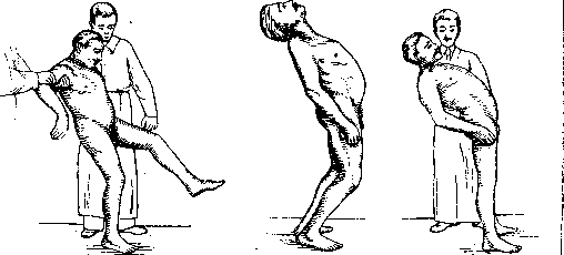
Figure 22.
Figure 23.
Figure 24.
The question arises as to what kind of coordinating action the cerebellum performs: does it itself send coordinating impulses, or, as a higher center, does it act through the mediation of other departments? Anatomy rather supports the second hypothesis, since in the cerebellum there is nowhere a grouping of cells or a special central apparatus that could be considered a coordination center. Is the cerebellum functionally homogeneous, or, on the contrary, can it be divided into strictly limited centers, each of which manages special functions? This question about the localization of functions is a controversial one. Some authors recognize in the cerebellar cortex the presence of the same separate centers as exist in the cortex of the large brain; others speak against it. The different relationship of parts of the cerebellum to afferent systems, the difference in the time of development of its lobules, as well as clinical data (the difference in symptoms observed when the vermis and hemispheres are destroyed) - all these facts indicate that the cerebellum is an organ not homogeneous in its entirety and that there must be some localization within it. The cerebellar cortex apparently possesses one function, and the localization consists only in attributing this function to one or another part of the body, to different muscle groups that act in one direction or another. The first attempt to clarify the localization belongs to Bolk. On the basis of comparative anatomical studies, he gave a new division of the cerebellum, and then by comparing the cerebellums of various animals, he found that there is a certain relationship between the development of the cerebellar lobules and the functional development in animals of various parts of the body or muscle groups: accordingly, with these observations, he distributed centers in the cerebellum (Bolk's scheme). Muscles that function together with muscles of the other side (muscles of the trunk, neck, larynx, tongue, and eyes) are served by unpaired centers located in the middle lobe. The limbs, however, which work both together and separately, are innervated by centers of two kinds - unpaired (in the middle lobe) and paired (in the lateral lobes). Bolk's research led to a whole series of experimental works. Experiments with electrical stimulation of the cerebellum were carried out by many researchers. Faradic stimulation of the lobi quadrangularis in a monkey caused movements of the fingers of the upper limb on the same side, and stimulation of the more anterior parts of this lobe caused extension, while stimulation of its posterior parts caused flexion of the fingers (Rothmann). Stimulation of the anterior lobe (according to Bolk) gave abduction of both upper limbs. In Bremner's experiments, stimulation of the cortex of the cerebellar hemisphere and the red nucleus had no effect on the tone of decerebrated animals, whereas stimulation of the cortex (vermis), especially at two points - at the level of lob. centralis and in the pyramis (sites of termination of fasc. spino-cerebellaris) - had an inhibitory, relaxing effect on the tone. However, all these experiments at the present time do not give the right to definite and final conclusions. - Experiments with destruction of the cerebellum gave more precise results. The data of experiments by Rynberk, Rothman, Tom, and others speak in favor of functional localization. They showed that removal of limited parts of the cerebellar cortex causes disturbance of coordination of movements, atonia and asthenia of muscles on the operated side in different, but definite parts of the body, corresponding to the removed area; if the destruction is very limited, then the function of one particular group - flexors, extensors, abductors or adductors - may be disturbed; this change in function manifests itself in a disturbance of tone - in hypotonia of muscles acting in one direction and hypertonia of others acting in the opposite direction, whereby during contractions there is a disturbance of equilibrium in the muscles and hence an incorrect position of the limb, which the animal either does not correct at all or corrects with great delay. With very limited damage, the animal does not correct displacements of the limb in one particular direction, but corrects it in others, for example, it does not correct when the limb is strongly abducted, and corrects when the limb is in a position of adduction; in this case, the experimenter encounters no resistance when setting the limb in a given position - it remains passive. Thus, with the destruction of a small area of the cerebellum, a double effect is observed: hyposthenia (decrease in resistance) in the muscles acting in one specific direction and hypersthenia (increase in resistance) in the muscles acting in the opposite direction, i.e., anisosthenia in antagonist muscles. This disturbance in the function of antagonists leads to abnormal positions that the limb assumes. Anisosthenia and passivity lie at the basis of the dysmetria observed in cerebellar disease. It was established (Rothman) that there exist in the cerebellum four separately located ('localized') centers for 4 groups of skeletal muscles, namely: for muscles of the neck, back, and anterior and posterior limbs. Further work in this direction led to the conclusion that destruction of the vermis causes disturbance of equilibrium in standing and walking, whereas destruction of the hemispheres gives ataxia, resp. asynergy in the movements of the corresponding limb; the anterior limb is dependent on lobus quadrangularis, the posterior on lobus ansiformis. Damage to the most anterior parts of lobus anterioris (lobus centralis) is accompanied by disturbance of movements of the jaw and larynx; destruction of lob. simplicis causes disturbance in movements of the head. Relatively recently, an attempt was made to determine the centers of the cerebellum in humans. Barany (Barany) in the study of nystagmus drew attention to reactive movements in the trunk, limbs, and sometimes even in individual joints of the limbs; these movements are analogous to those that appear in cerebellar diseases. These data led him to assume that in the cerebellum there must exist centers directing movements in the joints, or rather a collection of directing centers, since for each joint or even for each movement in the joint there is its own center. These are centers directing movements inward, outward, upward, and downward. They exert a constant tonic influence on the muscles. Under normal conditions they act simultaneously and with equal force, why their influence is mutually balanced; when any center is stimulated, the limb deviates in the corresponding direction, and when the center is lost, its antagonist takes over. The centers that influence the position of the trunk are located in the cortex of the vermis, while the movements of the limbs depend on centers located in the cortex of the cerebellar hemisphere and connected with the cerebral hemispheres. Barany tries to localize some centers of the limbs on the surface of the cerebellar hemispheres, and from the side of some clinical observations he found confirmation for himself, but this localization is disputed by other clinicians. Thus, the existence of localization in the cerebellum is beyond doubt, but the location of individual centers on the surface of the cerebellum and in depth, and the very nature of these centers, have not yet been finally established. V. Pathology of the cerebellum. Being a component part of the brain, the cerebellum can be involved in the pathological process simultaneously with other parts of the brain, which is observed in diffuse processes in the brain (e.g., in inflammations, arteriosclerosis, syphilis) or in processes arising adjacent to the cerebellum and with further development involving the cerebellum, but in addition the cerebellum has its own pathology, when the process arises primarily in the cerebellum and maximum changes are observed in it (various degenerative processes, tumors, abscesses, cysts, etc.), and other parts of the brain, if affected, are affected secondarily. All processes occurring in the cerebellum, in their nature, are analogous to those observed in the brain, and therefore their etiology, statistics, patho-anatomical changes, prevention and treatment are identical (see Brain). Symptomatology of cerebellar diseases. In cerebellar disease, regardless of the nature of the process, a number of symptoms develop, the combination of which forms the so-called cerebellar syndrome (syndrome cerebeHeux). Some of these symptoms are more constant and constitute as it were an integral part of the cerebellar syndrome, while others are rarer and are not observed in all cerebellar diseases. Variations in the intensity of symptoms, in their number and localization can be very great even in diseases known to be localized in the cerebellum. In some progressive diseases of the cerebellum, at the beginning of the disease it is sometimes difficult to find a symptom that would be pathognomonic; this is explained by many factors, and especially by the substitutive function of the brain, which, although not adapted to the regulatory function, but due to the fact that it receives numerous impulses from all parts of the body and from all sense organs, mainly sensations from the labyrinth and visual, takes upon itself to a certain extent the role that in normal conditions is performed by the cerebellum. Besides the motor zone, in this substitution, associative processes and some psychical functions, such as will, attention, memory, etc., participate.
With unilateral disease of the M., the function of the affected hemisphere can be partially replaced by the healthy hemisphere; many authors have noted that unilateral disease of the M. gives fewer symptoms than bilateral or diffuse disease. In recent times, in connection with more detailed knowledge of the physiology of the basal ganglia, the question arises about the possible replacement of lost functions of the M. by these latter. Thus, when examining a patient with disease of the M., all the above-mentioned possibilities must be kept in mind. The cerebellar syndrome is characterized by: 1) cerebellar ataxia, manifested by disturbance of equilibrium and disturbance of coordination of movements; 2) dizziness; 3) hypotonia of muscles; 4) involuntary movements, muscle weakness, disturbance of speech, writing, nystagmus. Disturbance of equilibrium, which is one of the characteristic and frequent symptoms in lesions of the M., can exist in isolation from all other disturbances of movement of the lower and upper extremities. It can be kinetic and static, i.e., observed during walking and during standing: the patient stands with feet widely apart, the head is bent forward, backward, or to the side, the upper extremities are in a state of abduction, as if regulating equilibrium, but there is no complete immobility, and the body sways either forward and backward or to the side; in rare cases these sways are so strong that the patient cannot maintain himself and falls to one side or the other. This inability to stand (astasia) depends either on the muscles of the trunk, or on the muscles of the lower extremities, or on both simultaneously. For a short time the patient can maintain equilibrium with feet shifted; closing the eyes does not affect the maintenance of equilibrium (consequently the Romberg symptom is absent), the swaying may be somewhat intensified; standing on one leg is impossible, already when attempting to lift a leg the patient falls. Disturbance of equilibrium is also manifested when sitting with arms crossed on the chest: the body sways in different directions depending on the localization of the process; this instability is clearer at the beginning of the test and with any change of position, which is performed very uncertainly, slowly; when moving from a sitting to a standing position the patient grasps surrounding objects until he is able to assume a more stable position. All these phenomena can vary in intensity depending on the disease. In disturbance of equilibrium, walking also suffers greatly—the patient walks slowly, uncertainly, cautiously, with feet widely apart, with very small irregular steps (marche a petits pas); he does not walk in a straight line, but deviates to the sides; in a zigzag pattern ('drunkard's gait'); with unilateral disease of the M., he deviates to the side of the affected hemisphere. When walking, the patient lifts his legs very high and jerkily and also lowers them quickly; swaying is noted in the trunk; the upper extremities are in a state of abduction and do not sway when walking. Uncertainty is especially pronounced at the beginning of walking, at the end, and when turning. Walking backward and to the side is even more difficult, the patient cannot run at all. Any unusual actions more clearly reveal disturbance of equilibrium. When walking, the patient fixes not his own feet, but the plane on which he is walking. Closing the eyes does not particularly worsen the gait. Very quickly, with lesions of the hemispheres, fatigue (asthenia) sets in.
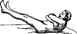
True cerebellar paralyses are observed very rarely; usually, in most patients with unilateral lesion, there is no noticeable difference in strength between the sides, and very rarely in some, a decrease in strength on the affected side is noted. The speed is reduced for most movements, especially for movements performed on command; to reveal this symptom, the patient is made to perform movements simultaneously with both hands, for example, to bring a finger to the nose: usually the finger on the affected side lags behind. The amplitude of movements, on the contrary, is increased on the affected side, and movements are performed with a certain impulsiveness, haste, and often go beyond the set goal. This lack of measure was described both in humans and in animals, and for it the name dysmetria (Luciani), hypermetria (Babinski) was proposed. Lack of measure in movements is observed not only in relation to space, but also to time—the extremity maintains the position it assumed for some act, and after the completion of this act. To reveal dysmetria, several tests have been proposed, for example, to touch the tip of the nose with a finger; usually the finger overshoots the goal, touches the cheek, but cannot remain in one place, and a whole series of oscillatory movements occur in it, which move the finger from one place to another; a similar test with touching the tip of the ear with a finger; this test is more complex and more difficult to perform; in the lying position of the patient there are even more errors. On the lower extremities dysmetria is manifested during walking; when attempting to place the heel on the opposite knee, the patient raises the leg very high and places it on the thigh; the same excessive range of motion is observed when the patient places a foot on a chair. Dysmetria worsens slightly with eyes closed, which has great diagnostic significance (see also Dysmetria). The phenomenon of 'pointing past the mark', 'missing the mark' (Vorbeizeigen) also belongs to the phenomena of dysmetria, which consists of the following: the subject is asked to touch with the index finger of an outstretched hand the finger of the doctor sitting opposite him and holding his own finger extended forward. At first this test is done with eyes open, and then with eyes closed; the patient usually misses, deviating to the healthy side. Preceding this test, the elicitation of nystagmus (by rotation or caloric stimulation of the labyrinth) does not have a great influence on the deviation of the finger, whereas if this test is performed with a healthy person, a very strong deviation of the finger will result. Sometimes dysmetria consists in that when performing some action the patient's extremity stops, not reaching the goal, and then slowly with oscillations reaches the goal; thus the act is performed in two moments; this phenomenon is called Bradyteleokinesis (Schilder) and is observed in both upper and lower extremities. Due to the disturbance of the ability to associate individual elementary movements in relation to each other in order, in time, and in space into one complex movement, the performance of a complex act is disturbed; this disturbance is called asynergy and dyssynergy, it is demonstrated by a series of tests. One of the tests, which is known under the name flexion combinee de la cuisse, consists of the following: the patient is placed on his back with arms crossed on the chest and asked to move to a sitting position—when attempting to sit, usually on the affected side (Figure 25) the leg is raised upward, whereas in a healthy person the legs remain immobile (Fig. 26) (see Asynergia).
In disease of the M., disturbance of complex sequential motor acts in the form of slowness or inability to alternate movements is also observed—adiadochokinesia (see). Dysmetria, tremor cause disturbance of writing—one of the frequent symptoms of cerebellar disease; the letters are very irregular, of different lengths and widths, the lines of letters are unequal, sometimes wide, sometimes narrow, with steps; a straight line cannot be drawn by the patient, it comes out winding; especially demonstrative is the disturbance of writing when drawing a zigzag line; if the patient writes with small letters, it is difficult to read anything; if the patient is asked to draw a line between two points, he either overshoots the marked point or falls far short of it. Tremor very often accompanies cerebellar diseases; it can be observed either only during movements or also in a state of rest, i.e., it can be kinetic and static. In milder cases only the smoothness of movements is disturbed: they become jerky, fragmented; in more severe cases of lesions of the M. the tremor resembles intention tremor, i.e., it intensifies when reaching the goal. Sometimes, if the patient performs the movement slowly and watches himself, the tremor may be absent. Tremor is very clearly revealed in the following test: take a glass, filled to the brim with water or shot, and carry it to the mouth. In the head and trunk the tremor is stronger when the patient performs some action standing; when sitting or lying down it decreases. Tremor is also explained by the impulsiveness of movements and lack of measure, which in normal conditions is corrected by antagonist muscles. Despite all desire and effort the patient cannot
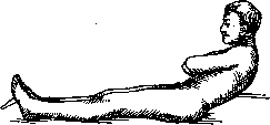
Figure 26.
it can stop tremor but can diminish it; distraction of attention increases the amplitude of tremor. Static tremor appears in some individuals always under the same conditions and in the same positions, e.g., when carrying a weight in an outstretched hand or with strong pronation of the hand, etc. In cerebellar patients, a special ability is observed to maintain a fixed position of the body or limb for a longer time than is possible for a normal subject; this condition is called cerebellar catalepsy. In markedly pronounced cases, the fixed part remains completely immobile until the researcher returns it to a normal position; absolute catalepsy is observed very rarely, but in most cases there are small oscillatory movements, especially at the beginning of the experiment at the moment of setting the limb. Dizziness is observed very frequently in diseases of the cerebellum; it has a rotational character, i.e., the patient feels that all objects are rotating around him or he himself is rotating around them. Dizziness can be constant and intensify with any movements, or it appears in attacks, often when changing position, e.g., from lying to sitting or from sitting to standing. Dizziness is always accompanied by nausea, and sometimes also by vomiting (see Dizziness). Speech is often impaired, a scanned monotonous speech with a nasal timbre is observed; each syllable is pronounced gradually; speech, like other motor symptoms, depends on dysmetria, adiadokokinesis, and the discontinuity of movements. Facial expression is very poor during speech and laughter, which is also a result of adiadokokinesis. Nystagmus is not particularly strong, horizontal, appears at extreme positions of the eyes; rotatory or vertical nystagmus occurs much less frequently; with unilateral lesion of the cerebellum, nystagmus appears when looking toward the affected side. In diseases of the cerebellum, changes in muscle tone are observed: upon palpation, they are flaccid, hypotonic; with passive movements, the muscles of the affected side do not offer any resistance, the amplitude of passive movements is greater due to the fact that the reflex of antagonists is diminished—there is hypotonia or hyposthenia, but sometimes, conversely, an increase in the reflex of antagonists is observed; then resistance with passive movements increases, and hypersthenia results. The state of tone is manifested not only with passive movements but also with reflex movements and with movements caused by mechanical irritation of the muscles; there are a number of experiments allowing to check the state of tone (see Tone). Tendon reflexes are altered, the responsive movements to irritations are faster, and their amplitude is increased; in some patients, a pendulum-like type of knee reflexes is observed, to reveal which the patient is seated on a table with legs hanging down and the ligamentum patellae is struck: the lower leg, instead of responding to this irritation once, describes several oscillatory movements of flexion and extension; on the affected side these phenomena are much more pronounced; the same applies to the reflexes of the upper limb with m. triceps; the plantar reflex is more lively on the side of the lesion. Postural reflexes fade. Movements caused by mechanical irritation of the muscle have a wider amplitude on the affected side, which is explained by stronger muscle contraction; it is especially noteworthy that the limb does not immediately pass into a state of rest, but describes several pendulum-like movements. The patient cannot have any influence on these involuntary movements, despite all efforts. With electrical examination by galvanic or faradic current, muscle contraction is stronger on the affected side. Superficial sensitivity is preserved, from deep sensitivity, sometimes the sense of weight, joint sensitivity is impaired: the patient orients himself less quickly in movements performed on the affected side; in the literature there are observations of injuries to the cerebellum, in which patients overestimated the weight, dimensions in space of some object; the threshold of irritation for differentiation of the sense of pressure increases. Finally, among the symptoms observed in diseases of the cerebellum (though rather rarely), associated movements should be noted, more strongly expressed in the lower limbs and sometimes spreading to all four limbs. To the symptoms of irritation of the cerebellum belong forced movements that appear suddenly: the body moves forward, backward, to the sides; sometimes such movements occur only in the limb: during some voluntary movement the hand is suddenly thrown aside; these movements can be explained by irritation of agonists or inhibition of antagonists; the phenomena when patients drop objects on the floor are explained by relaxation of agonists or irritation of antagonists. The above-described cerebellar syndrome develops either suddenly or gradually and remains more or less permanent depending on the process. Individual cases of periodic cerebellar ataxia have been described: in the patient, suddenly without any prodromal symptoms, disturbances of equilibrium, gait, writing, speech develop, nystagmus and other symptoms appear; after several hours everything passes, and the patient becomes completely healthy. There is a tendency to consider such a form of cerebellar ataxia as an equivalent of epilepsy with localization in the cerebellum. The combination of the above-mentioned symptoms varies depending on the localization of the pathological process, since anat.-physiol. data indicate different connections and functions in different parts of the cerebellum. A syndrome of lesion of the vermis and a syndrome of lesion of the cerebellar hemispheres are distinguished, which can be unilateral or bilateral. In diseases of the vermis, disturbances of equilibrium when walking, when standing, tendency to fall backward, strong dizziness, speech disturbances, nystagmus, tremor are observed. Since the functions of the upper and lower vermis differ, it can be assumed that the syndromes of their disease also differ. Loss of function of the upper vermis gives disturbances of equilibrium in the muscles of the head, speech disturbances, nystagmus, while lesion of the lower vermis leads to disturbances of equilibrium in the muscles of the trunk. The unilateral syndrome of the cerebellar hemispheres is characterized by disturbances of coordination in the limbs on the corresponding side, sometimes with predominant lesion of the upper or lower limb (adiadokokinesis, dysmetria, asynergy, tremor, changes in muscle tone, nystagmus). The cerebellar hemisyndrome may depend on lesion of the cerebellar cortex or its nuclei or may involve the fibers leaving or entering the cerebellum, most of which undergo decussation on their way; in cases when the cortex, subcortical nuclei and fibers before decussation are affected, the cerebellar hemisyndrome is observed on the same side; when fibers after decussation or those centers where they end or begin (nucleus ruber, thalamus opticus, gray matter of the pons) are affected, the hemisyndrome will be on the opposite side. All symptoms are more intensely expressed if the process involves the cerebellar subcortical nuclei. The participation of nucl. dentati in the myoclonic syndrome has been suggested. In diseases of the cerebellar peduncles, the cerebellar syndrome is almost always associated with other symptoms indicating simultaneous involvement of surrounding structures—cranial nerves or nuclei, motor, sensory pathways, sympathetic system—and these accompanying symptoms facilitate the topological diagnosis. Of the syndromes of the cerebellar peduncles, the syndrome of Wallenberg or the inferior cerebellar artery is most well known. In most cases, thrombosis of its branches is observed, which leads to softening in the retroolivary area. At the beginning of the disease—nausea, vomiting, disturbance of swallowing, headaches—then cerebellar symptoms join—disturbance of equilibrium, dizziness, dysmetria, adiadokokinesis on the side of the lesion; in addition, disturbance of sensitivity in the area of n. trigemini, crossed hemianesthesia, hemiparesis pharyngo-laryngo-palatina, sympathetic symptoms from the eye are observed. The cerebellar symptoms in this case depend on lesion of the nucleus of Monakow and the lateral nucleus of the medulla oblongata, sending fibers into corpus restiforme, fibers olivo-cerebellaris or of corp. restiformis itself, the bundle of Gowers or finally on lesion of the fibers or nuclei of the vestibular system (see Alternating syndromes). The syndrome of the superior cerebellar peduncle changes depending on whether the peduncle is affected before or after decussation. Before decussation, the peduncle can be affected simultaneously with tegmentum of Varolius bridge, and then cerebellar symptoms are present on the side of the lesion, while disturbance of sensitivity is on the opposite side. If the focus is very significant and extends low, it can simultaneously involve pedunculus cerebelli infer., and then cerebellar disturbances are expressed very strongly. Disease of the peduncle at the place of its exit from nucl. dentatus is often accompanied by lesion of n. trochlearis, so there is paralysis of m. obliqui super.
Lesion of the decussating pyramidal tracts is accompanied by disease of the red nucleus, so there is already a syndrome not of the pyramidal tracts, but of the red nucleus, or Benedikt's syndrome (see Alternating syndromes); when the process extends to the base of the pons, in addition to Benedikt's syndrome, there is Weber's syndrome. The syndrome of the middle cerebellar peduncle is observed with a focus in the base of the pons, and since pyramidal fibers are also affected, a very complex syndrome results, characterized by hemiplegia and bilateral cerebellar symptoms (due to damage to the pontine nuclei, pontine fibers, and peduncles). Under these conditions, the so-called pontocerebellar form of pseudobulbar paralysis often develops. Rotation around the axis is not noted in all cases. Diagnosis of cerebellar diseases. In the presence of a complete cerebellar syndrome, diagnosis does not present great difficulties, but in clinical practice one more often has to deal not with a pronounced syndrome, but with an incomplete one or with individual symptoms, and therefore the diagnosis, especially at the beginning of the disease when many of the characteristic symptoms have not yet appeared, often presents difficulties. At the same time, in some diseases of the cerebellum (tumors, abscesses, etc.), an early diagnosis makes it possible to save the patient through timely surgical intervention. Cerebellar ataxia must be differentiated from ataxias of other origins - ataxia in diseases of the frontal lobe of the brain, in disease of the labyrinth (see Labyrinthitis), tabetic ataxia (see Tabes), and finally from ataxia in general diffuse diseases of the central nervous system with localization in the cerebellum (multiple sclerosis, syphilis, encephalitis, etc.). The symptomatology of cerebellar disease very closely approaches that of labyrinth disease, in which there are also disturbances of equilibrium, coordination, gait, nystagmus, dizziness, etc., and therefore they are very difficult to differentiate (see Labyrinthitis, Labyrinth). Usually in labyrinth disease, all symptoms except disturbance of equilibrium are expressed less sharply, and in addition, phenomena from the side of hearing (hearing loss, noises) are observed; the main aid to diagnosis is the various methods of investigating vestibular functions (see Barany's method of investigation). The second question that must be decided is the localization of the lesion in the cerebellum itself, which also presents great difficulties, since rarely does the process involve only one part of the cerebellum, but usually extends from the vermis to the hemisphere or vice versa, and therefore a pure syndrome is not observed, and only through detailed questioning is it sometimes possible to determine where the process began. Pathology of the cerebellum. Developmental anomalies of the cerebellum. These include: 1) agenesis, 2) hypoplasia, and 3) dysgenesis. Under agenesis is understood the complete or partial absence of the cerebellum. Complete agenesis is a rather rare phenomenon. In place of the absent cerebellum, thickened meninges are found, which gather in folds and form the roof of the fourth ventricle; the vessels going to the cerebellum are very thin. Among the meninges in place of the cerebellum, there are mostly small formations somewhat resembling the cerebellum or one of its lobes (amygdala, flocculus, etc.). In partial agenesis, one or another part of the cerebellum is absent, more often a hemisphere, but cases of absence of the vermis have been described. The absent part is replaced by a stump, giving the impression of a miniature hemisphere, the size of a cherry pit or a nut; it is intersected by grooves and convolutions, the cortex is absent, but the white substance and nuclei are preserved. At the level of the absent parts, the meninges are thickened and form a pocket filled with fluid. When the hemispheres are absent, the adjacent parts of the vermis may also be affected, while when the vermis is absent, the cerebellum is as if divided into two parts. Agenesis of the cerebellum is sometimes accompanied by changes in other parts of the nervous system. Due to the rarity of observations, the clinical picture of agenesis is not sufficiently clear. In patients, cerebellar-type movement disorders are observed: the child starts walking late, walking is very difficult, all movements are awkward; at the same time there is also inadequacy from the psychological sphere - dementia, idiocy, and deficiency in physical development. Individual cases have been described in which the deficiency of the cerebellum manifested itself at a very late age (45 years, 53 years). In the anamnesis of such patients, syphilis is most often noted, which in the embryo causes meningoencephalitis, which can influence improper development. Hypoplasia of the cerebellum is characterized by the fact that the general configuration of the cerebellum is preserved, but its size is much smaller than normal. Two groups of hypoplasia are distinguished: in the first group, the general plan of structure is disturbed, different parts did not develop in parallel, some developed more strongly, others less. In the second group, the structure of the cerebellum is quite correct, all parts are developed proportionally, only its size is very small. When examining cases of the first group, traces of disease during intrauterine life are found (mostly meningitis): pia mater is thickened, injected; the cerebellum is very small, sclerotic, hard to the touch; sometimes only the peripheral laminae are affected, while in severe cases almost the entire cortex is affected, the laminae fuse together, their white substance disappears, while the subcortical nuclei remain normal; brachia conjunctiva retain their size, as they originate in the unchanged nucleus dentatus. This contrast between the state of the cortex and subcortical nuclei is characteristic of this form of hypoplasia. Very often this condition of the cerebellum is combined with changes in other parts of the nervous system - the meninges of the cerebral hemisphere are also thickened and injected, the ventricles are dilated, the cortex is thinned, the number of Betz cells is decreased; similar changes can be noted in the spinal cord, so in general the entire central nervous system is reduced in size. The second group of hypoplasia is characterized by a very small cerebellum, the weight of which may not exceed 20 g (normally 140-150 g), but all the grooves and convolutions of the cerebellum, as well as the subcortical nuclei, have a normal appearance: the meninges and vessels are unchanged; based on the weight, size, and general development, it can be determined at what month of intrauterine life the development of the cerebellum slowed down or stopped. Pure cases of hypoplasia are very rare. It is noted that other parts of the central nervous system are also significantly reduced in volume. Clinically, in such patients, cerebellar symptoms are observed - a staggering, unsteady gait, disturbance of equilibrium, disorder of coordination, speech disturbance, nystagmus; in most patients these symptoms are already revealed in early childhood, but cases have been described when they appear later - at 10 years, and finally there are indications in the literature of cases where during life the change in the cerebellum was not expressed in any way, but was discovered only at autopsy. Along with cerebellar symptoms, some others are observed, indicating a more extensive lesion of the nervous system; intelligence is greatly reduced, down to complete idiocy. Agenesis and hypoplasia can be combined with dysgenesis in the form of: 1) heterotopias - the presence in the white substance of accumulations of gray matter having the structure of cortex or subcortical nuclei, incorrectly positioned; 2) irregularities in the orientation of the convolutions and laminae, in some places giving the impression of their duplication. In addition to the cerebellum, heterotopias are also found in other parts of the nervous system. Dysgenesis can also be observed in isolation in a cerebellum that has a completely normal appearance. In developmental anomalies of the cerebellum, irregularities are also observed in the development of the skull, namely its posterior parts - the occipital fossae are reduced in size, larger on the side of the cerebellar lesion; protuberantia occipitalis interna is poorly developed and may be displaced more to one side, as well as other protrusions on the inner surface of the occipital bone. With unilateral lesion of the cerebellum, the difference between the two halves of the occipital bone is very significant: at first glance it seems that this bone deformation is the cause of the insufficient development of the cerebellum, whereas it is more likely that it is only a consequence. It should be noted that various developmental defects, as well as atrophies, are observed mainly in the hemispheres of the cerebellum (neocerebellum), while the vermis and flocculus (palaeocerebellum) are less affected or remain completely normal. This fact is explained by the fact that phylogenetically younger parts of the cerebellum are more vulnerable. Atrophies of the cerebellum (atrophia cerebelli) are divided into two groups: secondary atrophies, developing as a result of disease of the brain [see separate table (pp. 551-552), Fig. 6] and primary atrophies, depending on the localization of the pathological process directly in the cerebellum, in its meninges or vessels. Secondary atrophies of the cerebellum (atrophia cruciata cerebelli, atrophie croisée du cervelet) depend on the primary lesion of the opposite cerebral hemisphere; observed in hemiplegics. Atrophy of the cerebellum is most pronounced in cases where the brain disease developed in early childhood or intrauterinely; atrophies are also observed in adults, but very rarely: extensive lesions in the brain and a prolonged life after the cerebral process are necessary conditions for their occurrence.
In pathoanatomical examination, very extensive changes are found, both in surface and in depth (hemorrhages, softening on the surface of thrombosis) in the cerebral hemispheres, atrophy of the base of the corresponding half of the cerebral peduncle and the pons Varolii, and atrophy of the opposite pedunculus cerebelli ad pontem Varoli and the opposite cerebellar hemisphere; the vermis does not participate in the process due to the absence of connection with the brain. In the cerebellum, the atrophy affects the cortex and the subcortical white matter, as well as the subcortical nuclei—nucleus dentatus and emboliformis. The cerebellum atrophies unevenly, those of its lobules that have the closest connection with the cerebral cortex (lobus quadrangularis) suffer more; in the affected lobules, the convolutions are very thinned, mainly due to atrophy of the white matter; Purkinje cells are atrophied, less numerous or even completely absent; when nucleus dentatus is involved in the process, the pedunculus cerebelli ad corpora quadrigemina, which takes its origin there, is altered. Many theories have been proposed to explain the mechanism of these atrophies: 1) the myelogenous theory—reflection of brain disease on the cerebellum through the spinal cord; 2) the coincidence of brain disease and opposite cerebellar hemisphere—the simultaneous lesion of two structures cross-connected with each other; 3) the transneural or indirect action theory—brain disease through corticopontine and pyramidal pathways caused changes in the nuclei of the pons Varolii, leading to atrophy of the middle cerebellar peduncles, which originate there, and the opposite cerebellar hemisphere—the sites of termination of the middle cerebellar peduncles. The third theory is the most acceptable, which has been confirmed by experimental works (Monakov): in newborn animals, the brain was severely damaged, and after several years, autopsy revealed atrophy of the opposite cerebellar hemisphere. In these cases, the clinical picture of atrophy cannot be distinguished due to the presence of hemiplegia, which is usually very pronounced and masks the cerebellar symptoms. Since these cerebellar atrophies are discovered only at autopsy, there can be no question of therapy. - Primary atrophies of the cerebellum are also divided into two groups depending on the presence or absence of changes in addition to the cerebellum in the meninges and in the vessels. Cerebellar atrophy can be observed due to disease of its meninges or vessels. Meningitis arising on the basis of various infectious diseases, especially in childhood, can cause atrophy in the cerebellum; arteriosclerosis, especially early, by reducing nutrition, can lead to atrophy; syphilis can affect the development of atrophy either by direct action on the vessels of the cerebellum or by creating a certain predisposition, due to which the cerebellum becomes more vulnerable. Trauma can also be the cause of cerebellar atrophy through hemorrhage in the meninges. In autopsy of such cases, the cerebellum is found to be smaller in size, sclerosed, hard to the touch; the changes are unevenly distributed in the cerebellum: sometimes the hemispheres suffer more, in other cases the vermis; in this case, some lobules or parts of lobules suffer more than neighboring ones. The meninges are thickened, cloudy, and adhered to the cerebellar cortex. On section, it can be seen that the superficial convolutions are more atrophied than those lying deeper. The meninges, together with the vessels penetrating between the laminae of the cerebellar lobules, are greatly thickened and adhered to the laminae, which are sometimes severely destroyed and replaced by fibrous tissue and gliosclerosis. From the meninges, fibrous strands extend into the depth. In processes less intensely expressed, the structure of the cortex can already be seen in the convolutions, the presence of Purkinje cells, more or less strongly altered, intense glioproliferation. The vessels are greatly altered, and there is a certain parallelism between the intensity of changes in the vessels and meninges and the degree of atrophy and sclerosis of the cerebellar cortex. In the central nuclei of the cerebellum, secondary changes are observed in the projection fibers going to them, caused by lesions in the cortex or possibly also by disease of the vessels; the number of fibers in the nuclei is reduced, the cells are less numerous and smaller, in general the nuclei are reduced in size. If death occurs a long time after the onset of the disease, the lesion of the cortex affects not only the central nuclei but also the cerebellar peduncles. Along with changes in the cerebellum, changes are also observed in other parts of the central nervous system (hydrocephalus). In the acute phase of the disease, the involvement of the cerebellum in the process is characterized by a clearly expressed cerebellar syndrome, and then a gradual improvement of symptoms is observed, but complete recovery never occurs: gait remains unsteady, staggering, the patient walks with widely spaced legs; in the limbs there is a disturbance of coordination, tremor; difficulty of speech, etc. Very often there is a decrease in intelligence, which depends on the simultaneous lesion of the brain. The symptoms may be uneven: more pronounced on the side of the limbs or on the side of the trunk. Partial improvement of symptoms after the acute onset of cerebellar phenomena during an infectious disease is the main feature of cerebellar atrophy on the basis of meningo-cerebellitis.-When the cerebellum is in the stage of atrophy, any therapy is powerless. To the second group of primary atrophies of the cerebellum belong those cases in which there is a lesion of the cerebellar tissue itself, while the meninges and vessels are unchanged. Among parenchymal diseases of the cerebellum, several types have been distinguished: 1. Pure atrophy of the cerebellar cortex (atrophie lamellaire Thomas), in which the lesion affects exclusively the cortex: Purkinje cells in some convolutions completely disappear, while in neighboring ones they are more or less preserved; the disappeared cells are replaced by a dense network of glial fibers; in the molecular layer, the number of glial elements is increased. The disease is diffuse, both vermis and hemispheres are affected; meninges and vessels are normal. In general, the entire cerebellum is reduced in size sometimes by gi; clinically the disease is expressed by disturbance of equilibrium in standing and walking, tremor. To this form is added familial degeneration of the cerebellum (Holmes), characterized by primary and progressive degeneration of the nerve elements of the cerebellar cortex; the efferent system is excluded, while the afferent one remains normal; clinically the process is characterized by disturbance of gait, speech, uncertainty of movements, tremor of the head and limbs, nystagmus, absence of pyramidal and sensory disorders. Onset between 33 and 40 years. 2. Atrophia olivo-ponto-cerebellaris Déjerine, one of the most accurately described forms of atrophy; onset of the disease at the age of 40; course slow, gradual; with careful questioning it is possible to find indications that in younger years there was difficulty in walking, in performing certain movements. Anatomically this form is characterized by: 1) symmetrical atrophy of the cerebellar cortex, more pronounced in the hemispheres; 2) complete atrophy of the gray matter of the pons and complete degeneration of the middle cerebellar peduncles, with good preservation of brachia conjunctiva; 3) sharply expressed atrophy of olivae inferioris, olivae accessoriae and nucleus arciformis, degeneration of fibrae arcuatae externae and corpus restiformis. The atrophy of the cortex consists in atrophy of all 3 layers of the cortex, as well as the myelin fibers of the convolutions; Purkinje cells are unevenly distributed: in some places absent, in others very rare; nucleus dentatus is smaller in size, has fewer convolutions, but the cells are numerous and normal. The white matter of the cerebellum is greatly reduced in volume. Clinically, disturbance of movements of the whole body is observed, when transitioning from sitting to standing position and back; the whole body comes into oscillatory jerky movements at this time. When standing, the patient widely spreads the legs and is unable to maintain balance. When walking, all phenomena are intensified. Isolated movements of the limbs are relatively preserved, although slow, oscillating and uncertain. Strength and sensitivity are preserved, speech is scanned. Reflexes are somewhat increased. The development of the disease is slow but progressive; death usually occurs from accidental causes. 3. Atrophia olivo-rubro-cerebellaris (described by Lhermitte in 1909), a very rare disease, anatomically characterized by 1) sclerosis and complete demyelination of olivae infer., as a result of which fibrae olivo-cerebellares have disappeared in large numbers, corpus restiformis is partially reduced; 2) global atrophy of the cerebellar cortex, expressed in the disappearance of Purkinje cells and most of the granule cells with their replacement by glial elements; 3) atrophy of nucl. dentati and almost complete disappearance of brachium conjunctivum, as well as changes in nucl. ruber. Clinically the disease proceeds in the form of a clearly expressed cerebellar syndrome. 4. Atrophia olivo-cerebellaris (described by Holmes) very closely approaches the previous form.
Anatomically characterized by: 1) symmetrical and generalized atrophy of the cerebellar cortex with disappearance of Purkinje cells and cells in the molecular layer replaced by glial tissue, atrophy and sclerosis of the cerebellar white matter, 2) changes in the inferior olivary nuclei and accessory olivary nuclei with degeneration of cerebello-olivary fibers. The nuclei remain normal. The clinical picture resembles that of multiple sclerosis. 5. Senile atrophy of the cerebellar cortex is characterized by glial thickening in the molecular layer and glial proliferation in the Purkinje cell layer and around blood vessels. Such changes do not occur throughout the entire cortex, but are partial and selective with respect to certain gyri. 6. Cerebellar atrophy, or primary atrophy of the central nucleus, is characterized by: 1) small size of the cerebellum with preservation of the cortex and white matter, 2) atrophy of the dentate nucleus with reduction in the number of its convolutions and atrophy of its cells, 3) significant atrophy of the brachium conjunctivum, 4) reduction of the red nuclei, Forel's thalamic bundle, 5) preservation of the inferior olivary nucleus. 7. Hereditary cerebellar atrophy, or hereditary cerebellar ataxia (hereditary cerebellar ataxia, Marie's and Friedreich's disease)-see Ataxia.-Etiology. As seen from the above, some cerebellar atrophies are acquired and therefore can develop at any age-in the embryo, in childhood, adolescence, and in adults. Their development is associated with infection or trauma. Depending on the age at which the cerebellum was affected, there are various developmental anomalies or atrophies. Another group of atrophies is hereditary and specifically dominant diseases (hereditary cerebellar ataxia) with slow and progressive course. Special attention is given to atrophies that, by their slow and progressive course and anatomical changes, resemble the previous group, while indications of heredity and familial occurrence are absent; sometimes such atrophy is found in persons who have reached mature age (40-50 years), but can also occur in childhood; the late appearance of symptoms is explained by the wide compensatory function of the opposite hemisphere (in unilateral lesions) or other parts of the brain; in children, such compensation by neighboring structures occurs better. The question of the etiological factor remains open. It is possible that these sporadic progressive atrophies are only sporadic cases of hereditary forms. Thus, olivo-ponto-cerebellar atrophy was initially described as a sporadic disease, but later familial cases of this disease were described. Diagnosis. The symptom complex observed in cerebellar atrophies resembles the clinical picture developing in other cerebellar diseases-tumors and abscesses. Differential diagnosis is based on the presence of general cerebral symptoms in tumors, elevated temperature, changes in blood composition in abscesses; it is more difficult to differentiate these atrophies from multiple sclerosis and from various forms of atrophy among themselves; symptoms are more intensely expressed when the central nuclei are affected.-The course of the disease varies depending on the etiological factor: atrophies that developed on the basis of meningitis or encephalitis may gradually improve, but sometimes after improvement there is again a deterioration for some time and then improvement again, so that an intermittent course results. In atrophies of other etiology (hereditary abiotrophies) the disease slowly and gradually worsens.-The prognosis for life is favorable, for recovery hopeless.-Therapy-symptomatic treatment.-Prevention-see Infantile paralysis. Injuries to the cerebellum are very rare; during the last war according to Marie and Chatelin on 5,000 skull injuries there was only one cerebellar injury, and according to Chiray 2:4,100; more correctly, such patients rarely come to clinical examination, which is explained by the position of the cerebellum in proximity to formations very important for life (medulla oblongata), on which an injury to the cerebellum almost inevitably reflects and causes a fatal outcome, if not directly from the injury, then in a very short time, as a result of which only very mild injuries came under observation. The cerebellum can be damaged either directly by a penetrating object or by fragments of the skull that burst upon tangential injury. Often the tissue of the cerebellum and around the wound is affected due to hemorrhage, edema, nutritional deficiency or finally suppuration; part of the tissue under the influence of unfavorable factors may necrotize and be eliminated from the wound. Clinically, in cerebellar injury, the most typical picture of the cerebellar syndrome is observed, which varies depending on the location of the injury (see above). Gradually the cerebellar symptoms somewhat subside (due to the disappearance of edema, absorption of hemorrhage) and only those that depend on the destruction of the cerebellum remain; the exception is those cases where wound contamination and abscess formation occur, then the picture changes and general cerebral symptoms are added to the cerebellar symptoms.-Diagnosis does not present great difficulty in the presence of injury.-The prognosis remains uncertain for a long time and only after several months can a favorable outcome be spoken of.-Therapy-depending on the possibility of surgical intervention (see below).-In addition to injury, other traumatic factors can affect the cerebellum-blow, fall on the back of the head; even if they are not accompanied by skull fractures, they can still cause more or less extensive hemorrhages in the cerebellum, cerebellar concussion.-The clinical picture of the cerebellar syndrome depends on the intensity and location of the lesion. In favorable cases, gradual improvement occurs due to absorption of the hemorrhage; complete restoration of all functions can be expected.-Diagnosis presents no difficulties.-Therapy-rest, ice on the occipital region. Disturbances of circulation in the cerebellum are observed in different forms. In anemia, hyperemia, arteriosclerosis diffusely affecting the entire central nervous system, the cerebellum suffers along with other parts, and therefore its symptoms enter as part of the general picture of central nervous system damage. Hemorrhage into the cerebellum-a rather rare disease. The clinical picture is not especially clear, since it is difficult to determine which symptoms are due to cerebellar damage and which depend on damage to surrounding structures. Hemorrhages into the cerebellum are much less common than cerebral hemorrhages, their frequency is 1:12 (Rochoux), 1:30 (Hillairet) and they depend on the same causes as cerebral hemorrhages-arteriosclerosis, syphilis and other infections, intoxications, trauma (especially in newborns) (see Cerebral apoplexy). The highest frequency of hemorrhages is observed between the ages of 50 and 80 years; they occur due to rupture of miliary aneurysms. In size, large and small hemorrhages are distinguished. Hemorrhages from the artery of the dentate nucleus can be so extensive that they involve not only the corresponding hemisphere but also the vermis and the hemisphere on the opposite side, as well as the fourth ventricle. Small hemorrhages occur in the vermis and in the hemispheres-in the white matter, in the central nuclei and in the cortex. Multiple foci are not uncommon. Of the cerebellar peduncles, hemorrhages are more often observed in the middle one. The clinical picture varies greatly depending on the size of the hemorrhage, which is often preceded by prodromal symptoms in various combinations: headaches in the occipital region, vomiting, irregularity of pulse, dizziness, transient general weakness. The disease can begin suddenly, in a stroke-like manner; in these cases the patient usually dies immediately or after a short time. Less extensive hemorrhages do not proceed so violently-the prodromal symptoms intensify, especially vomiting, involuntary movements appear, disturbances of equilibrium, general malaise, then loss of consciousness; in severe cases, disturbances of respiration, pulse, appearance of nystagmus, conjugate movements of the head and eyes, tonic and clonic convulsions are observed; this complex of symptoms indicates rupture of the hemorrhage into the fourth ventricle and medulla oblongata; in more favorable cases consciousness is restored and then a number of cerebellar symptoms appear, which either gradually improve or remain forever, gradually decreasing in intensity. Cerebellar symptoms are usually observed on one side. Improvement progresses and sometimes all symptoms as if completely disappear-only very fine examination reveals one symptom or another; some of the above symptoms do not belong to the cerebellum but indicate involvement of the medulla oblongata, meninges and fourth ventricle.
Differential diagnosis must be made 1) with hemorrhages in the brain; in the latter, paralysis, pyramidal symptoms, pathological reflexes are observed; 2) with hemorrhages in the meninges, accompanied by rigidity of the neck and limbs, convulsions; 3) with hemorrhages in the brainstem, giving alternating syndromes (see) and 4) with various processes in the C. itself. - The prognosis depends on the intensity and localization of the hemorrhage: almost instantaneous death after severe hemorrhages or complete recovery after small hemorrhages, and between these two extremes various outcomes of the disease. - Treatment and prevention-see. Apoplexy of the brain. Softening of the C. is observed even less frequently than hemorrhages because the cerebellar arteries depart from the a. basilaris at a large angle, which prevents an embolus from entering them, while softening due to thrombosis as a result of arteritis (arteriosclerosis, syphilis) is also very rare due to anastomosis between the arteries. Softening can be observed in all parts of the C., more often in the cortex than in the white matter or in the nuclei; foci can be single or multiple. In large, long-standing softenings, deformation of the cerebellum is observed; a cyst may form in the place of softening [see separate table, figures 2 and 4]. On autopsy of the C. with fresh thrombosis, it can be noted that the softened tissue sharply differs from healthy tissue by its pale color and its softness; the pia mater is infiltrated, edematous and as if raised above the underlying tissues; later the softened tissue takes on a yellowish color and sinks in; at the border, hemorrhagic foci are very often found; in softening of the cortex, only a framework of neuroglia remains from the nervous tissue. Symptoms vary depending on the localization of the foci and their spread. The onset of the disease is preceded by a more or less prolonged prodromal period (up to several days), but a sudden onset is also observed, with the patient immediately falling into a comatose state. The prodromal period is characterized by headaches in the occipital region, dizziness, involuntary movements of the head. Clinically, in softening, a typical cerebellar syndrome is observed, the intensity and character of which vary depending on the localization of the focus- sometimes mental disorders, deviation conjuguee des yeux et de la tete toward the side of the disease are observed. Cases of fairly extensive softening of the hemispheres of the C. without involvement of the vermis have been described, which occurred without any cerebellar symptoms. In apoplectic onset, the outcome is most often fatal-the patient dies in the first days of the disease, being in a comatose state before death-loss of reflexes, paralysis of sphincters, convulsive movements on the affected side. In small softening, symptoms gradually disappear; when the subcortical nuclei are involved, improvement is very slow and some cerebellar symptoms always remain. In acute cases with sudden onset, it is difficult to determine whether we are dealing with a hemorrhage or softening of the C., but the further course of the disease sometimes decides the question. Softening can also simulate multiple sclerosis, chorea, and in this case too only the course can decide
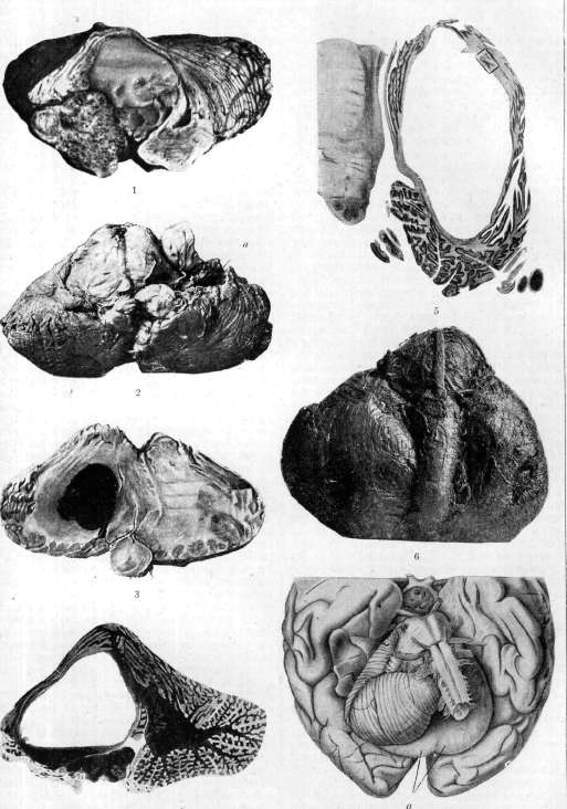
Figure 1. Tumor of the cerebellopontine angle, having grown into the cerebellum and undergone colloidal degeneration in it. Figure 2. Cyst (a) of the left hemisphere of the cerebellum, formed as a result of softening. Figure 3. Hemorrhage into the white matter of the right hemisphere of the cerebellum. Figure 4. Cyst of the right hemisphere of the cerebellum. Figure 5. Cystically degenerated cerebellar tumor. Figure 6. Atrophy of the left hemisphere of the cerebellum. Figure 7. Cyst (a) of the left hemisphere of the cerebellum.
See article Cerebellum. Treatment. In a young patient, antisyphilitic treatment must be carried out; in more mature age, such treatment should be carried out with caution. There is no specific treatment for softening. Abscesses of the Cerebellum. Among purulent diseases of the brain in general, abscesses of the Cerebellum rank second in frequency (first place is occupied by brain abscesses). They are observed: 1) in head injuries in the occipital region with damage to the bones of the skull and with penetration of a wounding object or bone fragments into the skull cavity; they are significantly less common with simple concussion of the occipital region without violation of the integrity of the coverings. 2) In diseases of the bones of the skull in the same area (osteomyelitis of various origins). 3) In diseases of the middle ear (otitis media), an abscess is a relatively rare complication; according to French statistics, out of 1100 cases of purulent disease of the ear, one case of an abscess of the Cerebellum occurs; in chronic otitis media, an abscess is observed more often (80-88% of all cases of ear-derived abscesses of the Cerebellum) than in acute cases (10-12%) (Neumann, Heiman); abscesses are rare before the age of 10 and after 30 years; the period of development of the abscess after the onset of the ear disease is very uncertain - from several days to several years; some infectious disease, injury can activate the process in the ear and cause the formation of an abscess in the Cerebellum. Usually the abscess develops on the side where there is an ear disease. 4) Abscess of the Cerebellum of metastatic origin - in septicemias, pyemias, endocarditis, in broncho-, pleuropneumonias, in some infectious diseases. -Pathological anatomy. When opening the skull box, some changes can be noted: dura mater is hyperemic, edematous, strongly tense in the posterior parts due to the bulging of the underlying parts; when opening the dura mater, the changed Cerebellum can be seen; in it, significant asymmetry is observed - since the abscess is localized mostly in the white matter of the hemisphere of the Cerebellum, the corresponding hemisphere increases in size not only due to the presence of pus but also due to edema; vermis deviates to the healthy side; the contours of the medulla oblongata and the pons of Varolius are also greatly changed. The gyri located above the abscess are strongly stretched, the grooves are smoothed out; when the abscess is located directly under the cortex, the color of the cortex of the Cerebellum also changes, which takes on a greenish-yellowish tint; the size of the abscess varies greatly - from a small hazelnut to the complete replacement of the hemisphere of the Cerebellum, and the Cerebellum itself can be reduced to a very thin shell, in which elements of the cortex of the Cerebellum can hardly be found; in most cases, the abscess is single, rarely there are two or more; simultaneous development of an abscess in the brain is also observed. Sometimes the presence of a fistula can be noted, which leads from the abscess to the surface of the Cerebellum, and then in this place there are adhesions of the Cerebellum with the dura mater. The cortex can be covered with a shell, more or less infiltrated with pus. The pus does not always have the same appearance: it can be thick or more liquid, yellowish or greenish, sometimes with a very strong repulsive odor; among microbes in the pus, staphylococci, streptococci, diplococci are found, but the pus can also be sterile. Two anatomical forms of abscess are described - diffuse abscess and encapsulated. In the first case, the abscess does not have its own capsule, but infiltrates the nervous tissue; in the second case, it has a very dense capsule, giving it the appearance of a purulent cyst. The nervous tissue at the site of the cyst is destroyed, around the cyst it is edematous, softened; cells near the abscess are also greatly changed, up to their complete disintegration; neuroglia is greatly proliferated. -Symptomatology. Despite the almost constant localization of abscesses in the hemisphere of the Cerebellum, still in addition to the syndrome of the hemispheres, the syndrome of the vermis is also observed, which suffers due to edema, compression, displacement; moreover, in an abscess of ear origin, the vestibular system also suffers, so that in general a very complex syndrome is obtained. If the abscess develops as a complication of otitis media, then to the symptoms of the labyrinth, general cerebral phenomena usually join: headaches in the occipital region, vomiting, dizziness, increased temperature, general weight loss, fatigue, lethargy, nystagmus; later, stagnant papilla and cerebellar phenomena join, which are sometimes difficult to detect due to the severe general condition of the patient. When the general symptoms somewhat subside, the symptoms of the disease of the Cerebellum appear, changing depending on the localization of the process and on the greater involvement of one or another part of the Cerebellum (see above). To them are added rigidity of the neck, pyramidal symptoms and other symptoms depending on the compression of the medulla oblongata or the pons of Varolius; all these symptoms can be observed on the other side, while the symptoms of the Cerebellum are observed on the side of the lesion. Examination of the cerebrospinal fluid may not reveal anything pathological, sometimes hyperalbuminosis, hyperglycemia are noted; in superficial abscesses, cellular elements (polynucleosis, lymphocytosis, etc.) are found in it; the fluid usually flows out under pressure (see Cerebrospinal fluid). In later stages of the disease, mental phenomena are observed - confusion, stupor, etc. The development of symptoms is gradual or sudden, depending on the severity of the infection. -Diagnosis of an abscess of traumatic origin does not present great difficulties - trauma in the anamnesis, severe general condition, cerebellar symptoms; diagnosis of an abscess of ear origin is usually facilitated by the presence of otitis media, to which cerebellar symptoms are added; it is also necessary to differentiate with sinus thrombosis, with meningitis (see). -Prevention consists in the careful treatment of skull wounds, ear diseases and other purulent inflammatory processes in the body. -The prognosis is very serious. -Treatment in case of recognition of an abscess of the Cerebellum is only surgical (see below). With timely intervention, recovery is possible; statistics of results are contradictory: according to Bourgeois, recovery in 21 cases out of 38; according to Wildenberg - in 32 out of 63; according to Neumann in 40 cases out of 196. -Inflammatory processes in the Cerebellum are not observed in isolation, and the cerebellum is affected along with other parts of the central nervous system, but sometimes the cerebellum is affected more severely than other parts and then they speak of encephalitis with localization in the cerebellum (see Encephalitides). Tumors of the Cerebellum [see separate table III (art. 551-552), fig. 1 and 5] are one of the frequent diseases of this organ; they are especially common in children, in whom among all parts of the central nervous system, tumors in the Cerebellum rank first in localization. (Statistics and structure of tumors - see Brain.) In their structure and origin, tumors of the Cerebellum do not differ from tumors of the cerebral hemispheres. They are divided into true tumors and false ones (pseudotumor). The former can be primary and secondary (metastases). Primary tumors originate in the Cerebellum itself or in its surrounding membranes; they include: 1) gliomas, the size of which can vary from that of a small pea to a chicken egg; they develop either in the tissue of the Cerebellum itself or from the ependyma of the fourth ventricle, and then grow into the Cerebellum. Sometimes in gliomatous elements of the tumor, myelin fibers and nerve cells are found (neurogliomas). 2) Sarcomas develop either from the dura mater, or from the periosteum, or from the soft meninges and the adventitia of vessels and reach very large sizes; they begin on the periphery of the Cerebellum and then grow into it; they are quite common (13 cases of sarcoma out of 86 cases of tumor of the Cerebellum, according to Starr), especially in children (10 cases out of 35). Psammomas, endotheliomas, lipomas, myxomas, cholesteatomas, angiomas are also encountered. -Secondary tumors are also not uncommon (in 13 cases out of 63); they can be single or multiple; their primary focus is different: lungs, stomach, kidney, adrenal gland, etc. To pseudotumors belong tubercles, gummas, aneurysms, encapsulated meningitis and serous cysts of the soft membranes. Tubercles are found in the Cerebellum more often than in the cerebral hemispheres, their size is very diverse, they can be single or multiple, with localization in the cortex and in the white subcortical substance; they are usually well demarcated from the surrounding brain tissue; initially they have a grayish-rose color, later yellowish due to caseous disintegration. -Gummas are rarer, can be located either in the Cerebellum or in the membranes, from where they penetrate into the cortex of the Cerebellum. -Aneurysms of the cerebellar arteries are rare. Symptomatology. The symptoms observed in tumors of the Cerebellum can be attributed to three groups: 1) general cerebral symptoms due to increased intracranial pressure; 2) cerebellar or focal symptoms and 3) symptoms of compression. First, general cerebral symptoms usually appear, which are not characteristic only for tumors of the Cerebellum, but are observed in all tumors inside the skull box, but in cerebellar tumors they are usually more intense and appear earlier, which is explained by the location of the cerebellum in the posterior cranial fossa - a very narrow space, tightly covered above by the tentorium cerebelli.
The slightest change in the size of the cerebellum can compress the vena Galeni magna and the nearby sinuses (sinus rectus, transversus), which causes the early appearance of general cerebral symptoms and their rapid development. Among the general cerebral symptoms, the following are observed: headaches in the occipital region, spontaneous or under the influence of external irritations (percussion, movement); they radiate throughout the back of the head and into the neck muscles; they can be constant or appear in the form of attacks, accompanied by dizziness, vomiting; vomiting generally appears quite early and is very frequent. Stagnant papilla in tumors of the C. appears almost always early, intensifies very quickly and leads to blindness. Dizziness exists in all cerebellar tumors, even in the absence of other cerebellar symptoms and thus is a symptom depending on increased intracranial pressure, not a focal symptom; dizziness can be observed both when changing positions and in all positions, it depends on the edema of the inner ear and compression of the labyrinth; it is often accompanied by vasomotor reactions. With lumbar puncture due to increased intracranial pressure, fluid flows out under very high pressure (30-45 cm, sometimes up to 100 cm in the recumbent position instead of the normal 15 cm). Generally, in case of suspicion of a tumor in the posterior cranial fossa, puncture is contraindicated, because when pressure decreases, displacement of the formations in the posterior cranial fossa (medulla oblongata, Varolius bridge) may occur, which can lead to sudden death. In small children, increased intracranial pressure can cause divergence of sutures, expansion of fontanelles. - Cerebellar symptoms at the beginning of the disease can be very inconspicuous and be lost among the general cerebral symptoms caused by the tumor, which, once appearing, gradually increase; the character of cerebellar symptoms changes depending on the localization: in disease of the vermis, there is a disorder of gait, balance, whereas in tumors of the hemispheres - disorder of coordination of movements (see above cerebellar syndromes); gradually developing, the tumor from the vermis spreads to the hemispheres or vice versa, due to which the purity of syndromes is lost - symptoms of disease of the vermis are joined by symptoms of the hemispheres and vice versa. Only through very strict analysis is it possible to establish which symptoms appeared first. - Symptoms appearing due to compression are very numerous due to the large number of surrounding formations that can be compressed; most cranial nerves are affected, primarily the VI*-VII-VIII cranial nerves as being in the closest proximity to the C.; their damage causes paralysis or insufficiency of musculus rectus externi, paralysis or paresis of facial muscles, decreased hearing or, conversely, with irritation of n. VIII - noise in the ears; IV pair, V - disorder of sensitivity on the face, especially on the mucous membranes, X - change in pulse, vomiting can also be involved; the other nerves are also affected. Compression of the pyramidal pathways is also observed - paralysis, paresis with pathological reflexes on the opposite side or on the same side; sometimes the combination of observed symptoms takes the character of alternating syndromes. - The onset of the disease is usually slow and inconspicuous - with general cerebral symptoms, to which cerebellar symptoms gradually join; their increase can be gradual or in the form of attacks (tumors of vascular origin); in the development, very brief remissions can be observed, after which the progressive development continues. The duration of the disease is several months (as an exception - several weeks) or the disease drags on for a year or more. Death is mostly sudden, from hemorrhage into the tumor, but it can also depend on compression or on gradually increasing cachexia. - Diagnosis is made on the basis of the presence of cerebellar symptoms and symptoms of increased intracranial pressure. In the presence of only the latter and in the absence of focal symptoms, diagnosis is made on the basis of the localization of headaches, presence of severe dizziness, frequent vomiting, early appearance and rapid development of stagnant papilla. If these symptoms are joined by cerebellar ones, then differentiation with an abscess of the C. is necessary. The presence of temperature, polynucleosis in the blood, corresponding etiological moment (otitis media, skull injury) speak for an abscess. Extracerebellar tumor (cerebellopontine angle) is characterized by the onset with auditory phenomena, involvement of a large number of cranial nerves and significantly less pronounced symptoms of the C. Tumors of the frontal lobe can be accompanied by symptoms of cerebellar ataxia, but the presence of other focal symptoms allows for a correct diagnosis (see Frontal lobe). Establishment of the localization of the tumor in the C. itself is based on physiological data (see above physiology and symptomatology). The nature of the tumor in some cases can be suspected (e.g. in gumma, tbc, metastatic tumors) due to the presence of symptoms from other organs and results of some laboratory investigations (RW, chemical change of cerebrospinal fluid, reaction to tuberculin, etc.), but in all other cases, the character of it is concluded from the course, intensity of symptoms, etc. - Treatment - symptomatic; lumbar puncture can for a short time improve the condition - reduce headache, dizziness, cerebellar symptoms; but since it represents the danger of sudden death, it is recommended not for therapeutic purposes, but only for diagnosis. Radical treatment - surgical (see below). Cysts of the C. in their symptomatology approach tumors, from which they differ in their origin and course. Cysts of the C. can be a consequence of developmental anomaly and come from normal cavities of the brain and from diverticula of the ventricles - these cysts are called primary by some authors. Others are formed either due to softening of tissue, resorption of hemorrhages or are a result of atrophic or inflammatory processes (e.g. encapsulated meningitis - meningite enkyste, arising on the basis of chronic inflammation of the membranes; its favorite place is the cerebellopontine angle; some authors connect its origin with tbc, syphilis, ear disease - see Meningitis). The greatest interest is presented by the so-called primary cysts as a completely independent and definite form. There is no firmly established view on the origin of these cysts; they are explained by developmental anomalies: 1) during the development of the C., a temporary protrusion of the IV ventricle and formation of His-Bolkowski's cleft occurs; separation of this cleft leads to the formation of a preformed cavity, which, stretching with accumulating fluid, turns into a cyst (Henschen); 2) penetration in the embryonic period of life into the substance of the C. of parts of the soft meninges, which then separate off and form the cavity from which later, due to accumulation of fluid in it, a cyst is formed (Vershilov); 3) congenital disease of the ependyma of the IV ventricle and surrounding neuroglia - congenital ependymitis with sclerosis of neuroglia; similar changes explain hydrocephalus and syringomyelia, uniting these three diseases into one nosological group (Muratov); thus, from the point of view of this doctrine, a cyst of the C. is nothing other than a syringomyelic cavity in the C. The cavity of the cyst can have a connection with the IV ventricle or be independent; sometimes in the C. new formations are simultaneously observed. The size of the cyst is very different; it can occupy the entire hemisphere of the cerebellum, due to which it is stretched like a bag [see separate table (art. 551-552), figure 7]; its configuration is changed, the gyri and sulci are smoothed, compressed; sometimes the cyst occupies only a part of the substance of the C., but from the main cavity small cysts can extend deep into the substance; cysts are also observed in the hemispheres and in the vermis. The cavity of the cyst is lined with a smooth shiny membrane; the contents of the cyst can be liquid or semi-liquid (colloid); the amount of fluid can reach up to 30 cm3; specific gravity of the fluid about 1.006, it contains small amounts of protein. In very large cysts, displacement of the medulla oblongata and Varolius bridge is noted. The walls of the cyst of the C. consist of highly proliferated neuroglia; glial fibers are intertwined in all directions; between the fibers, Deiters' spider cells are visible; on the inner wall, in places, single cuboidal cells, apparently ependymal, poorly preserved their structure; a complete continuous ependymal covering is rarely observed; usually, hyperplasia of the ependymal covering is also noted in the ventricles, which in the form of massive protrusions extends into the cavity of the ventricles and the underlying white matter. Sometimes near large cavities, small independent closed cavities or islands of ependymal cells are found. Very often, cavities in the spinal cord are also found simultaneously, having the same structure as the cavity in the C.
In the vicinity of the cyst, the white matter is softened and loosened, with a reduction in nerve fibers; the cortex near the cyst is thinned, and the number of Purkinje cells is reduced. The pia mater shows no particular changes. Thus, cysts of the cerebellum can be a congenital condition that remains hidden for a long time; they are discovered in young age-between 20 and 30 years. Of 37 cases of cerebellar cysts, 19 cases fell into this age group; up to 10 years there were 4 cases; from 10 to 19 years-7 cases; between 30 and 50 years-9 cases and over 50 years-3 cases" (Versilov).-The disease begins gradually with general cerebral symptoms, to which cerebellar symptoms soon join depending on the localization (see above). Sometimes cerebellar symptoms appear simultaneously with general cerebral symptoms, and sometimes they precede them. Later, symptoms of compression of surrounding structures appear. During the course of the disease, short-term remissions may be observed. Death occurs from the same causes as in tumors; the duration of the disease ranges from 2 months to 2-3 years.-Differentiating cerebellar cysts from cerebellar tumors is almost impossible; some authors allow for the possibility of a diagnosis based on the course of the disease with fluctuations and remissions. A diagnostic puncture by Neisser-Pollack was proposed as an auxiliary means for differential diagnosis (see below).-Therapy-only surgical intervention. Parasitic cysts of the cerebellum are a very rare disease; among parasites, the cysticercus (cysticercus cellulosae) and echinococcus (echinococcus) should be noted. The cysticercus resembles a blister the size of a millet seed but can reach the size of a walnut; in rare cases there is only one such blister, but more often there are very many; sometimes the blisters do not sit separately from each other but, developing from the main one, remain connected to it (cysticercus racemosus). The cysticercus can develop anywhere in the cerebellum-in the cortex, in the white matter, in the peduncles, in the walls of blood vessels, as well as in the meninges, mainly on the base of the cerebellum near the pons of Varolius.-The echinococcus resembles a blister or tumor the size from a pea to a man's fist; it can be solitary, multiple, and multilocular; it is located either on the surface or in the depth of the brain tissue. The development of the cysticercus and echinococcus blister is accompanied by a reaction from the surrounding tissue-local encephalitis, softening (when a nearby vessel is blocked), small hemorrhages; when localized in the meninges-fibrous meningitis; in echinococcosis this reaction is more pronounced.-The clinical picture is not well defined due to the rarity of the disease. When parasites reach large sizes, they give a clinical picture of a tumor or cyst of the cerebellum (see above), sometimes during the patient's life they do not manifest themselves in any way and are found only at autopsy; cases with mental phenomena have been described.-Diagnosis is very difficult, sometimes facilitated by the presence of eosinophilia. The duration of the disease is assessed differently by various authors, from 3 to 20 years.-Therapy-exclusively surgical. Prevention is of great importance for preventing infection with these parasites (details see respective parasites). Syphilis, tbc besides infectious granulomas can also give diffuse diseases, but* then the cerebellum suffers along with other parts of the nervous system (see Nervous system, pathology). Lesion, in general infectious diseasesAt the height of many infectious diseases or during the convalescent period, disorders of the nervous system are observed, with cerebellar symptoms playing no small role. Among these diseases should be mentioned-malaria, typhus (relapsing, spotted and abdominal), pneumonia, diphtheria, rheumatism, etc. Attacks of malaria in severe cases are accompanied by nervous symptoms, which can give a picture of the cerebellar syndrome; gradually the symptoms subside to reappear with a new attack; sometimes the syndrome is not particularly typical and is expressed by unsteadiness when walking, tremor in the hands, speech disorder. In mild cases, symptoms disappear with the use of quinine, in more severe cases they are persistent and do not respond to treatment. In spotted typhus, complications from the cerebellum have also been observed; after the disappearance of general cerebral symptoms, symptoms of localization of infection in the cerebellum come to the fore; after recovery from the main disease, the symptoms begin to improve very slowly but progressively; in mild cases, complete recovery is possible, in more severe cases a number of cerebellar symptoms remain for life-unsteadiness when walking, when performing various actions, difficulty speaking, nystagmus. In relapsing typhus, cerebellar symptoms may increase after each attack, and then> with improvement of the disease sometimes disappear completely, sometimes some of them remain-indicating disease of the cerebellum. The same is observed with other infectious diseases. Pure cerebellar syndromes, which are observed quite rarely, are mostly accompanied by symptoms indicating the involvement of other parts of the nervous system in the process (details-see Acute ataxia and corresponding infectious diseases).-In intoxications, both acute and chronic, the central nervous system is affected, in particular-the cerebellum. The external manifestation of acute alcohol poisoning somewhat resembles the picture of cerebellar disease-unsteady, swaying gait, unsteady, uncoordinated movements, tremor in the limbs, in the tongue, speech and writing disorders; in addition to these symptoms, others are observed indicating that besides the cerebellum, other parts of the nervous system are affected; in chronic alcoholism, more or less persistent changes in the cells of the cerebellar cortex have been noted. Cerebellar symptoms are also observed in uremia (in which besides the moment of intoxication, edema and changes in blood vessels play a role), as well as in other intoxications. Thus, cerebellar syndromes have been described* in poisoning with castor bean seeds. Lesion of the cerebellum in diseases of the central nervous system. The involvement of the cerebellum has also been noted in many diseases of the central nervous system. In multiple sclerosis, plaques are almost always found either in the cerebellum or in its peduncles, which is why this disease is so rich in cerebellar symptoms and some of them are even characteristic of the classical form of multiple sclerosis. In Sydenham's and Huntington's chorea, along with other symptoms, some cerebellar symptoms are observed, indicating the involvement of the cerebellum in the process. At autopsy in Sydenham's chorea, minor changes were found in the peduncles of the cerebellum or diffuse hyperemia throughout the cerebellum; autopsies in this disease are very rare. In Huntington's chorea, small areas of softening were observed in the cortex of the cerebellum, reduction in the size of Purkinje cells, their deeper position in the granular layer; reduction in the size of the cerebellum itself.-In tabetic changes were observed mainly in the cortex, in the molecular layer, reduction in the number of myelin fibers and increase in glial fibers; in the nucleus dentatus-shrinkage, deformation of cells and presence of pigment. These changes are not considered specific for tabes.-In progressive paralysis, various changes were found in the cerebellum: reduction in the number of Purkinje cells, their atrophy, changes in their neurofibrillary apparatus, atrophy of nerve fibers, reduction of the granular layer, proliferation of glial elements. In addition, as in other parts of the nervous system, changes in blood vessels, meninges were noted; some authors want to explain these changes by such symptoms as tremor, speech difficulty, etc. Developmental anomalies of the cerebellum (agenesis, hypoplasia, atrophies) and the brain are very often associated with idiocy. Changes in the cerebellum were noted in amaurotic idiocy; they appear quite early and are characterized by the disappearance of the granular layer, small cells of the molecular layer, basket cells, mossy and climbing fibers; Purkinje cells remain more or less normal, but their processes change, giving atypical collaterals; due to the strong proliferation of neuroglia, the entire cerebellum acquires a denser consistency; clinically these changes were expressed by cerebellar gait and disturbance of equilibrium. In dementia praecox, very pronounced hemiatrophy of the cerebellum or diffuse atrophy of both hemispheres was found, reduction in the number and size of Purkinje cells, as well as cells of the granular layer; clinically, besides catatonia, disturbance of equilibrium, tremor, adiadokokinesis, asynergy, speech disorder, etc. were observed.-In diseases based on thyroid insufficiency (myxedema), cerebellar symptoms are often observed in various combinations, indicating that the disorder of thyroid internal secretion affected the cerebellum; improvement from hormone therapy confirms this. At autopsy in myxedematous patients, atrophy of the cerebellar cortex, a gelatinous appearance of the cortex of the vermis is found. There are no professional neurotoxins that would affect exclusively only the cerebellum. Usually the entire central nervous system suffers, and cerebellar symptoms are included as separate components in the general picture of nervous system poisoning.
Sometimes with certain professional poisonings (mercury, lead), cerebellar symptoms may predominate and are distinguished as the 'cerebellar form' of this or that poisoning.
E. Kononova. VI. Surgery of the cerebellum. Surgical intervention on the cerebellum is indicated in cases of trauma, certain forms of inflammatory processes (mainly abscesses), and finally, tumors in the broad sense of the word (cysts, parasites, neoplasms). This category also includes surgical intervention in the area of the cerebellum in cases of internal hydrocephalus, as well as the incision and removal of hemispheres in tumors of the auditory nerve. - In cases of hydrocephalus, two types of surgical intervention are used. For hydrocephalus of the absorptive type (hydrocephalus resorptivus) and hypersecretory type (hydrocephalus hypersecretorius), the cavity of the cisterna cerebellomedullaris is opened through an incision in the posterior atlanto-occipital membrane (operation of Anton-Schmieden). The edges of the opened cystern wall are sutured to the occipital muscles, or a broad or narrow rhomboid opening is made in the membrane, thereby creating the possibility for cerebrospinal fluid to seep into the intermuscular spaces of the cervical musculature. This operation is not complicated. A longitudinal incision (10-12 cm long) is made along the midline of the neck, starting slightly below the occipital tubercle to the spinous process of the seventh cervical vertebra. The muscles along the entire length of the incision are separated from the underlying bony structures: above from the occipital bone, below from the arches of the cervical vertebrae. Thus, the posterior edge of the foramen magnum, the arch of the atlas, and the taut atlanto-occipital membrane between them are exposed, in which the opening is made. Bleeding in this case is very insignificant. Through this approach to the cisterna cerebellomedullaris, the cavity of the fourth ventricle can also be opened, which is necessary in cases of obstructed hydrocephalus (hydrocephalus internus) and particularly when the hydrocephalus is caused by the closure of the foramen of Magendie and both foramina of Luschka. In such cases, the dilated fourth ventricle bulges into the cavity of the cistern and can be easily opened. However, this cannot always be done with certainty, and sometimes it is necessary to resort to exposure of the hemispheres, vermis, with resection of the occipital bone. The hemispheres are separated, the vermis is lifted, and the bulging wall of the dilated fourth ventricle is exposed and opened. In isolated cases, the cavity was opened by incising the vermis. - The second group of cerebellar diseases requiring surgical intervention are traumatic injuries and their complications (mainly abscesses of the cerebellum). The third group of cerebellar diseases are abscesses, which occur either as a result of trauma to the posterior cranial fossa or are of otogenic origin. - Treatment consists of draining the abscess cavity, wide opening of the abscess with subsequent drainage - with gauze, rubber, or glass (according to Witzel) or by the method of Lemaitre - without wide opening by draining the abscess cavity with gradual expansion of the drainage opening, by using drains of larger diameter or by repeated punctures (Spasokukotsky). The outcomes of surgical treatment of cerebellar abscesses, as of brain abscesses in general, are gradually but progressively improving; in general, recovery is observed in 65-70% (Delvoi, Razumovsky); for otogenic cerebellar abscesses, the figures are less favorable - from 10.45% (Nussmann; 1920) to 52.8% (Köppen). The fourth group of cerebellar diseases requiring surgical intervention are tumors of the cerebellum. Surgical intervention in diseases of parts of the brain located in the area of the posterior cranial fossa (subtentorial) is divided into operations 1) for diseases of the cerebellum itself - cerebellar diseases, 2) for extracerebellar structures located in the area of the posterior cranial fossa; these include a) tumors of the acoustic nerve and b) diseases of the meninges in the form of tumors or circumscribed meningitis, of the bones, such as cholesteatomas, further aneurysms, hematomas, parasitic formations, granulomas, syphilis, tuberculosis and finally abscesses. All these formations, different in their histogenesis, according to the general opinion of neurologists and surgeons, should be subjected to surgical treatment. Access to subtentorial tumors is made through the occipital bone either by resection of it or by the osteoplastic method. The bone flap is fashioned with the base downward. It can be made wider or narrower depending on whether it is necessary to expose and make accessible one or both hemispheres of the cerebellum. Corresponding to the different methods of creating access to the posterior cranial fossa, i.e., with removal of the occipital bone or with preservation of it, the skin incisions are also made. At present, two types of incisions are practiced - in the shape of the letter Pi over one or both hemispheres or an incision according to Cushing in the shape of a bow. With both types of incisions, very good access to the area of the occipital bone is created. If it is necessary to perform a resection, the skin-muscle-periosteal layer is carefully separated from the bone to the foramen magnum. Then an opening is made in the bone with a large trephine corresponding to the most convex posterior part of the hemisphere, and from here the bone is resected to the desired length with Luer's forceps: upward to the exposure of the transverse sinuses, and sideways to the location of the mastoid cells, which it is better not to open in order to avoid possible infection of the operative field. When removing a section of bone where the occipital protuberance lies, there is significant bleeding from the sinuses, which is successfully stopped by applying pieces of muscle. It should be said that when the periosteum is detached from the bone, sometimes significant bleeding occurs from the emissary foramina, which is easily stopped by means of muscle tissue. The exposed area of bone allows easy orientation in the location of the cerebellar hemispheres, vermis, occipital sinus, in the tension, pulsation, and in the presence of inflammatory changes of the meninges. The opening of the dura mater over both hemispheres can only be done with preliminary ligation of the occipital sinuses. The ligation is made through two incisions parallel to the sinus, preferably in its upper part. Then a flap is fashioned from the dura mater with the base downward. The upper edge of this flap passes 1/2-2/3 cm below and parallel to the transverse sinus, bending at the sides parallel to the descending part of the transverse sinus. With the osteoplastic method, unilateral or bilateral, there are a number of significant disadvantages: when the flap is turned downward, the foramen magnum is usually opened. This moment is very dangerous, as shock often occurs (apparently from concussion). Therefore, it is recommended not to violate the integrity of the foramen magnum when fashioning the bone flap. Not without reason, some authors point to a higher percentage of mortality with the osteoplastic method. In 75 cases by Burdenko in operations in the area of the posterior cranial fossa, the plastic method was used in only 5%. The osteoplastic unilateral or bilateral method in the area of the posterior cranial fossa usually bears the name of the Krause method, although the principle of osteoplastic and plastic operations on the skull was proposed by Wagner and used before the description by Krause (1906) by a number of surgeons (e.g., Eiselsberg in 1894). - After exposure of the hemispheres, the surgeon proceeds (depending on the purpose of the operation) to the examination and investigation of various parts of the cerebellum, nerves, or areas of the posterior cranial fossa. In this, it must be remembered that any examination of the cerebellar hemispheres is easily accompanied by bleeding. This is especially observed when the hemispheres are carelessly moved in the caudal direction and when a retractor is inserted between the tentorium and the upper surface of the cerebellar hemisphere. The bleeding depends on the rupture of veins going from the cerebellum to the veins of the tentorium. To avoid sometimes very severe bleeding, they should be ligated beforehand. Ligation makes it possible to perform a complete and perfect examination of the various parts of the cerebellum and mainly the course of the nerves. In addition to the general technique of operations on the posterior cranial fossa, it is necessary to mention the method of preparation for the operation, the choice of the patient's position during the operation, the performance of the operation in one or two stages, anesthesia, and the control of bleeding. The methods of preparation for brain operations should be simple and should not exhaust the patient - laxatives should be given in very limited quantities. During the operation, patients are placed in various positions - Cushing considers the prone horizontal position particularly advantageous: the patient lies on his abdomen and chest on the table, the shoulders rest on supports attached to the table, and the head rests on the forehead on a special support that can be placed in various positions; of the many models, the simplest are the Cushing model and the model of the Leningrad Orthopedic Institute (Polenov). Other authors place patients in a sitting position (de Martel), others in a lateral position. Cushing considers the prone position particularly advantageous. Burdenko believes that each position has its advantages and disadvantages. A very important question is anesthesia; while some authors use exclusively local anesthesia (de Martel, Forster), others (Cushing) use both local and general anesthesia or a combination of both.
In some cases, especially in patients exhausted by pains, vomiting, and consciousness of the severity of their condition, it is difficult to do without general anesthesia. Recently, Burdenko tried using avertin anesthesia for brain operations; this type of anesthesia has many positive data behind it. Whatever type of anesthesia is chosen for operations in the posterior cranial fossa, it is necessary to carefully observe the blood pressure, pulse, and respiration. It is best to observe the blood pressure by instrumental measurement. Often during operations on the posterior cranial fossa, with a good pulse, breathing stops either gradually or suddenly. Large doses of caffeine, inhalation of carbon dioxide in most cases help restore breathing. Sometimes it is necessary to resort to artificial respiration for a long time (almost up to 6 hours). If the fight against bleeding is a general surgical rule in relation to all operations, then in brain operations and particularly in subtentorial operations, it must be carried out with special care. In these operations, even the initial acts, such as the detachment of the periosteal-musculo-cutaneous flap, resection of the occipital bone over a certain extent, cause a catastrophic drop in blood pressure, so the question arises of the need to interrupt the operation and divide it into two stages. These operations have mainly raised the important principle of the question of one-stage and two-stage methods of performing brain operations. Two-stage methods cannot be elevated to a rule - they are not safe in terms of infection and one must only put up with them. Cushing explains his brilliant achievements mainly by careful attention to stopping bleeding. In this regard, enormous importance must be given to preliminary puncture of the posterior horn of the lateral ventricle in order to reduce intracranial pressure and create more favorable conditions for blood circulation and reduce the phenomena of stasis, and thereby also bleeding. Ventricular puncture must be done before opening the dura mater. It is useful, as already mentioned above, to use tamponade with muscles, and in case of bleeding from bone - with wax or wooden plugs. Sometimes it is very useful in bleeding to give the patient an elevated position if the operation is performed with the patient in a horizontal position. During the operation, it is recommended to repeat intravenous administration of glucose when pressure drops. In case of bleeding from the bed of the removed tumor or from the tumor itself if it has to be removed in parts, the fight against bleeding is best achieved with the help of electrocautery. In its absence, muscle tamponade, irrigation with saline solution, tamponade with saline compresses from gauze in many cases allow to cope with bleeding. Some authors recommend vivicol, but it has not found wide application. In operations on the posterior cranial fossa, postoperative care is also very important in terms of maintaining blood pressure and very strict control of respiratory movements. It is very useful to repeat inhalation of CO2 and permanent intravenous administration of glucose in small portions (Forster). From these general rules, deviations are made in some cases, particularly in pontocerebellar tumors or, more precisely, in tumors of the n. acustici. Panse (1904) proposed access through the labyrinth to the n. acusticus, with proximity to the n. facialis and exposure of the a. carotis int., bulbus v. jugularis, sinus sigmoideus, sinus petrosus superior. This method has the advantage of providing the shortest path to the tumor; a small tumor can be easily removed this way. In practice, however, it turned out that part of the tumors remained overlooked, and the postoperative period gave fistulas and secondary infection. The petro-occipital combined method with partial resection of os. petros. and ligation of the sinus sigmoideus (Eisberg, Borchardt, Eiselsberg) also proved little practical. Both of these methods aim to make access to the tumor shorter, but they have so many disadvantages that this advantage is not worth the risk and the small number of positive results after these operations. The different nature of tumors, their number and location create specific indications for each intervention, which is why it is impossible to give general rules for surgical technique. A method suitable for a well-demarcated tumor will be completely unsuitable for an infiltrating tumor. The same is true for cysts of various origins: in one case, for example, with a dermoid cyst, its complete removal is possible, while with cysts representing hydrocephalus of the fourth ventricle, the operation should be limited to opening it and partial removal of the wall. A detached cyst can be removed entirely. - In the absence of obvious changes on the surface of the cerebellum, it is sometimes necessary to perform a puncture. With a positive finding, the properties of the punctate allow easy orientation in the situation and development of a plan for further surgical intervention. When accessing the tumor, it is necessary to excise one or another part of the cerebellum - hemispheres or vermis. This excision is relatively well tolerated. Hemispheric sectioning was performed either only on one side or simultaneously on both sides. Few cases of such interventions have been published, with no mention of any severe symptoms of loss (for example, the case of Krause, verified by section 4 years after hemispheric section). Hemispheric sectioning was also proposed for access to tumors of the cerebellopontine angle. For tumors involving one of the cerebellar hemispheres, it is necessary to perform resection of large areas of it, up to complete removal with opening of the fourth ventricle. Interestingly, no severe symptoms of loss develop in this case. It is also necessary to proceed with the vermis - its sectioning must be performed in case of suspicion of cysticercus in the fourth ventricle or suspicion of tumor in the ventricle or in one or another part of the vermis itself. Sometimes a tumor develops in the vermis itself, in its posterior part or in its upper part. In these cases, it is necessary to completely remove the affected part until the entire cavity of the fourth ventricle is exposed and with removal of the velum medullare. The gaping wound easily closes with the cerebellar hemispheres (case of lymphangiosarcoma plexiforme - Krause). In tumors with cystic degeneration or in serous cysts, one of the hemispheres and the vermis may be affected to almost complete disappearance of the brain substance. In cases of tumors of significant size in one of the hemispheres and simultaneously the vermis, it is necessary to perform resection of both parts of the cerebellum. These severe interventions were undertaken for myxosarcoma, sarcoma with secondary cystic degenerations. Most interventions ended fatally. Surgical intervention in tuberculomas is in essence no different from the methods of removing solitary tumors in the stage when there is not yet their melting or when their petrification has already occurred. In these cases, the tuberculoma is removed with exceptional ease. When removing solitary tuberculomas, especially those located superficially, bleeding is very insignificant. It is more difficult to remove infiltrating tumors of tuberculous nature. Here it is necessary to perform resection of areas of brain tissue without sufficient certainty, without exact anatomical orientation regarding the boundaries of healthy and affected tissue. Tuberculous tumors are sometimes arranged in conglomerates, affecting the surface of the cerebellum, the dura mater. - Gummas were less often the object of intervention; in appearance and in relation to adjacent areas of brain tissue, these granulomas differ little from some forms of tumors. The prognosis of surgical intervention depends on many conditions that can be subjected to more or less precise analysis - duration of the disease, nature of the disease, structure of the tumor, its consistency, size, malignancy or benignity. The most favorable predictions and outcomes are given by serous inflammatory extramedullary cysts and further serous cysts. Borchardt cited statistics of 36 cases with a good outcome in 35 cases. Tumors of significant size, whose removal requires sacrificing large areas of one or other parts of the cerebellum and opening the ventricle, give a severe prognosis. Cases with secondary cystic degeneration of the cerebellar tumor also have an unfavorable outcome in the final result. In these cases, if complications are not always obtained directly during the operation, as a rule, recurrences appear. The most severe prognosis is for angiosarcoma. Very often due to various complications from important vital centers located near the area of surgical intervention, or due to bleeding, or due to the extent of pathological changes, or due to the inability to find the tumor or determine the cause of severe symptoms from the posterior cranial fossa, the operation is not carried to completion and in essence turns into a palliative operation, i.e., decompressive trepanation.
Sometimes this operation is undertaken deliberately, wishing to limit it, for example, in very exhausted patients. Decompression operations as palliative measures sometimes alleviate the severe condition for a longer or shorter time, but sometimes only worsen the condition. Operations performed for tuberculous granulomas are often very effective at first, but in the majority of cases, they subsequently end in tuberculous meningitis (Cushing, Krause, Burdenko). However insignificant the surgical achievements in this field may be, one must acknowledge one thing: the condition of patients with disease of the Cerebellopontine Angle is so severe and hopeless that if anything can create any chance of improvement, it is only the operation. Attempts to treat with X-rays or radium have not yet gone beyond the experimental stage, and the results of these attempts are very modest and questionable.
I. Burdenko.
associate professor at the Military Medical Academy. In 1902, he was elected professor of pathological anatomy at the Military Medical Academy and held this position until 1930, when he retired due to length of service.-M. was one of the founders of the Society of Pathologists in Leningrad and served as its chairman on many occasions.-M. created his own school of pathologists, from which came a whole number of people holding positions as professors of pathological anatomy and pathological physiology in the USSR. The subject of M.'s main scientific research was infectious diseases: glanders, diphtheria, pneumonia, intestinal infections; in this field, M.'s work contributed greatly to the clarification of the pathological anatomy of these diseases. Of very great importance is the method for the experimental reproduction of arteriosclerosis developed by M. and his school, by feeding animals with cholesterol-containing material; the related works, which placed the experimental study of arteriosclerosis on a firm footing and greatly contributed to clarifying the essence of this disease of the arterial system, brought world fame to M.'s school. In recent years, M. and his school have developed a number of questions relating to the pathological anatomy of alcoholism, as well as diseases of the heart, liver, and blood-forming organs.
Related articles
Mentioned in
Cite this page
“Cerebellopontine Angle.” Soviet Medical Encyclopedia. English translation of Bolshaya Meditsinskaya Entsiklopediya, 1st ed. (Moscow, 1928–1936), ed. N. A. Semashko. https://sovietmedicalencyclopedia.pages.dev/article/cerebellopontine-angle/