Meninges
Historical document, translated for reference. It reflects medical knowledge of the 1920s–30s and is not medical advice.
Summary
The meninges are membranes that cover the brain and spinal cord, consisting of dura mater, arachnoid mater, and pia mater. They contain cerebrospinal fluid and have specific anatomical relationships and functions in protecting and nourishing the central nervous system.
Encyclopedia article (1928–1936)
MENINGES (meninx), membranes that cover the brain and spinal cord on all sides. They are divided into dura mater (see) (dura mater) and leptomeninx, or soft meninges, which in turn consist of arachnoidea (see) (arachnoid mater) and pia mater (proper soft meninges). (For their histological structure, see Dura mater, Arachnoidea and Pia mater.) Between pia mater and arachnoidea is the subarachnoid space, containing subarachnoid tissue, consisting of connective tissue trabeculae connecting both membranes; between the trabeculae circulates cerebrospinal fluid (see). The space between arachnoidea and dura mater is called subdural, and between the two layers of dura mater-epidural or intradural. The outer layer of dura mater directly adheres to the bones of the skull, replacing the periosteum; in the spinal cord, the dura mater sac is separated from the periosteum by a layer of fatty tissue and numerous veins. Both hard and soft membranes send sheaths to the exiting roots of cranial and spinal nerves and to the filum terminale. Pia mater closely adheres to the surface of the brain, lining all its depressions, and, penetrating into the ventricles of the brain, forms telae chorioideae (choroid plexuses). Blood vessels that nourish the brain branch in pia mater, and together with it they penetrate into the brain tissue. Arachnoidea, like pia mater, covers the surfaces of the gyri and here merges with pia mater into one membrane due to the shortness and density of the subarachnoid trabeculae; over the sulci, however, both membranes separate: pia mater penetrates into the depths of the sulci, while arachnoidea passes over them, whereby the subarachnoid spaces expand; at this point, the trabeculae connecting both membranes become longer. In the spinal cord pia mater closely covers
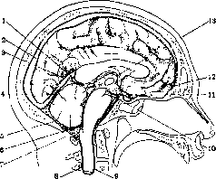
Subarachnoid spaces of the brain: 1- v. cerebri magna; 2-sinus sagittalis superior; 3-cisterna venae magnae cerebri; 4- sinus rectus; 5-cavum subarachnoidale encephali; 6-foramen Magendii; 7-cisterna cere-bello-medullaris; 8-cavum subarachnoidale medullae spinalis; 9-arachnoidea spinalis; 10-cisterna interpeduncularis; 11-chiasma opticum; 12-pia mater encephali; 13-foramen interventriculare (Monroi).
the spinal cord, while arachnoidea and dura mater form a wider sac. The more significant expansions of the subarachnoid spaces in the brain are called cisterns (cisternae subarachnoidales) (see figure); they are filled with cerebrospinal fluid. The largest cistern (c. magna) lies between the medulla oblongata and the cerebellum and is formed because the membrane from the spinal cord is thrown over the cerebellum; it connects the cranial subarachnoid spaces with the spinal ones, as well as with the ventricles of the brain through foramen Luschka and Magendii. The second large cistern (cisterna basalis) is located at the base of the brain and extends forward from the anterior edge of the Varolius bridge to the optic chiasm, and laterally to the Sylvian sulcus, forming a very large cavity, subdivided into a number of smaller ones (cisternae chiasmatis, interpeduncularis, laminae terminalis, etc.). Along the upper surface of the corpus callosum is the cisterna corporis callosi, around the cerebral peduncles-cist. ambiens, near vena Galeni magna-cist. venae magnae cerebri. The subarachnoid spaces communicate with the lymphatic spaces of the adventitia of intracerebral vessels (Virchow-Robin spaces).-The meninges receive blood from all three meningeal arteries (Meningeae arteriae, venae, see), especially from art. meningea media (a branch of art. carotis externa). All art. meningeae leave on the bones of the skull strictly defined grooves. Large blood vessels pass inside the subarachnoid spaces; thinner ones penetrate into pia mater, and some of them together with pia mater- into the brain tissue.-Nerve fibers of the meninges come from the n. sympathicus (cervical part), from nn. trigeminus, vagus, hypoglossus; some of these fibers (non-myelinated) are contained in the vessels of pia mater; others (nn. proprii) form with their ramifications a network in the meninges and end between its cells.
E. Kononova. Pathology. The meninges are involved in the process in diseases of the skull, spine, and brain. In addition, they are affected independently, and some diseases of the meninges do not spread beyond the membranes, while others extend to the brain or skull. Among the diseases of the meninges, the following should be noted: developmental anomalies, circulatory disorders, inflammatory processes, tumors, parasites. I. Developmental anomalies of the meninges occur simultaneously with underdevelopment or malformations of those parts of the central nervous system that they cover (brain, spinal cord, cerebellum), or with defects of those structures in whose cavities they are contained (skull, spine) (see the respective articles and Spina bifida). II. Circulatory disorders in the membranes of the brain and spinal cord are observed in the form of venous and arterial hyperemia, edema, anemia, and hemorrhages, thrombosis of the sinuses of the dura mater and veins of the pia mater. The edema and venous hyperemia of the pia mater, often found on autopsies, are the result of autonomic nervous system disorders, as well as changes in vascular permeability. This process is sharply expressed in diseases of the heart (cardiosclerosis, decompensated defects, myodegeneration, etc.), kidneys, lungs (in tuberculosis, emphysema, pneumonia, and especially in Spanish influenza), and in asphyxias-croup, tumors of the larynx, chronic inflammatory processes in it, foreign bodies entering the larynx, hanging, strangulation, etc., and in "asphyxia of newborns." - Arterial hyperemia of the dura mater and pia mater accompanies acute infectious diseases, sunstrokes. In the pia mater, this is accompanied by strong transudation. This process is particularly intense in inflammation (see Meningitis). Disorders of the activity of vasomotor nerves leading to hyperemia are the cause of the so-called neuritic form of hyperemia. They can be caused by paralysis of vasoconstrictors (neuroparalytic hyperemia) or irritation of vasodilators (neuroirritative hyperemia). If a rabbit's skull is trephined and then Claude Bernard's classic experiment is performed, after cutting the cervical nerve on the same side, hyperemia of the meninges can be observed along with redness of the ear and pupil constriction. The same is observed in humans after injury to the sympathetic and vagus nerves or damage to them by tumors, scars, etc. Hyperemia in some infectious diseases (croupous pneumonia, typhus, diphtheria, scarlet fever, etc.) is due to pathological changes in the autonomic nervous system (see Neuritis). In unilateral diseases (e.g., in pneumonia), during autopsy, hyperemia of the meninges on the corresponding side is sometimes striking. Periodic irritation of vasomotor centers causes hyperemia of either certain areas (migraines) or all meninges (climax, Basedow's disease). - Edema of the pia mater, arachnoidea (hydrocephalus externus) accompanies stagnation, inflammatory processes, nephritis (hydremia), and also occurs ex vacuo due to partial or complete reduction of the brain. Fluid accumulating in the subarachnoid spaces and in the cisterns between arachnoidea and pia mater expands them and compresses the gyri. At the same time, there is replenishment of serous transudate in the lateral ventricles of the brain (hydrocephalus internus). The arachnoid membrane bulges in the area of sulci and over the surface of the gyri. Hemorrhages into the meninges are most often of traumatic origin. In newborns, traumatic meningeal hemorrhages occur both as a result of operative procedures and due to compression of the fetal head in narrow birth canals. In infectious-toxic diseases, numerous hemorrhages can develop on the basis of thrombosis, thromboangiitis, degenerative fatty changes, necrosis, increased vascular wall permeability (typhus and relapsing fever, comatose malaria, sepsis, etc.); the same can occur in intoxications (phosphorus, mercury, snake venom), including of bacterial origin (diphtheria, tetanus). Processes of an autointoxication nature include meningeal hemorrhages in jaundice, eclampsia, leukemia, nephritis, scurvy. In nephritis, the increase in blood pressure undoubtedly has a certain influence, and in blood diseases (along with changes in vessel walls) - changes in the properties of the blood itself. Spontaneous hemorrhages are often in the form of haemorrhagia intermeningealis. Significant hemorrhages are observed in pachymeningitis haemorrhagica interna (see Hematoma). In traumatic hemorrhages of the meninges, damage to the soft tissues and bones of the head is not necessary. Often extensive hemorrhage, ending fatally, occurs with their complete integrity. Hemorrhages into the membranes are observed in fractures of the convex surface and base of the skull; most often they occur from the a. meningea media, cerebral sinuses, veins flowing into the sinus longitudinalis, and vessels of the pia mater, arteries of the base of the skull, diploe. Hemorrhage from the sinus longitudinalis, art. meningea media and diploe is localized on the outer surface of the dura mater; with rupture of veins flowing into the longitudinal sinus - under the dura mater; with rupture of vessels of the pia mater, it is distributed in the subarachnoid space and under the pia mater. In newborns, the source of hemorrhage is often the ruptured tentorium cerebelli. Hemorrhages correspond to the site of injury; however, in some cases, ruptures and hemorrhages develop on the opposite side in the order of contre-coup. The spilled blood on autopsy is partly liquid and partly in the form of red and mixed clots. The amount ranges from small ecchymoses to 100-300 cm3. Small, often numerous hemorrhages into the tissue of the dura mater are found in strangulation (dural hemorrhages). Hemorrhages between the skull and dura mater (epidural or extradural hemorrhages) occur due to rupture of vessels (arteries), for example, as a result of a skull contusion. In elderly people with calcified, brittle arteries (art. mening. media and anterior) and deeply located sulci meningei, even a light blow to the head can lead to the formation of extradural hematomas. Epidural extravasates in burn patients have been described by Strassmann, explaining them by the fact that heat changes the permeability of vessel walls, and blood from the diploe penetrates into the cranial cavity. Hemorrhages between dura and arachnoidea are called subdural hemorrhages, into the cells of arachnoidea - subarachnoid. In injuries, hemorrhages can also occur due to rupture of veins of the pia mater (subpial and pial). - In birth injuries, intracranial hemorrhages are observed: a) Due to rupture of veins in the area of open pial venous trunks flowing into the sinus sagittalis due to detachment of skull bone plates, more rarely - due to rupture of venae Galeni magnae at the point where it flows into the sinus rectus, or due to rupture of sinus transversi. b) Due to rupture of the layers of the tentorium cerebelli, either both layers or the free thickened edge of the tentorium are damaged. The hemorrhages lie on the upper layer or under the lower, which at the same time is the dura mater of the cerebellum (peribulbar, infratentorial hemorrhages). The latter, due to compression of the medulla oblongata, can cause fatal asphyxia. c) Large subdural hemorrhage is observed in ruptures of the intervertebral joint capsule. Hemorrhages during childbirth, by traumatizing the brain, often cause atrophy, sclerosis, cysts, porencephaly, and cortical epilepsy. - Thrombosis of sinuses - see Sinuses. Immediately after traumatic hemorrhage, due to simultaneous concussion of the brain, faintness occurs; unconsciousness lasts from several minutes to several hours; in severe cases, consciousness is not restored until death. The so-called free intervals (Horsley) are very characteristic: a short return to normal consciousness is followed by clouding of the latter and gradually increasing other symptoms characteristic of meningeal hemorrhage. - In dyscrasic conditions of vessels, the clinical picture of the disease usually develops gradually, with the appearance of a number of new symptoms. In general, meningeal hemorrhages should be classified as severe diseases, usually ending fatally. The outcome depends on the degree of brain compression and the speed of surgical intervention. Statistics show that with conservative treatment, 90% of patients die from meningeal hemorrhage, with surgical treatment the mortality does not exceed 33% (Darkshevich). - The symptomatology of traumatic and spontaneous meningeal hemorrhages is somewhat different. In the first, symptoms develop slowly after a free interval and are due to increased intracranial pressure and pressure on the brain. Severe headache, dominating throughout the disease, is often accompanied by vomiting and slowing of the pulse. Often, as in other hemorrhages, temperature rises. Consciousness ranges from mild clouding to deep coma. From the side of the eyes, congested discs and pupil inequality can be observed.
In addition, focal symptoms are often observed: hemiplegias, hemianesthesias, paralysis of cranial nerves, local and general hyperkinesias. If the clinical picture of spontaneous hemorrhages sometimes resembles that of traumatic ones, more often it resembles the picture of acute cerebrospinal meningitis. - In the differential diagnosis of both forms, the so-called free intervals, the presence of erythrocytes in the cerebrospinal fluid upon repeated punctures, the presence of trauma, infectious, toxic, dyscratic and other factors in the anamnesis are of great importance. - When discussing the topography of the process, it is necessary to take into account the possibility of lesion par contre-coup. - T r e a t m e n t. After determining the size of the hemorrhage, in the case of the slightest significant process, immediate surgical intervention with stopping the bleeding and removing the spilled blood is necessary. III. Inflammatory processes of the M. o.: 1) inflammations; 2) sclerotic processes; 3) infectious granulomas of the M. o. 1. Inflammations of the M. o.-see Meningitis, Pachymeningitis. 2. Sclerotic processes. In kidney diseases, alcoholism, arteriosclerosis and in old age, diffuse focal cloudings of the soft M. o. are observed, caused by the proliferation of connective tissue and bearing the incorrect name of chronic meningitis. Most of these phenomena have nothing in common with inflammation. In these cases, strong adhesions of pia mater with the surface of the brain and spinal cord often form. With superficial spreading softening, fibrous, fused with the brain thickenings of the membranes form, known under the name plaques jaunes, having the appearance of yellow dense scars. If in these processes adhesions of the soft M. o. with dura mater and obliteration of the subarachnoid and subdural spaces develop, then stagnant hydrocephalus of the brain and spinal cord may arise. In the hard and soft M. o., the presence of tooth-like bone plates (sometimes called meningitis ossificans) can sometimes be established, which apparently form here either through metaplasia or through heterotopic pathways. 3. Infectious granulomas: tuberculosis and syphilis-see Meningitis. Actinomycosis (see) of the hard M. o., as well as actinomycotic encephalitis, are the result of 1) direct transition of the process from the basal part of the skull and 2) hematogenous metastases from various organs. They occur both in the form of separate granulomas and in various parts of the brain, as well as in the form of basal fibrinopurulent meningitis. In the exudate, a significant number of plasma cells and lymphocytes are present. IV. Tumors of the M. o. 1. Tumors of the hard M. o.: a) endotheliomas- tumors consisting mainly of endothelial cells; b) fibro-endotheliomas- tumors with a predominance of connective tissue; c) alveolar fibro-endotheliomas- have a cellular structure; complexes of endothelial cells occupy the cells formed by connective tissue; d) pure fibromas, sometimes originating from the periosteum; e) osteomas, sometimes reaching significant sizes (17 x 7 cm in the case of Stano); f) dermoids, developing from misplaced epithelial rudiments of the skin coverings of the skull, are mainly found at the base of the brain in the form of one or multiple cystic tumor formations; g) sarcomas, originate from the periosteum or from the inner part of the dura mater. Most often they belong to the spindle-cell fibrosarcoma and osteosarcoma type, less often to polymorphous-cell and round-cell types. Some of them have the appearance of alveolar tumors (sarcoma alveolare), others belong to the hemangiosarcoma type. As rare forms, perivascular sarcomas [sarcoma cylindromatosum (Kaufmann) and sarcome angioleptique (Cornil and Ranvier)] are described. The most common are limited fibro-endotheliomas. They never penetrate into the lymphatic pathways and are delimited from the brain substance by a connective tissue capsule. Some of these tumors are rich in polymorphous endothelial elements and morphologically resemble sarcomas. They can be both single and multiple. Single endotheliomas reach a significant size (hen's egg), multiple ones show a tendency to group into small conglomerates. Often they undergo central softening. Upon histological examination, these tumors appear extremely diverse depending on the amount of connective tissue, vessels and endothelial cells. The following forms of tumors of the dura mater are distinguished: fibromas and fibro-endotheliomas, the tissues of which, undergoing degenerative-atrophic processes, are incrusted with calcium salts, bear the name psammomas. If this process occurs in sarcomas, they are designated as psammo-sarcomas. 2. Tumors of the soft M. o. Among the tumors of the soft M. o., the most common form (even more often than in the hard M. o.) are endotheliomas. Cushing gives the name 'meningiomas' to the endotheliomas of the M. o., considering the most characteristic feature of these tumors to be that they do not infiltrate the brain, but gradually displace and compress it. In rare cases, diffuse endotheliomas simulating chronic meningitis are observed. Among other tumors, the following are found: a) Fibromas; these tumors compress the brain but do not grow into it; b) Lipomas are found in the membranes of the basal parts of the brain base: at the infundibulum, substantia perforata anterior. Usually not large. c) Cavernous hemangiomas. d) Cavernous lymphangiomas. e) Dermoid cysts. f) Cholesteatomas-epidermoids in the form of cysts, filled with solid masses of keratinized scales, giving the tumor a special shine and whiteness. The favorite location of these tumors is the soft M. o. in the area of the bulbi olfactorii, tuber cinereum, corpora candicantia. Sizes-from a wheat grain to an orange.-Among malignant* tumors, the following are observed: a) round-cell sarcomas, myxosarcomas and hemangiosarcomas. b) From the pia mater, more often in the area of the medulla oblongata, melanosarcomas arise. They form from the pigment cells normally present here. In diffuse sarcomatosis, flat sarcomatous formations grow along the entire length of the spinal cord from the cauda equina to the medulla oblongata, and sometimes higher-up to the base of the brain. The tumor, covering the entire spinal cord with a thick sheath, sometimes also penetrates into its substance. Histologically, it presents the picture of a round-cell sarcoma. Clinically, it manifests as meningomyelitis (see Myelitis).-Among secondary metastatic tumors, in addition to various sarcomas, carcinomas (metastases of hypernephroma, chorionepithelioma, bronchial carcinoma, prostate carcinoma, etc.) are also found. Malignant neoplasms of the bones of the skull and spine often grow into the membranes of the brain and spinal cord. From the plexus choroideus, epithelial tumors consisting of ependymal epithelium form: plexus-epitheliopapillomas or neuro-epitheliogliomas and ependymal carcinomas. From the connective tissue of vessels, the following arise: a) fibromas, which upon incrustation of the tissue with lime take the appearance of psammomas; b) hemangiomas; c) lipomas (rarely); d) gliomas (myxogliomas); e) ganglioneuromas; f) fusocellular sarcomas (Hirsch) and g) perivascular sarcomas; h) angioma arteriosum, s. arterioma. - The clinical picture of tumors is characterized, besides general cerebral phenomena (headache, vomiting, stagnant discs, slowed pulse, epileptic seizures), also by local symptoms depending on the location of the tumors. In the same area, vessels enter the thickness of the cerebellum. Topographical boundaries of the M.-m. u.: posteriorly medially-the oblongata of the medulla oblongata, posteriorly laterally-the anterior edge of the tonsillae and lobuli biventeris and semilunaris inferior.; anteriorly laterally-the edge of lobuli quadrangularis, anteriorly medially the boundary is the Varolian bridge. Here the bottom of the area from below is the middle cerebellar peduncle. Nn. abducens and trigeminus-are located more to the side, but they must also be considered in connection with the topography of the area. On the skull, the M.-m. u. corresponds to the anterior part of the cerebellar fossa and lies medially from the posterior-superior surface of the pyramid of the temporal bone, the upper edge of which at the apex is crossed by the trigeminal nerve. Along the same edge, the tentorium cerebelli is attached. In this small space, a number of cranial nerves (including the vestibular tract), the cerebellum and the brainstem are found. In certain (mainly extracerebral) pathological processes (especially tumors), in this narrow area, due to anatomical conditions, the n. acusticus (with disturbance of cochlear and vestibular functions), n. trigeminus (in the early stage it gives weakening of the corneal reflex) and the cerebellum (nystagmus, corresponding coordinatory-static deficits) are usually affected first. To this can be added conduction phenomena from the nn. abducens, facialis,* often difficult to explain due to homolaterality. The latter feature is explained by the fact that a formation developing in a narrow bony sheath compresses the relatively narrow in diameter brainstem to the opposite side of the bony canal, thereby causing compression phenomena more on the opposite side.
And since this compression occurs above the decussation, phenomena of conduction deficit may often predominate on the same side. In connection with these different conditions of compression of the trunk on one side and the other, one can point to another sign—qualitatively different pyramidal phenomena on different sides in tumors of the angle: for example, flexor pyramidal phenomena on one side, extensor on the other, increased Achilles reflex on one side, plantar phenomena on the other, etc. Tumors of the M.-m. u. can have various origins. Less frequently, the compressive formation may originate from the brain substance itself, but much more often it is located extracerebrally. The extracerebral mechanism of the M.-m. u. syndrome can also depend on lesions associated with the bones of the skull. In the immediate vicinity of the formations that constitute the M.-m. u. is the base of the skull, and therefore processes on the base of the skull can give a picture resembling a tumor of the M.-m. u. The pyramidal bone is located further away, but it has a more intimate connection with the n. acusticus, and since lesions of it in combination with some general V. Of the parasites in the soft M. o. are found: 1. Cysticercus cellulosae. It is observed with varying frequency in different countries. It is more common where the population consumes pork in a raw form. It spreads under the arachnoid membrane or between pia and the brain substance, indenting the latter. Their number can be very diverse (sometimes whole hundreds). The size usually does not exceed a pea. It should be noted that sometimes despite the large number of cysticerci, their presence does not give any clinical phenomena and they are discovered accidentally only at autopsy. The parasites are surrounded by a connective tissue capsule and a band of thickened fibrous glia. In addition to the usual form, branched forms of cysticerci (Cysticercus racemosus) are occasionally encountered, the morphological feature of which is the formation of lateral branches resembling cysts. Cysticercic meningitis is characterized by extreme variability of symptoms in its clinical course. Meningeal phenomena (headache, epileptic seizures) fluctuate. It is often the cause of sudden death. Diagnosis during life is very difficult. It is facilitated by finding calcified vesicles under the skin. Since cysticerci rarely limit themselves to the membranes, in the clinical picture sometimes in addition to meningeal symptoms (see Meningitis), phenomena from the brain and spinal cord also appear (see Encephalitis and Myelitis). Pathologically-anatomically, cysticercic meningitis can simulate gummatous lesions of the brain. 2. Unilocular echinococcus. As for the multilocular echinococcus, it is an exceptional rarity (Roth). 3. Blastomycosis is extremely rare, giving the picture of a cyst or tumor.
Related articles
Mentioned in
- Acervulus Cerebralis
- Alternating Syndromes
- Anatomy
- Brain (cerebrum, a comprehensive term for the entire)
- Cerebellopontine Angle
- Cerebro-Cranial Topography
- Cerebrospinal Fluid
- Cholesteatoma
- Corpora Arenacea
- Dural Sinuses
- Embryoma
- Endothelioma
- Endothelium
- Exencephaly
- Gliosarcoma
- Gout
- Hydrocephaloid
- Hyperesthesia
- Kushev, Nikolai Egorovich; Cushing, Harvey Williams
- Meningioma
Cite this page
“Meninges.” Soviet Medical Encyclopedia. English translation of Bolshaya Meditsinskaya Entsiklopediya, 1st ed. (Moscow, 1928–1936), ed. N. A. Semashko. https://sovietmedicalencyclopedia.pages.dev/article/meninges/