Radiculitis
Historical document, translated for reference. It reflects medical knowledge of the 1920s–30s and is not medical advice.
Summary
Radiculitis refers to inflammatory and degenerative diseases of the spinal nerve roots, often associated with infections such as influenza, tonsillitis, and syphilis. The condition typically affects multiple adjacent nerve roots and presents with sensory, motor, or mixed symptoms, including pain and sensory disturbances in specific dermatomal patterns.
Encyclopedia article (1928–1936)
Radiculitis (from Latin radix-root), diseases of the roots of the spinal nerves, a term that became established in the early 20th century thanks to the work of Déjerine and his school. The basis of R. is an inflammatory-degenerative process in the nerve roots [see separate table (pp. 255-256), Fig. 2], sometimes small hemorrhages are added to it, and later a sclerosing process and the formation of small cysts may occur. Most, following Déjerine, call radiculitis a disease of the nerve root only along its course from the spinal cord to the point where it passes through the dura mater (Fig. 3), others include the continuation of the nerve root up to the spinal ganglion. Due to the intimate connection between the meninges and the nerve roots, inflammatory processes usually involve both. Thus, clinical R. in its patho-anatomical essence are meningo-radiculitis. Having passed to the nerve root, the pathological process more often spreads toward the peripheral nerve than passes to the root fibers going to the spinal cord. The posterior root is accompanied by the meninges on a much longer course than the anterior root - sometimes all the way to the ganglion itself. The meninges form along the posterior root a deep and wide blind pouch filled with cerebrospinal fluid. This creates more favorable conditions for the infection to pass from the cerebrospinal fluid to the posterior root, which is affected more frequently and more severely in radiculitis than the anterior root. Various infections serve as etiological factors for R. Thus, R. occur during or more often after influenza, tonsillitis, typhoid fever, pneumonia, epidemic meningitis, and encephalitis. In a number of cases of R., a primary infection of still unknown nature must be assumed. Among chronic infections, syphilis and tuberculosis have special importance in the etiology of R. Predisposing factors include cold, trauma, and overexertion. For this reason, R. more often occur in persons engaged in heavy physical labor. R. are localized at various levels of the spinal cord, most often in the lower cervical and lumbosacral regions. R. is extremely rarely limited to one nerve root; as a rule, it involves two or three or more adjacent nerve roots. Polyradiculites involving a large number of nerve roots are rare, but even in these cases the pathological process is not expressed uniformly everywhere, but is localized in individual nerve roots. According to their course, R. are divided into acute, subacute, and chronic. The latter often exacerbate. According to symptomatology, R. are distinguished as sensory, motor, and mixed. Sensory and mixed forms of R. are most common. Purely motor R. are rare and are found almost exclusively in the lower cervical region. In the symptomatology of sensory R., caused by damage to the posterior roots, disturbances in cutaneous sensitivity deserve special attention. A characteristic feature of R. is the localization of sensory disturbances in root zones, limited on the extremities by longitudinal lines, and on the trunk by transverse lines. Examples of the root distribution of sensory disturbances are shown in Fig. 1 and 2). In the vast majority of cases in radiculitis, there is only a decrease, not a loss of sensitivity, since every area of skin is innervated from three or more nerve roots. In milder cases, instead of hypesthesia, there is hyperesthesia. In R., tactile and pain sensitivity suffer more than deep, temperature, and stereognostic sensitivity. The opposite relationship is observed only rarely. In the affected area of skin, changes in sensitivity are more or less evenly distributed, sometimes they are arranged in islands. In addition to objectively demonstrable disturbances of sensitivity in sensory and mixed R., there are paresthesias and pains. The pains sometimes have a constant, sometimes paroxysmal character. Mostly they are of considerable intensity, boring, aching, sometimes shooting. Sometimes they are felt in the back over the site of anatomical damage, sometimes they radiate to the root skin zones. The pains usually increase with coughing, sneezing, movements of the spine, and changes in meteorological conditions. When the sensory form of R. is exhausted only by pains, they speak of radiculalgia. Many cases of intercostal neuralgia, sciatica, and Roth's disease are in essence radiculitises. Motor disturbances in R. are more often expressed in paresis than in paralysis of the musculature. The pareses are peripheral.
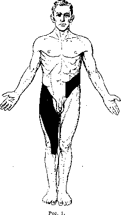
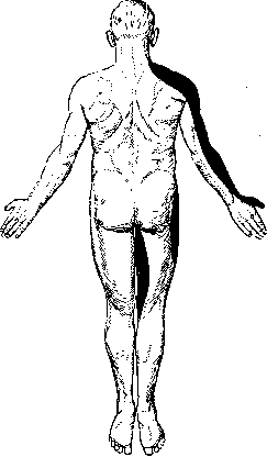
Figure 2.
Figure 1 and 2. Diagram of root-type sensory disturbances (areas of sensory disturbance are shaded black). (Author's own observations.) On the front surface of the right thigh, anesthesia is observed in the area of innervation of the skin by the first, second, and third lumbar roots (I-II-III L). On the left side of the abdomen, disturbance of sensitivity is observed in the area of the tenth, eleventh, and twelfth thoracic roots (X-XI-XII D). On the back surface, disturbance of sensitivity involves the fourth, fifth, and sixth cervical roots (IV-V-VI C). A small black line on the right thigh corresponds to the third lumbar root, which slightly extends to the back surface from the front. (Depending on the intensity of the process, all types of sensitivity are usually affected.)
Rys.
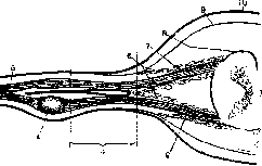
Figure 3. Diagram of the spinal cord with roots: 1-spinal cord with anterior horn (at the top of the figure) and posterior horn (at the bottom); 2-posterior root, originating from the spinal ganglion (4) and extending to the posterior horn and posterior columns; 3-root nerve; 4-spinal ganglion; 5-lymphatic space; 6-pia mater plexus surrounding the internal parts of the roots; 7-anterior root, emerging from the anterior horns of the spinal cord and connecting with the posterior root outside the ganglion; 8-pia mater, surrounding the spinal cord and internal parts of the roots; 9-arachnoid membrane, extending somewhat further; 10-dura mater, surrounding the spinal cord with roots. Radiculitis has a specific character, i.e., it is accompanied by atrophies, hypotonia, hypo- and areflexia, and changes in electrical excitability. The distribution of pareses in radiculitis is peculiar. In each spinal root, fibers pass that subsequently enter various peripheral nerves. Each muscle is innervated not by one root, but by at least two, sometimes three. For this reason, pareses are rare in radiculitis. Of the muscles innervated by any peripheral nerve, only those that receive innervation from the affected roots are affected by paresis in radiculitis. Other muscles innervated by the same nerve do not suffer, since their root fibers pass through adjacent healthy roots. For example, in radiculitis of the C5 and C6 roots, among others, the long extensors of the wrist and fingers, innervated by the radial nerve, are affected by paresis, since the nerve fibers for these muscles are formed in the aforementioned roots. However, in this localization, the long supinator remains unaffected; although it is also innervated by the same radial nerve, the fibers for it originate from the C5 and C6 roots. The peculiar nature of root paresis is even more evident in the rectus abdominis muscle, innervated from C8 to T12 roots. In radiculitis, a dissociated paresis of only one part of this muscle is sometimes observed. - Tendon reflexes in radiculitis are elevated only rarely and then only at the beginning; mostly they are diminished or disappear due to damage to one or both parts of the reflex arc. Skin reflexes are also diminished, which is especially evident in abdominal reflexes, which disappear early in thoracic radiculitis. Since sympathetic fibers also pass through the roots, autonomic disorders are not uncommon in radiculitis. They manifest as cyanosis, cooling of the skin, its dryness, less often sweating, hypo- or hypertrichosis. Autonomic disorders are especially prominent in radiculitis of the C8-T1 and T1 roots, when atrophy and paresis of the small muscles of the hand and fingers are joined by Horner's syndrome (see Horner's symptom complex) and thus the Dejerine-Klumpke symptom complex is created (see Dejerine-Klumpke paralysis). It is probable that shingles may also depend on the disease not only of the spinal ganglion but also of the roots. - The pressure of cerebrospinal fluid is usually slightly elevated in radiculitis. In it, a mild lymphocytosis and hyperalbuminosis are often present in radiculitis. Xanthochromia is rare; in individual cases, protein-cell dissociation was observed: a large amount of protein with a small number of white cells. In the diagnosis of radiculitis, the main difficulty lies in determining its primary or secondary nature. The presence of signs of spinal cord damage (tabes, meningomyelitis), tumors of the membranes, spondylitis speaks for the symptomatic nature of radiculitis. Changes in the spine, both congenital (spina bifida, sacralization, lumbarization) and acquired (spondylosis, spondyloarthritis), often occur together with radiculitis, but cannot be considered as their cause. In our Union, the diagnosis of radiculitis has received excessive prevalence. It often and incorrectly serves to denote lumbar pains of various and non-neurogenic origin. The incorrect use of this term leads, as a result of statistical calculations, to a false representation of the widespread prevalence of radiculitis among our working population. The clinical outlines of radiculitis should be clear enough for this diagnosis to be made only in the presence of the main components of the disease. - Prevention of radiculitis lies in the fight against infectious diseases (syphilis, tuberculosis, etc.) that cause it. - Therapy in the acute stage consists of bed rest, thermotherapy, and analgesics. When syphilitic origin is established, specific therapy is in the first place. In subacute and chronic stages, the most successful is course treatment at mud and sulfur resorts (Odessa limans, Saki, Matsesta, etc.). The percentage of significant improvement in radiculitis with mud treatment reaches 84.5-97 (Cherkes).
Related articles
Mentioned in
Cite this page
“Radiculitis.” Soviet Medical Encyclopedia. English translation of Bolshaya Meditsinskaya Entsiklopediya, 1st ed. (Moscow, 1928–1936), ed. N. A. Semashko. https://sovietmedicalencyclopedia.pages.dev/article/radiculitis/