Typhoid Fever
Historical document, translated for reference. It reflects medical knowledge of the 1920s–30s and is not medical advice.
Summary
This article from the 1928–1936 Soviet Great Medical Encyclopedia covers typhoid fever, detailing its etiology, pathology, clinical picture, and historical understandings of the disease. It describes the typhoid bacillus discovered by Eberth and Gaffky, its biological properties, and methods of laboratory isolation.
Encyclopedia article (1928–1936)
TYPHOID FEVER. Contents: Etiology................... 160 Epidemiology................. 164 Statistics................... 167 Pathological anatomy........... 187 Pathogenesis................... 193 Clinical picture............. 195 Complications.................. 201 Diagnosis.................... 204 Prognosis.................... 206 Treatment.................... 206 Prophylaxis................. 208 Vaccination against typhoid fever....... 209 Typhoid fever (from Greek typhos - smoke, mist), a general acute infectious disease characterized by a typical temperature curve, phenomena of general intoxication, and a peculiar lesion of the lymphatic apparatus, especially the intestinal tract. Under the general, anciently existing name of "typhus", which designated a febrile state with clouding of consciousness ("feverish" state), typhuses were formerly grouped together: typhoid, typhus, and relapsing. The anatomical changes characterizing typhoid fever, known still to Morgagni, were in 1829 laid by Louis as the basis of the clinic of typhoid fever. To Hildebrand and Griesinger in Germany, Jenner and Murchison in England, and Gerhard and Pennock in America belongs the credit of the clinical and epidemiological separation of typhoid fever as a nosological entity distinct from typhus and relapsing fevers (around the middle of the 19th century). The greatest merits in elucidating the clinic and pathogenesis of typhoid fever must be accorded to Curschmann, Liebermeister, S. P. Botkin, Schottmüller, Jürgens, and others. Murchison (1857) saw the cause of typhoid disease in a "putrid principle" developing in decomposing feces of various origins (miasmatic theory). Budd, through a series of observations and logical conclusions, came to the hypothesis of a peculiar living contagious principle and constructed a theory (contagious) of infection from a typhoid patient through his excreta. In contrast to this theory,
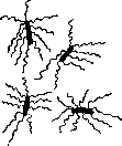
Figure 1. Typhoid bacilli.
Buhl and Pettenkofer put forward a theory (localistic) connecting the occurrence of typhoid fever with the level of ground water. Eberth in 1880, by discovering the typhoid bacillus, studied in detail and described by Gaffky (1883), established the bacterial nature of the causative agent of typhoid fever. Etiology. The causative agent of typhoid fever is a special bacillus, 1-2 µ long, growing on ordinary nutrient media and possessing motility thanks to 10-12 peritrichously located flagella (see figure 1). On media, the bacillus often grows into long threads. On gelatin, it forms characteristic delicate colonies with scalloped edges and a darker center, covered with wavy striations and resembling a grapevine leaf. On potato, it gives an invisible growth on the surface. On agar, it grows in the form of a bluish coating. On broth, it does not form indole, and on sugar broth, no gas bubbles. On blood serum with litmus, it gives only a slight reddening. Media with Neutral Red are not changed. In a litmus solution of nutrose with grape sugar, it causes acid formation and coagulation, and in a litmus solution of nutrose with milk sugar, it gives no changes. It grows on bile. It is agglutinated by a specific agglutinating serum and gives phenomena of bacteriolysis in the abdominal cavity of immune animals. The listed characteristic properties of the typhoid bacillus serve as identifying features to distinguish it from typhoid-like organisms. - Viability in the external environment. The typhoid bacillus, getting into water and soil, can persist there for weeks. Putrefaction processes unfavorably affect the viability of the typhoid bacillus, which dies due to competition with saprophytic microbes. In cesspools and latrines, the typhoid bacillus can persist for months, especially in winter. Light and drying kill the typhoid bacillus slower than the cholera vibrio. Heating to 60° for an hour kills the typhoid bacillus. In rotting corpses, the typhoid bacillus survives for no longer than a month, in frozen corpses - significantly longer. The typhoid bacillus getting with sewage onto irrigation fields can survive there in summer for up to two weeks, and in winter significantly longer. In river water, the typhoid bacillus disappears due to the action of light (Buchner) and competition with water bacteria and, especially, protozoa, which explains the self-purification of rivers. Chemical influences (sublimate 1:1,000 and carbolic acid 5%) kill the typhoid bacillus in a few minutes, if it is not protected by particles of organic substances contained in feces. The typhoid bacillus can inhabit as a saprophyte in the human intestine and, 161
TYPHOID FEVER especially, in the gallbladder. Thus, the primary source of typhoid infection is man, in whose intestines typhoid bacilli are located. Typhoid bacilli are pathogenic for animals upon subcutaneous, intravenous, and intraperitoneal administration of a culture. Infection per os succeeds in rabbits and monkeys upon using large doses or after removing the spleen from the animals (Chantemesse, Mechnikov, Bardakh, Besredka, and others). The typhoid bacillus produces endotoxins (Pfeiffer), which can be liberated thanks to various lytic procedures, such as, for instance, mechanical grinding and crushing (Buchner, Hahn), dissolution by alkalis (Lustig and Galeotti), grinding after freezing with liquid air (MacFadyen), or digestion by digestive and serum enzymes (Besredka). Certain strains of the typhoid bacillus are capable of producing toxic substances in filtrates (Kraus and Steinitzer), which, however, differ by their lability from the true exotoxins obtained from cultures of the diphtheria bacillus, tetanus bacillus, and botulinum bacillus. The technique of isolating the typhoid bacillus and its differentiation from typhoid-like ones is of great importance. Since Löffler, typhoid-like bacteria have been divided into three groups: Typhaceae, Colaceae, and Josarceae. To isolate the typhoid bacillus, the enrichment method is usually applied at the beginning of the study, through which the accumulation of typhoid bacilli to the detriment of other microbes is achieved. Formerly, carbolized broth (Chantemesse), potato gelatin with potassium iodide of Elsner were used for this purpose; currently, bile and broth with malachite green are used. Blood is usually inoculated into bile, while feces are inoculated into broth with malachite green. For further isolation and differentiation of the typhoid bacillus, differential media serve: blue - Conradi and Drigalski, red - Endo, green - Padlevsky. On Conradi-Drigalski medium, the typhoid bacillus does not change the medium, unlike colonies of Bact. coli, which cause reddening of the medium. On Endo and Padlevsky media, typhoid bacilli give colorless colonies, whereas Bact. coli gives red on Endo medium with fuchsin and green on Padlevsky medium with malachite green and bile. The isolation of the typhoid bacillus from water encounters great difficulties in view of the fact that it is contained in water in small quantities. Most methods of its isolation are based on seeding flakes of organic substances precipitated by Reduction of mortality from typhoid fever in Italy from 79.0 per 100,000 population (annual average) for the period from 1886 to 1890 to 23.9 for the period from 1921 to 1925 (see table on p. 168). An increase in mortality is noted from 1915 - during the imperialist war.
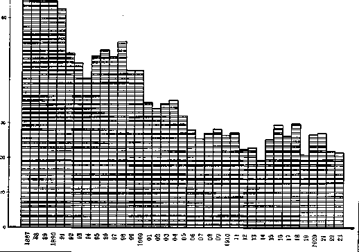
Figure 2. Mortality from typhoid fever in Italy (per 100,000 population).
with iron sulfate or alumina, onto appropriate media. Agglutinating serum was also used to precipitate typhoid bacilli. Another series of isolation methods is based on collecting water bacteria by filtering large quantities of water through a Chamberland candle and subsequently inoculating bile and selective media with the bacterial deposit removed from the candle. An indirect indicator of possible water contamination Figure 3. Mortality from typhoid fever in with typhoid bacilli is the determination of the colon bacillus titer (Bact. coli) in water using the Eijkman and Bulir tests. To separate the typhoid bacillus from other water microbes, researchers also used its faster passage through a sand filter and a tube filled with broth (Gabrichevsky). Immunity against the typhoid bacillus has been studied thanks to the works of Pfeiffer, Isaev, Kolle, Besredka, and others. Immunization is most easily achieved using cultures killed by heating to 58°, by chloroform, or by ether. This produces antibacterial immunity, with the formation of specific antibodies in the blood and organs (agglutinins, bacteriolysins, bacteriotropins, or opsonins). The agglutination titer in highly immunized animals reaches several tens of thousands, i.e., the serum of such animals agglutinates the typhoid bacillus at a dilution of Germany (per 100,000 population). 1 : 10,000, 1 : 20,000, and more. In addition, the serum of immune animals possesses bacteriolytic and bactericidal properties both in vivo and in vitro. When injected into a guinea pig, the serum of immune animals induces passive immunity and phenomena of bacteriolysis of typhoid bacilli upon their introduction into the guinea pig's peritoneal cavity. Typhoid bacilli turn into spheres within 20–30 minutes and dissolve (Pfeiffer's phenomenon). Epidemiology. Epidemics of typhoid fever develop most frequently in the summer, although autumn and winter outbreaks are not excluded. Decline in mortality from typhoid fever in Germany from 17.0 per 100,000 population in 1892 to 1.8 in 1926. Sharp rise in mortality starting in 1914 during the imperialist war. Typhoid fever is a disease of the prime of life (20–25 years). Migrants, seasonal workers, students, and others living in abnormal conditions are most frequently affected. Regarding the nature of disease sources. The so-called early contacts in the incubation period and at the beginning of the disease are the most dangerous. According to Klinger's statistics, out of 812 typhoid fever patients, 183 contracted the infection from people in the incubation period. From patients in the 1st week of the disease—187, Decline in mortality from typhoid fever in England from 17.4 per 100,000 population (annual average) for the period from 1891 to 1895 to 1.2 for the period from 1921 to 1926. Absence of a rise in mortality from typhoid fever during the imperialist war.
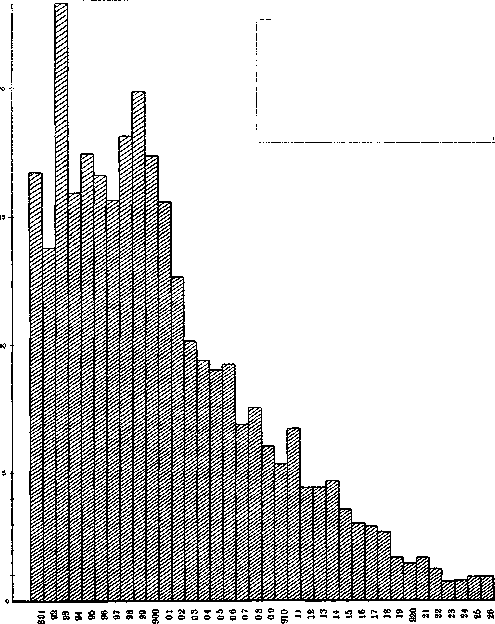
Figure 4. Mortality from typhoid fever.
of the epidemic are most frequently contact infections, followed by water-borne and food-borne ones. According to Prigge's study, out of a thousand cases of the disease, 70% are attributed to contact infections, 14% to water, and almost as much to milk and other food products. Contact infections occur both from patients and from bacillary carriers, in England (per 100,000 population). in the 2nd—158, in the 3rd—116, in the 4th—59, in the 5th—34, and in the 6th–10th—75. Infection occurs most frequently via hands; according to Klinger, out of 1,397 infections, in 1,315 cases the infection was transmitted by hands. Churilina found typhoid bacilli on the hands of nurses twice out of 23 cases. Besides patients, bacillary carriers play a tremendous role in the spread of typhoid fever, 6 among whom the typhoid bacillus can be excreted for years. Bacillary carriers are mobile foci of infection and can transmit it over long distances when moving. Female bacillary carriers who have a direct relation to food preparation are the most dangerous (see Bacillary carriage). — Food products (milk, salads, vegetables). Milk is a convenient medium for the development of typhoid bacilli introduced from hands during milking by carrier women or when rinsing a contaminated milking pail. Typhoid bacilli can get onto vegetables when watering garden beds with sewage water. When preparing salads and cold dishes, the latter can be contaminated by bacillary carriers or flies, and the typhoid bacillus gives abundant growth, since salads and jellied dishes are usually not consumed immediately, but after some standing time, which is sufficient for the microbe to multiply. — Water-borne epidemics, described many times, most frequently break out when water is contaminated with sewage. In this regard, typhoid bacilli have been found in water supply sources. A characteristic feature of water-borne epidemics is the rapid rise of the morbidity curve (see Water-borne infections). Improvement of water supply in cities, installation of filters, and chlorination of water are usually accompanied by a drop in the morbidity rate. Recent years have been marked by an increase in typhoid fever morbidity not only in our country but also abroad. Particularly large typhoid fever epidemics occurred in 1926–27 in Hanover, Montreal, Rostov-on-Don, and Krasnodar (see below—typhoid fever statistics). The sources of infection served as: in Hanover, apparently, Flutgrundwasser; in Montreal, contamination of dairy products by a bacillary carrier at a milk-canning factory; and improvement of sanitary and hygienic conditions, primarily water supply and sewerage, mortality from typhoid fever has been gradually declining. This can be noted if we trace the trend of typhoid fever over a long period of years (see Table 1). Table 1. Mortality from typhoid fever in different countries (annual average). Countries Italy Spain Germany Netherlands Switzerland Years Mortality per 100,000 1886-90 79.0 1896-900 49.7 1911-15 23.5 1916-20 26.7 1921-25 23.9 1896-900 61.4 1911-15 25.4 1916-20 29.0 1921-25 24.5 1891-95 14.7 1896-900 11.0 1901-05 7.6 1906-10 4.8 1911-15 6.2 1916-20 6.2 1921-25 3.2 1891-95 12.4 1911-15 4.0 1916-20 5.9 1921-25 2.4 England and Wales Australia . -9 5 -15 -20 -2 5 -75 -9 5 -15 -20 -25 -15 -20 -25 -15 -20 25
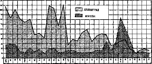
Figure 5. Mortality from typhoid fever in Moscow and Leningrad (per 100,000 population).
in Rostov-on-Don, contamination of the water supply system by seeping sewage water. In recent years, views on the mechanism of development of typhoid fever epidemics have been undergoing some changes.
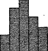
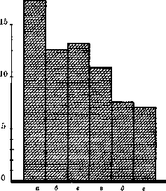
D. Zabolotny. Statistics. 1. Mortality from typhoid fever. As a result of the rising cultural level of the population, the cited figures, as well as the attached diagrams (see figure 2, 3, and 4—mortality from typhoid fever in Italy, England, and Germany), give a clear idea of the decrease in mortality from typhoid fever in various states, although in some of them (Italy, Spain, Germany, Holland) an increase in typhoid fever mortality can be noted in the period from 1916 to 1920, which was a result of the influence of the imperialist war. To give an idea of the change in typhoid fever mortality in the USSR, the following typhoid fever mortality rates in Leningrad and Moscow are presented (per 100,000 population, average annual mortality): Table 2. Number of deaths from typhoid fever in St. Petersburg-Leningrad and Moscow. Years: St. Petersburg-Leningrad, Moscow. 1888-92: 58.0, 96.7. 1893-97: 69.0, 124.2. 1898-902: 77.0, 11.8. 1903-07: 74.5, 15.5. 1908-12: 52.9, 12.9. 1913-17: 47.6, 9.9. 1918-22: 29.2, 13.2. 1923-27: 17.2, 46.8. These figures (see figure 5), despite rather significant fluctuations in individual years (epidemic outbreaks), definitely indicate a steady decrease in typhoid fever in both of these cities, which is the result of improvements in the public services and amenities of these cities. In Leningrad, for almost all of these years, typhoid fever shows a significantly higher mortality rate than in Moscow; this is mainly explained by the better water supply and sewerage conditions in the latter. The imperialist war, and especially the civil war, caused a significant increase in typhoid fever mortality. This was particularly sharp in Moscow, where in 1919-22 the typhoid fever mortality was even higher than in Petrograd; starting from 1924, Moscow again shows a lower typhoid fever mortality. With a general downward trend, typhoid fever mortality continues to remain at varying levels in different countries, which is one of the indicators of the sanitary well-being of these countries (see table 3). Table 3. Mortality from typhoid fever in various countries (per 100,000 population). Countries: 1924, 1925, 1926. Chile: 47.3, 42.8, 21.5. Spain: 21.3, 19.1, 15.6. Italy: 19.3, 18.7, 14.3. Japan: 24.8, 17.5, 8.3. Hungary: 17.0, 12.8, 6.7. Czechoslovakia: 8.0, 8.0, 5.0. Estonia: 6.7, 8.0, 3.0. Canada: 6.0, 6.2, 4.2. France: 4.3, 4.5, 3.8. Austria: 3.7, 4.4, 3.0. Belgium: 3.5, 4.3, 1.8. Irish Free State: 4.1, 3.5, 1.5. Romania: 3.7, 3.5, 1.3. Australia: 3.0, 2.9, 1.4. Germany: 2.2, 2.7, 0.9. Holland: 1.0, 1.8, 0.8. Switzerland: 1.0, 1.2, ... Denmark: 1.5, ..., ... New Zealand: 1.3, ..., ... England: 0.7, ..., ... Scotland: ..., ..., ... In individual states, typhoid fever mortality for these years ranges from 1-1.5 (England, Scotland, New Zealand, etc.) to 15-20 (Spain, Japan) per 100,000 population, while for any given country it generally remains at approximately the same level over the past three years, despite rather major outbreaks in individual regions of these countries (for example, in Germany). The picture of typhoid fever mortality by individual states presented here is significantly supplemented by information on typhoid fever mortality in cities of various countries. If we take groups of large cities in different countries, a comparison of mortality in them gives the following figures: Table 4. Mortality from typhoid fever in groups of cities of various countries (per 100,000 population). Cities in thousands: 1925, 1926. 4 cities of India: 29.1, 33.8. 2 cities of Egypt (1,351): 32.9, 32.4. 21 cities of Japan: -, 26.3. 49 cities of Spain: 20.9, 25.4. 7 cities of Italy: 14.0, 18.7. 4 cities of Poland: 12.8, 15.4. 79 cities of Ukraine: 16.9, 15.3. 8 cities of Belarus: 14.0, -. 2 cities (...): 12.7, 11.3. 4 cities of Czechoslovakia (1,176): 8.2, 7.1. 5 cities of France: 5.6, 5.4. 59 cities (...): 3.4, 2.8. 48 cities of Germany: 2.0, 2.8. 2 cities of Belgium: 3.5, 2.0. 14 cities of Holland: 2.4, 1.8. 30 cities of Switzerland: 1.4, 1.1. 16 cities of Scotland: 1.0, 1.0. 3 cities of Scandinavia (1,300): 0.8, 0.8. 107 cities of England: 0.9, 0.7. The highest mortality is noted in the cities of India, Egypt, and Japan; among European states, in the cities of Spain, Italy, etc. Typhoid fever mortality in individual cities of various countries (per 100,000 population) for 1925 and 1926 is expressed by the following figures: Table 5. Mortality from typhoid fever in individual cities (per 100,000 population). Cities: Rostov-on-Don, Calcutta, Hanover, Cairo, Tula, Tokyo, Novorossiysk, Dnepropetrovsk, Alexandria, Madrid, Kharkov, Warsaw, Kherson, Rome, Nizhny Novgorod, Leningrad, Budapest, Poltava, Sofia, Krasnodar, Krakow, Kiev, Moscow, Minsk, Odessa, Paris, Dublin, New York, Brussels, Hamburg, Oslo, Edinburgh, Chicago, Copenhagen, Vienna, Stockholm, Berlin, London, Dresden. Due to extremely large outbreaks of typhoid fever, Rostov-on-Don and Hanover in 1926 occupied one of the highest places in terms of typhoid fever mortality (see map, fig. 6). If all cities are divided according to the level of typhoid fever mortality into four groups: low mortality—up to 1.0 per 100,000 population, medium—from 1.0 to 5.0, high—from 6.0 to 10.0, and very high—over 10.0 per 100,000 population, then among the cities of the USSR listed in Table 5, only Kiev, Moscow, Minsk, and Odessa fall into the group of cities with high mortality, while the rest fall into the group of cities with very high typhoid fever mortality. Mortality from typhoid fever in cities shows fairly large fluctuations depending on the population of these cities. For the RSFSR (for 1925) and Ukraine (for 1926), for individual groups of cities, we have the following typhoid fever mortality indicators (see table 6): In the RSFSR, typhoid fever mortality is inversely proportional to the population of the cities. Particularly high typhoid fever mortality is shown by RSFSR cities with a population of 50 to 100,000. In Ukraine, the lowest mortality is shown by cities with a population of over 100,000 inhabitants, and the highest by those with a population of 30 to 50,000. The lower typhoid fever mortality in large cities should be explained by their better public utilities and amenities (water supply, sewerage). Materials on typhoid fever mortality in French cities (according to Dopter; the data refer to the period 1906–13) show the opposite picture—typhoid fever mortality increases with an increase in the population of the cities (see figs. 7 and 8). 2. Mortality by sex and age can be illustrated by the following data for England (see table 7). At the age of 5 to 14 years, female mortality in the last two periods is higher than male mortality; at older ages, as well as in early childhood, females show lower mortality than males. The highest mortality among both sexes for all indicated periods is observed at the age of 15 to 44 years. This same table provides an idea of the decline in typhoid fever mortality across individual age groups, with mortality in childhood falling somewhat faster than in older age groups.
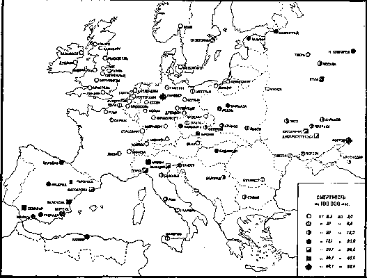
Figure 6. Mortality from typhoid fever in major European cities in 1926. Table 6. Mortality from typhoid fever in cities of the USSR depending on population size. Cities of the USSR: Number of inhabitants in thousands. Mortality per 100,000: a) 2 cities with a population above 1,000,000; b) 20 cities with a population above 100,000; c) 40 cities with a population from 50 to 100,000. Ukraine: a) 5 cities with a population from 100 to 500,000; b) 10 cities with a population from 50 to 100,000; c) 12 cities with a population from 30 to 50,000; d) 7 cities with a population from 20 to 30,000; e) 7 cities with a population from 10 to 15,000. Table 7. Mortality from typhoid fever by sex and age in England (per 100,000 population of the corresponding group).
must be concluded based on the following materials from Denmark (for the period from 1921 to 1924): Table 11. Mortality by age group in Denmark. Age: 0-4 years, 5-14 years, 15-64 years, 65 and above. Fell ill:

5. The morbidity of typhoid fever (number of registered patients) for the period from 1919 to 1926 in individual countries is presented in the following absolute figures (see Table 12):
Table 12. Number of registered typhoid fever patients by individual countries. A noticeable rise in typhoid fever after the imperialist war is noted in the USSR, Germany, Lithuania, and Poland. Subsequently, typhoid fever in them gradually declines. The morbidity of typhoid fever over the last 3 years in individual states per 10,000 population is presented in the following figures: Table 13. Number of registered typhoid fever patients per 10,000 population. States...

Figure 9. Morbidity of typhoid fever in various states in 1926 (see Table 13). ...and the height of both indicators. Figure 10 shows the morbidity indicators for the USSR (Russia) over the last 37 years (data for 1917-18 are incomplete and are omitted here). During this period, three rises in typhoid fever are noted: in 1892 in connection with the famine, in 1906-10 associated with the Russo-Japanese War, and in 1919-22 as a reaction to the imperialist and civil wars. In recent years (starting from 1923), typhoid fever has been registered lower than in the pre-war period. Comparative morbidity by regions of the RSFSR is presented in Table 14. In 1913, the highest morbidity of typhoid fever was observed in the Western, Central Black Earth, and Ural regions and in Crimea. In 1926, typhoid fever is registered lower across all regions of the RSFSR than in 1913 (except for the Far East); it gives high morbidity in the Western region, in Crimea, and in the Far East. Comparison of morbidity by individual gubernias for recent years and pre-revolutionary times presents great difficulties due to changes in the borders of these gubernias and sharp fluctuations in the number of inhabitants. The diagram (see Figure 11) provides a comparison of morbidity coefficients for 1913 and 1926 for those gubernias where such a comparison is possible. Table 14. Number of registered typhoid fever patients per 10,000 population by individual regions of the RSFSR...
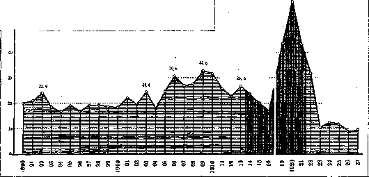
Sharp rise in typhoid fever morbidity in the USSR during the civil war and famine (dark shading - war, famine).
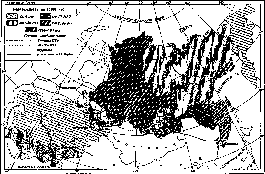
Figure 10. Morbidity of typhoid fever in the USSR (Russia) for 1890-1927 (per 10,000 population). Figure 11. Morbidity of typhoid fever in individual gubernias of the RSFSR in 1913 and 1926. Gubernias: 1-Voronezh, 2-Oryol, 3-Tambov, 4-Pskov, 5-Kursk, 6-Yaroslavl, 7-Ulyanovsk, 8-Leningrad, 9-Tula, 10-Saratov, 11-Arkhangelsk, 12-Penza, 13-Smolensk, 14-Ryazan, 15-Kostroma, 16-Moscow, 17-Vladimir. In all these gubernias in 1926, typhoid fever remains below the pre-war level. The height of...
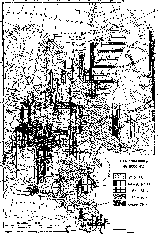

Figure 12. Morbidity of typhoid fever in the Asian part of the USSR in 1926.

Men
The greater incidence of typhoid fever in cities is possibly explained by a weaker accounting for these patients in rural areas. 8. Incidence of typhoid fever by months of the year. Typhoid fever in the USSR, as in other countries, is registered throughout the year; it is registered most highly everywhere in the autumn months, with the maximum for any given region generally falling on a specific month. In the RSFSR, in particular, this maximum, as the table shows, holds lowest from April to June; from July it begins to rise, in order to give the highest figures in September (in Ukraine, in October); in the following months it gradually declines. The correct monthly distribution of typhoid fever throughout the year is observed everywhere. The provided diagrams (see Figure 15) illustrate the movement of typhoid fever by months of the year in certain countries, and it can be seen that these curves almost everywhere coincide. When summing the number of cases by trimesters, it can be seen that in most countries the maximum number of cases falls in the third or fourth trimesters. For example, let us cite the corresponding figures for 1926 (see Table 17): Table 17. Number of cases by trimesters in 1926 in various countries (absolute figures). States: England... Bulgaria... Hungary... Germany... Italy... Poland... Romania... France... Czechoslovakia... RSFSR... Ukraine... Japan... Korea... Trimesters 1... England, Germany, the RSFSR, and Japan, on the one hand, and Bulgaria, Italy, Ukraine, and the United States of America, on the other hand, which differ sharply in their climatic conditions, give maximum figures for typhoid fever in the very same trimesters. 9. Duration and movement of individual outbreaks of typhoid fever. The given curves of typhoid fever for individual localities are often disrupted by epidemic outbreaks of typhoid fever; the duration of some of the epidemics observed in recent years and the time of their development are as follows (see Table 18): in the disease, the swollen plaques and follicles of the intestine undergo necrotization (2nd stage of necrosis, see Figure 18), and in some cases the necrosis encompasses the main, Table 18. Character of certain typhoid fever epidemics in recent years. Years, Place of outbreak, Population (in thousands), Beginning and end of outbreak, Number of cases, Number of deaths, Presumed cause. 1925: Ahlem-Germany, 420, End of June-early September, 303, 36, Milk. 1926: Hannover, Germany, 305, May-September, 2,224, 282, Water. 1926: Rostov-on-Don, RSFSR, 627, February-June, 1,199, 131, Water. 1927: Montreal, Canada, 162, December-January, 4,846, 485, Milk. 1927/28: Krasnodar, RSFSR, -, -, -, -, Water. * Hospitalized in medical institutions. The movement of these typhoid fever outbreaks by weeks can be seen from the provided diagrams (see Figure 16). Their duration depends both on the number of residents of a given city and the cause that triggered the outbreak, as well as on the speed of implementing measures to eliminate this cause. But all outbreaks are characterized by a rapid rise and a significantly slower decline (the so-called "tail" of the epidemic).
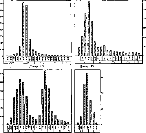
Figure 16. Typhoid fever epidemics: diagram I - epidemic in Hannover (Germany), 1926; diagram II - epidemic in Montreal (Canada) in 1927; diagram III - epidemic in Rostov-on-Don in 1926; diagram IV - epidemic in Krasnodar in 1927-28.
of the swollen plaques, and the latter return to normal through involution. Along with the indicated special changes in the lymphoid apparatus of the intestine, there also exist vulgar, diffuse changes in it in the form of catarrh of the mucosa, which underlies typhoid diarrhea. In the lymph nodes of the mesentery, phenomena of acute lymphadenitis are noted: the nodes are succulent, grayish-red; microscopically, the same process as in the intestine. Among the rare outcomes of typhoid lymphadenitis may be perforation (with peritonitis) and calcification, especially in the presence of extensive necroses analogous to the necroses and ulcerations in the intestine. Other lymph nodes usually do not experience special changes—reduction of follicles and hyperplasia of pulp cells, causing the cut surface of the organ to assume a homogeneous dark appearance of raspberry paste; sometimes infarctions are noted, associated either with the formation of granulomas in the walls of the sinuses and large veins with the closure of their lumen or with vascular thrombosis. On the part of the most important parenchymatous organs (e.g., heart, liver, kidneys), various degrees of degenerative processes are noted (cloudy swelling, degenerative fatty infiltration), in rarer cases—a picture of inflammation, e.g., hepatitis, myocarditis, glomerulonephritis. In the rectus abdominis muscles, as well as in the adductor muscles of the thigh, phenomena of Zenker's necrosis of entire bundles of muscle fibers are sometimes noted (the muscles then look like fish meat), which is usually accompanied by hemorrhage into the intermuscular or retroperitoneal tissue, sometimes quite significant. Suppurations (e.g., in muscles, bones), associated with the presence of typhoid bacilli,

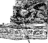
Figure 17. Peyer's patch in the 1st week of the disease.
Hyperplasia of the plaque cells; strands and nodules a) from typhoid cells (low magnification). are rare and usually occur after the expiration of the acute period of the disease. Laryngeal perichondritis belongs to equally rare but severe complications. Spielmeyer described focal inflammatory changes in the molecular layer of the cerebellar cortex in typhoid fever. Among atypical and rare localization forms of typhoid fever, mention should be made of the lesion of the gallbladder (typhoid cholecystitis) and bile ducts (cholangiotyphoid - Posselt, Abrikosov), sometimes in the absence of usual intestinal lesions. The independent existence of so-called meningo-nephro-pneumotyphoid cannot yet, apparently, be considered proven; on the other hand, inasmuch as there are cases of pure typhoid sepsis without any localization or with atypical localization (see above), one can also conceive of a selective lesion of the meninges, kidneys, lungs, and other organs.
I. Davydovsky. The pathogenesis of typhoid fever cannot yet be considered definitively elucidated. The results of bacteriological studies show with certainty that the flooding of the blood of typhoid patients with typhoid fever pathogens—bacteria—occurs from the very first days of the manifestation
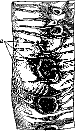

Figure 19. Typical typhoid ulcers with a clean floor; the circular layer of the intestinal musculature shines through at the bottom; the edge of one ulcer has already subsided, that of another (larger) is still clearly thickened; 4th-5th week of the disease.
of the disease, being discovered sometimes even in its latent period (Conradi). In accordance with this fact, the initial, undoubtedly erroneous, conception of typhoid fever as a primary disease of the intestinal walls was abandoned, and instead the theory was advanced that the spread of typhoid bacteria by the bloodstream and general intoxication of the organism are the main factors in the pathogenesis of typhoid fever, leading to the successive lesion of the lymphatic apparatus of the intestinal mucosa. According to the theory of Schottmüller, who did so much in the field of the bacteriological study of typhoid fever, the causative agent of the latter, having penetrated from the intestine into the lymphatic vessels of the mesentery, settles and multiplies in them. Its further spread proceeds along the lymphatic pathways centripetally (into the mesenteric lymph nodes and further into the thoracic duct) and centrifugally, which leads to the lesion of the entire lymphatic apparatus and, in particular, the intestines. According to Schottmüller, the roseolae of typhoid fever are also the consequence of the carriage of bacilli via the lymphatic pathways (retrograde) to various areas of the skin. He sees confirmation of his view in the works of E. Frankel, who, studying the structure of roseolae, found typhoid bacilli not in the capillaries, but in the lymphatic clefts surrounding them. Obtaining indisputable evidence regarding the pathway of the spread of typhoid fever in the organism is hindered by the impossibility of the experimental study of this complex problem, since although the infection of laboratory animals with typhoid fever leads to their disease and death from general intoxication, it usually does not cause the processes and changes characterizing human typhoid fever. However, upon the introduction of bacilli into the gallbladder (e.g., in a rabbit), it is sometimes possible to induce characteristic changes in the animal both in the gallbladder and in the mesenteric lymph nodes. This emphasizes the importance in the pathogenesis of typhoid fever of the system of the biliary tract and of the bile itself, which, moreover, follows from the fact of constant bacteriacholia in typhoid fever. It should apparently be considered established that the moment of absorption of typhoid bacilli into the blood (typhoid sepsis) and the moment of their excretion by the bile are indispensable factors in the mechanism of the development of the most important clinical and anatomical symptoms in typhoid fever. Be that as it may, there is no doubt that typhoid fever is a vivid example of a general infectious disease, the local manifestations of which develop only successively; being an expression of the reaction of the body's tissues, mainly the reticulo-endothelial apparatus, to the pathogenic agent that has invaded them and the poison secreted by the latter, they can achieve extremely varying degrees of development independently of the degree of general intoxication (see below—features of the course). Sharp microscopic changes in the lymphatic apparatus of the intestine, leading at times to severe clinical phenomena, should not distract attention from the changes scattered in various organs and tissues, associated both with general intoxication and with local tissue reaction. The correlation of these changes with various clinical manifestations of typhoid fever and its complications still awaits clarification to a large extent. The site of invasion of the typhoid pathogens into the body's tissues has also not been established to date; along with the assumption that the portal of entry of the infection should be sought in the intestine, there is an opinion that they are located in the tonsils, proof of which is seen in the frequency of angina at the onset of the disease, especially in laboratory infections. The excretion of bacilli occurs not only through the biliary tract system and the intestine (with feces), but also through the kidneys and through the lungs (with urine and sputum). At the basis of the pathogenesis of local specific complications of typhoid fever lies the ability of the typhoid bacillus to cause local inflammatory, degenerative, and even suppurative processes at the sites of its settlement, from which processes associated with the addition of secondary infections should be distinguished. All pathogenetic processes determining the development and course of typhoid fever are the consequence of a specific reaction between the human organism and the typhoid bacillus, infection with which, although in much rarer cases, can also be expressed in various local diseases (acute gastroenteritis, cholecystitis, etc.). Clinical picture of a typically proceeding typhoid fever is very characteristic and regular, representing a series of successive stages. The incubation period, ranging from a few days to three weeks, is assumed to be two weeks on average. Proceeding from the correspondence between the development of the clinical manifestations of pathogenetic processes and the temperature reaction, the entire course of typhoid is divided into the stage of growth of morbid phenomena and temperature (st. incrementi), usually occupying about a week, the stage of greatest intensity (st. acmis), lasting on average about two weeks, but sometimes greatly protracted, and the stage of regression (st. decrementi), lasting from a week to two, characterized by the gradual fall of temperature and the fading of all symptoms characteristic of typhoid fever and passing into the period of convalescence, i.e., the general restoration of the normal state and functions of the organism. At the basis of the syndrome of the first and second stages lie predominantly processes dependent on general intoxication and the body's reaction to it. The specificity of the intoxication corresponds to a significant constancy and character of these syndromes. By the end of the second stage, manifestations of local processes come to the foreground. The increase in the phenomena characterizing typhoid fever is preceded by the prodromal stage of the disease, which often passes unnoticed. Frequently, patients only later recall the apathy, heaviness in the head, slight physical and mental fatigue, irritability, sleep disturbance, loss of appetite, etc., that overtook them before the disease. Corresponding to the rise in temperature, the general condition of the patient so characteristic of typhoid gradually develops (status typhosus), expressed in general weakness, lethargy, loss of interest in surroundings, loss of appetite, need for rest and sleep, and in progressive clouding of consciousness. The skin is dry, the mouth dries out, the lips crack, the tongue is covered with a coating which can then become dark, fuliginous; extremely painful erosions can form on the tongue, the mucosa of the fauces and cheeks; the patient is tormented by intense thirst. Herpes, as a rule, is absent. The pulse quickens somewhat, but significantly lags behind the temperature, for example, at a temperature of 40–41°, the pulse is 85–100—a symptom extremely characteristic of typhoid fever. In children, it is absent. Often, the pulse has a dicrotic character early on. Headache can reach extreme degrees. Nosebleeds are observed. Stool is usually delayed at the beginning of the disease, liquid at the end. Phenomena of dry bronchitis are sometimes observed. The abdomen is usually bloated, sometimes, upon palpation, slightly tender in the right iliac region, while gurgling (gargouillement) is heard and felt. The spleen, as a rule, is palpable with a rounded edge, sensitive. Blood examination reveals a short-lived initial leukocytosis, quickly replaced by leukopenia. Eosinophils disappear until recovery, neutrophils decrease in number until the temperature falls, the number of lymphocytes is relatively somewhat increased, especially towards the end of the disease. The stated syndrome reaches its greatest severity and persists throughout st. acmis. The clouding of consciousness sometimes reaches loss of consciousness, delirium, comparatively rarely violent. More often, patients lie in a stupor, sleep calmly or toss about, catching at the air with their hands, making no demands, not even asking for water, despite the fact that when it is offered, they sometimes drink greedily. A decrease in hearing is often noticed, depending on a transient lesion of the auditory nerve. The amount of urine is reduced, it is saturated, and often contains protein. The diazo reaction is positive in many cases at the beginning of the disease. A new symptom, usually appearing from the 9th to 11th day, is exanthema in the form of roseola: pink spots, sometimes elevated, about 3–4 mm in diameter, disappearing upon pressure with a finger or upon stretching the skin. Their appearance depends on the formation of inflammatory foci around accumulations of typhoid bacilli in the lymphatic clefts. In the greatest numbers, roseolae are located on the skin of the abdomen (from 2–3 to 20–30), chest, and back; not so infrequently, in smaller numbers, they are also observed on the extremities. In some cases, roseolae quite densely cover the entire body; as an exception, the exanthema has a papular or vesicular character. With a corresponding diathesis, hemorrhagic rashes on the skin and bleeding from the gums, tonsils, etc., are observed. The eruption of roseolae occurs periodically: as some fade (in 4–5 days), new ones erupt. Besides the specific exanthema, occasionally on the skin, most often of the chest and abdomen, an abundant eruption of small, pinhead-sized vesicles is observed, the so-called sudamina, miliaria crystallina, while its appearance is not necessarily associated with profuse sweating. Temperature in st. acmis, as illustrated by Wunderlich's schematic curve (see Figure 21), has a continuous character (continua) at a height of 39–40°, with fluctuations per day from 0.5 to 1°. The higher the temperature, the more sharply expressed is usually the lesion of the nervous system.
The pulse remains slowed during the first 10-14 days, and dicrotism increases. By the end of the second week, the previous constipation is often replaced by 2-3 loose stools per day, without pain. More frequent stools are characteristic of large intestine involvement. The accumulation of gas (meteorism), which greatly disturbs patients and threatens serious complications, leading to abdominal distension and pain, can develop regardless of the nature of the stool. In the event of a favorable course of the disease, usually from the end of its third week, the fading of symptoms of general intoxication begins. Consciousness clears, the patient becomes capricious, reacting vividly to everything around them. A great appetite quickly develops, prompting even the theft of foreign or forbidden food. The temperature drops, as a rule, with large swings (2-2.5°), with the morning temperature dropping faster than the evening one. The corresponding period, lasting usually about a week, is called the amphibolic stage. The roseolous rash ceases. The spleen contracts, hiding behind the costal margin. The stool returns to normal. Diuresis in typhoid fever, according to Wunderlich, increases sharply, reaching very high figures (2-4 liters), with a low specific gravity of pale urine. By this same time, the strong emaciation of the patient, the anemic coloration of the skin and mucous membranes are especially noticeable; the face becomes covered with wrinkles, especially during mimicry, which appears exaggerated. Sharp hair loss, replaced by indistinct dry down. In the absence of complications, with proper nutrition, full restoration of the patient's strength, weight, and psyche takes from 4 to 6 weeks. The adynamia expressed in the recovery period, great lability of cardiac activity, sweating, and rapid fatigue of the psyche require protecting the patient from an enthusiasm for work, both physical and mental. Deviations from the above occur frequently even in uncomplicated typhoid fever and can be extremely diverse. In addition to differences in the intensity of individual temperature
symptoms, the duration of the disease is subject to large fluctuations: cases of typhoid fever lasting for months have been described, and any stage of the disease can drag on or shorten. Quite often, after the temperature drops to normal, it continues to give repeated jumps for a long time without exact explanation, and usually these jumps cause neither chills nor sweats. In forms with severe damage to the central nervous system, mental disorders can drag on for a long time. As a rule, typhoid fever proceeds worse in infants under one year old and in the elderly. In childhood, the lymphatic apparatus is affected much less. In contrast
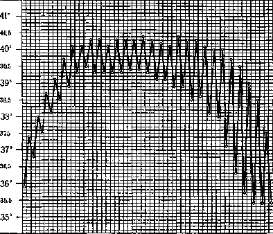
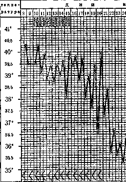
Figure 22. Typhoid i -roseolae; h----st. typho
to strongly toxic forms leading to death in a comatose state already by the end of the second week, the so-called ambulatory typhoid is observed, borne by patients on their feet and sometimes manifesting as fatal complications (hemorrhage, perforation). Abortive typhoid fever is called the form that begins as a well-expressed disease and suddenly, critically, breaks off. Features of the course of typhoid fever include so-called returns, or relapses. Their frequency varies greatly according to epidemics: after several days of normal temperature and good well-being, as a rule no later than two weeks, the temperature rises, often very quickly, to febrile figures and, accordingly to the repetition of the typhoid curve in a shortened form, the characteristic syndrome is repeated (see figure 22). Sometimes a sufficiently pronounced temperature wave is accompanied by barely noticeable subjective and objective phenomena. Blood cultures reveal a new invasion of the blood by Eberth's bacilli. Very rarely, cases of extremely severe septic course of typhoid fever, representing immense theoretical interest, are encountered, showing no changes in the lymphatic apparatus of the intestine upon autopsy (typhus sine typho). The course of typhoid fever in vaccinated individuals is extremely peculiar (see below). Along with alleviated forms of the normal course of the disease, sharp deviations are frequently observed. typhoid with relapse. iosus; < - enlargement of the spleen. The temperature throughout the entire disease, sometimes protracted, does not reach febrile figures (subfebrile-subchronic forms). Accordingly, st. typhosus is absent. Sometimes a paroxysmal course of the disease is observed with attacks of violently growing phenomena resembling influenza or malaria. Roseolae are rare. Instead of leukopenia, leukocytosis is observed. Pain in the joints, bones, and muscles is very common; forms fully analogous to paratyphoid gastroenteritis are frequently encountered. Oeller, who studied the course of the disease in the vaccinated, makes a number of conclusions concerning the pathogenesis of typhoid fever, suggesting that the atypia depends on changes in both "the mechanism of poison production and changes in susceptibility to it under the influence of vaccination."
A
E
3
N
And the complications of typhoid fever, as of a general infectious disease accompanied by the spread of a specific pathogen throughout the entire organism and strongly undermining the resistance of its tissues to the introduction of secondary infections, affect various systems and organs. Skin lesions are most often expressed by the appearance of multiple boils. A frequent, and sometimes very severe, complication is bedsores. Their formation can proceed both from the surface of the skin and from its depth, by virtue of necrosis of the subcutaneous tissue. Following the ulceration of the skin, foci of deeply penetrating necrosis of the underlying tissues can rapidly form, sometimes leading to sepsis or finding their clinical expression; sometimes the enlargement and tenderness of the liver persist even in the recovery period. Disease of the gallbladder and bile ducts is encountered much less frequently than one might expect based on the property of bile to promote the growth of the Eberth bacillus, which often settles in the gallbladder for long periods (years). The phenomena of cholecystitis usually develop in individuals who have previously suffered one or another lesion of the gallbladder, most often on the basis of gallstone disease, and therefore are encountered more frequently in women. Complications of typhoid fever on the part of the small intestines are of particularly great importance. The destruction of the vessels of the intestinal wall by the ulcerous process can lead to intestinal hemorrhage, sometimes profuse. Small hemorrhages can proceed completely unnoticed, expressed only by a dark color of the stool, depending on the combination of hemoglobin with hydrogen sulfide. More significant blood losses produce a number of characteristic symptoms: a drop and quickening of the pulse, pallor of the integuments, a sudden decrease in t° (see Figure 23). In the excreta—unchanged blood, liquid or in clots; nausea and vomiting are frequent. With hemorrhage in the upper section of the intestines, blood may not appear in the excreta immediately. With profuse hemorrhages, severe collapses are observed, rapidly leading to death. Hemorrhages occur most often in the third or fourth week, sometimes earlier, sometimes suddenly disrupting the convalescence that has begun. A formidable complication is ulcerative perforation of the intestine; without being preceded by intestinal adhesions, it leads to general peritonitis (see) and usually to death. Main symptoms: a sharp drop in t° and pulse, quickening of the pulse, abdominal pains, pallor, cyanosis of the lips, nose, and ears, sunken eyes, dry tongue, cold sweat, voiceless voice, a feeling of mortal anguish, extreme weakness, nausea, vomiting, abdominal distension and tension of its walls, disappearance of liver dullness; the stool is more often delayed. With severe damage to the psyche, subjective complaints may be absent. Symptoms that force one to watch alertly for the manifestation of the first hints of peritonitis are meteorism, which contributes to the thinning of the intestinal wall, and hemorrhage. According to Lowman, hemorrhages precede perforations in 28.2%. Perforations occur most often at the end of the 3rd week, in the 4th week, but are also encountered much later, up to the 110th day, according to Madelung. Their frequency varies across epidemics, on average 2 to 3% of cases. The complication of typhoid fever with peritonitis can also be a consequence of a ruptured suppuration of the mesenteric glands or a spleen abscess. Changes in the heart, bearing the character of acute infectious myocarditis, can lead, depending on their intensity and spread, to an expansion of the borders of the heart, quickening of the pulse, arrhythmia, and the appearance of murmurs. Phenomena of acute endocarditis are not so rarely observed. Of vascular lesions, thrombophlebitis is encountered most frequently, especially on the lower extremities (great saphenous and femoral veins), expressed by pain in the legs and a painful swelling at the site of thrombus formation, the formation of dense cords along the course of the vessels, edema of the extremities, persisting sometimes for weeks and longer, easily resuming upon walking. Thrombosis of the arteries, leading to gangrene, is observed much more rarely. On the part of the respiratory organs, ulcerative lesions of the laryngeal mucosa are noted; deepening, they lead to damage of the cartilages, their destruction and sequestration; the edema accompanying these processes can cause laryngeal stenosis. Pain sensations and voice changes are optional and depend on the location of the ulcers. A very frequent complication of typhoid fever is pneumonia. A predisposing factor to it is the bronchitis often accompanying typhoid fever, the phenomena of atelectasis and hypostasis in the lower lobes, depending on circulatory disorders and the reduction of respiratory excursions of the lungs due to the patient's position, meteorism, the state of the psyche, etc. Pneumonias caused by secondary infections (the etiological role of the Eberth bacillus cannot be considered proven) more often bear the character of confluent lobular bronchopneumonias. Encompassing entire lobes and accompanied by the phenomenon of sharp condensation of the lung tissue, they proceed according to the type of croupous pneumonia, sometimes terminating in gangrene. Pulmonary infarctions, accompanied by bloody sputum and the sudden appearance of pain, are relatively rarely observed. Pleural involvement with serous and purulent effusions is encountered much more frequently. The organs of the urinary system, despite the ability of the typhoid pathogen to linger in them for a long time, are relatively rarely affected to a degree leading to a sharp clinical manifestation. However, in individual cases, kidney damage can be very severe. The developed pyelitis and cystitis can be very prolonged and produce repeated relapses. Lesions of the genital organs have been described: testes, epididymis, ovaries, fallopian tubes, vulvitis, etc. Lesions of the central nervous system, aside from the indicated mental changes, can be organic in nature, expressed in the forms of meningoencephalitis, hemiplegia, and myelitis. Along with the phenomena of meningeal irritation ("meningism") that frequently accompany typhoid fever, purulent typhoid meningitis is also observed. Of the peripheral nerves, the branches innervating the toes are most often affected, which is expressed in agonizing pains in them. Polyneuritides have also been described. Relatively rarely do the changes in the bone marrow specific to typhoid fever attain clinical expression, with which complications in the form of osteomyelitis of both long and flat bones are associated. Periostites are observed more frequently, but are often overlooked. Likewise, due attention is not paid to the changes in the muscles characteristic of typhoid fever, especially of the abdomen (Zenker's degeneration), which only in rare cases reach the point of necrosis and suppuration breaking outward. Finally, it should be noted that typhoid patients have a tendency to react with the formation of abscesses to various therapeutic injections, with the pus turning out to be either sterile or containing Eberth bacilli. The degree of development of some of the described complications can reach dimensions that overshadow the syndrome characteristic of typhoid fever, which gave rise to the terms: pneumo-, nephro-, and laryngo-typhubs. Considering that the essence of the pathogenesis of typhoid fever, defining it as a nosological entity, does not change from the brightness of expression of one or another complication, these terms should be discarded. The diagnosis of typhoid fever is, as a rule, uncomplicated; in some cases, not to mention vaccinated individuals, it is very difficult, and at times impossible without laboratory assistance. The syndrome pointing to the diagnosis of typhoid fever is outlined in the description of its clinical picture of development. One has to differentiate it from a number of diseases.
Distinctive signs may serve: for typhus—absence of a prodromal period, more rapid development of the disease, temperature curve, appearance of roseolous-petechial rash on the 5th–6th day of the disease over the entire body, excited and irritable state of the patient, positive Weil-Felix reaction; for relapsing fever—suddenness of onset, with chills, sometimes vomiting, pain in the calf muscles, sharp enlargement and tenderness of the spleen, often enlargement of the liver, leukocytosis, presence of Obermeier's spirochetes in the blood; for influenza—presence of runny nose, herpes, chills and sweating, etc.; for central pneumonia—general appearance of the patient, rapid rise in temperature with chills, leukocytosis, characteristic sputum; for meningitis—marked nuchal rigidity, Kernig's sign, changes in cerebrospinal fluid; for sepsis—bounding temperature, chills, sweats, corresponding bacteremia, septic exanthems. Most difficult is distinguishing typhoid fever and the typhoid form of miliary tuberculosis, which must always be kept in mind; pointing toward miliary tuberculosis are: paleness, cyanosis, and sharp dyspnea not corresponding to the degree of cardiac failure and changes in the lungs, frequent pulse, absence of leukopenia, typhoid bacteremia, presence of tubercles in the retina upon examination of the fundus oculi, Koch's bacilli in the blood. The bacteriological diagnosis of typhoid fever is established by blood, feces, bile, and urine cultures. For greater success in blood cultures, so-called enrichment methods are used; blood extracted with a syringe from the veins of the cubital fossa is inoculated into bile, bile broth (Kaiser-Conradi), or water (Klodnitsky); the inoculate is placed in a thermostat at 37° and after 12–24–48 hours, subcultures are made onto differential media (see above—Etiology). The best results are obtained by inoculating 5–10 cu. cm of blood into 50 cu. cm of broth with 10 cu. cm of bile. To determine the degree of bacteremia and to limit the influence of accidental contamination, blood inoculation is performed by mixing it (without shaking) with agar melted in a water bath and cooled to 40° (in a 1:5 ratio). This mixture is immediately poured into Petri dishes. Colony growth is sometimes noted only after 2 days. The number of colonies will determine the intensity of bacteremia. The earlier the culture is made, the higher the continuous-type temperature, the more often the culture result is positive (up to 90%). Isolation of the typhoid pathogen from feces is significantly more complex, succeeds relatively rarely, because typhoid bacilli perish, unable to withstand the competition of B. coli and other flora of the large intestine, and finally, it is not always conclusive (carrier state). More consistent results are yielded by cultures of duodenal contents obtained with a thin probe (V. A. Waldman, Schotter). The significance of both methods increases in the late stages of the disease, after the cessation of bacteremia. The simplest serological method for diagnosing typhoid fever is testing the ability of the patient's blood serum in known dilutions to agglutinate Eberth's bacillus, the so-called Widal reaction (see Widal reaction). Evaluation of this reaction must be carried out with caution. The widespread opinion that a positive result at a serum dilution of 1:50–100 determines the diagnosis cannot be considered well-founded. Vaccinated individuals may also give a reaction of this titer, and it may be group-specific. Of greater importance are the rise and fluctuations of the reaction when compared with the serum's relationship to other microbes of the typhoid group. The Widal reaction usually appears from the end of the first week. It is interesting to note that earlier than this, the ability of the serum to agglutinate a culture of so-called paratyphi No. 2 is observed (M. N. Fischer). The rise in the Widal titer occurs by the third stage, after which the reaction usually slowly fades, sometimes persisting for months and flaring up later under the influence of certain other infections, especially typhus. Prognosis of typhoid fever must always be made with caution, since fatal complications can develop suddenly and in the late stages of the disease. Sharply worsening the prognosis are: prolonged high temperature, a gradually progressive drop and acceleration of the pulse (more than short-term collapses), stubborn meteorism, advanced age and age under 1 year, general exhaustion, hemorrhagic diathesis, lesions of the large intestines. Mortality varies greatly depending on epidemics and the environment in which the patient is located. On average, it is now estimated at 5–10%, instead of the 20–25% usual for the last century (see above—Statistics). Frontline epidemics yield up to 40%. Among the vaccinated, mortality is very low, about 1%. Complication by perforation, with rare exceptions, leads to death. Mortality from hemorrhages, estimated by Liebermeister at 38%, is now significantly lower. Early hemorrhages are more dangerous. Treatment. Specific methods of treatment for typhoid fever have not become widespread. The attempt at specific serotherapy made by Chantemesse as early as 1901 did not yield conclusive results. In recent years, the use of serotherapy in the form of antitoxic and bacteriotropic sera in amounts of 20–40 cu. cm has been warmly recommended by R. Kraus (Vienna). Schottmüller considers the use of convalescent serum more reliable. Vaccine therapy for typhoid fever is well developed. Practically, the simple application of the subcutaneous method does not yield any striking results. The intravenous method, first used by Ishikawa in 1914, yields somewhat better results. According to the work of A. L. Lavrinovich, a single administration of 100–200 million bacterial bodies in 25% of cases aborts the course of typhoid fever, in 18.5% accelerated lysis is observed, and in 47.5% the effect of vaccination is not detected. Similar results were obtained by E. A. Granstrom and others. Intravenous administration of a vaccine, not only typhoid but also heterogeneous, causes a rather violent reaction. In view of such a reaction, the method has many contraindications, especially in case of weakness of the cardiovascular system, and its use can be permitted only in a clinical setting, with careful selection of patients. According to Glukhov, the abortive effect of the vaccine is obtained in individuals who yield the skin reaction developed by him: the formation of a limited infiltrate at the injection site into the thickness of the skin of 0.1 cu. cm of typhoid toxin. The greatest clinicians have based the care of a typhoid fever patient on proper maintenance, nutrition, and attentive nursing aimed at easing the general condition and preventing possible complications. Excessive activity in the desire to help a patient who needs rest above all else was condemned by Griesinger as early as then. Extreme infatuation also touched at one time (Brandt) upon the powerful method of hydrotherapy. The desire at all costs to lower the temperature to normal gave rise to the prescription of baths with a temperature of 4–6°. S. P. Botkin, who appreciated the significance and clinical effect of hydrotherapy, introduced the now generally accepted use of cool baths at 26–28°, which are most useful and pleasant for high-fever patients, especially with gradual cooling of the water from 36° to 26°. Baths can successfully be replaced by cold wet packs, which are especially indicated for weak patients. A striking example of the harmful narrowing of the task of helping a typhoid fever patient is the mandatory and forced use of antipyretics (Liebermeister), after which patients, in the words of S. P. Botkin, look like "poisoned flies." The opposite extreme should be considered the refusal to prescribe, under certain indications, small doses of pyramidon to relieve headaches and the general well-being of patients. The hopelessness of using so-called "disinfectant" agents for the complete eradication of the typhoid pathogen residing in the intestines in no way eliminates the expediency of prescribing salol, urotropin, benzonaphthol, etc., to prevent excessive fermentation processes leading to meteorism, diarrhea, etc., as well as to infection of the urinary tract. Individualization of each case, careful consideration of both the intensity of individual symptoms and their connection with the underlying pathological processes must lie at the basis of all measures. Special attention is required by the complication of the disease with intestinal hemorrhage: complete rest is required, an ice pack suspended on the abdomen, an injection of pantopon for patient restlessness, sterile gelatin internally or subcutaneously (30–40 cu. cm of a 10% solution). Intravenous or subcutaneous administration of some serum; intravenous infusion of 10–15 cu. cm of a 10% NaCl solution, calcium chloride solution, and finally, blood transfusion often save a seemingly hopeless patient. With large blood losses, one should by no means fear the subcutaneous administration of physiological saline, which is also indicated in all hypertoxic forms. It can also be used in the form of drip or repeated, small (200–300 cc) enemas of it. Early recognized perforations require transferring the patient to surgeons. The significance of surgical assistance increases toward the third period of the disease in connection with its various complications (Madelung).
In cases of severe damage to the central nervous system, agonizing headaches, phenomena of increased intracranial pressure, and irritation of the meninges, a wonderful effect is produced by Quincke's lumbar puncture, with the release of cerebrospinal fluid until it drips out. The basis of patient care includes: the arrangement of the bed giving the opportunity to approach the patient from all sides, a good bed, fresh linen; careful and thorough care of the mouth: wiping with gauze moistened in a boric solution, lubricating the tongue with glycerin with wine, lemon juice, etc.; care of the patient's skin: wiping with camphor alcohol, dusting with talc; placing on rings or water mattresses when there is a threat of bedsores; offering drinks: fruit drinks, lemonade, cold tea with lemon, etc. The amount of fluid introduced into the patient must be commensurate with the state of the heart. - Diet. The fixation of attention on bowel lesions led to the restriction of patients' nutrition to the limits of a starvation diet. Meanwhile, a typhoid fever patient very much needs a supply of fuel material. The dispute over the correct diet continues to this day. Its correct solution lies in the same individualization of individual cases and in the prescription of a high-calorie diet that does not lead to the development of violent fermentation processes and does not clog the intestines. In addition to milk, acidophilus milk, cream, egg yolks, butter, kissel, porridges, compotes, jelly, white croutons, one can give scraped meat, ham, broth, fish, caviar, pureed vegetables and other easily digestible, even if not liquid, predominantly vegetable dishes. The greatest caution must be exercised in the 3rd to 4th week of the disease (ulcerative process in the intestine). The diet must correspond to the task of giving the patient about 130-150 g of protein, 200 g of carbohydrates, and 150 g of fats. In total, about 2,500-3,000 calories. In accordance with the patient's weight, one can proceed from the need to supply approximately 40 calories per kg of weight (Schottmüller). It must be emphasized that in the matter of nutrition, the method of preparation and the order of intake are of greater importance than the composition of the food. It is necessary to reckon with the patient's appetite, exciting and maintaining the secretion of the gastrointestinal tract, which is achieved, again, not so much by medicinal means as by general proper care and hydrotherapy.
G. Ivashentsov. Prophylaxis. The basis of preventive measures is the improvement of general hygienic conditions of populated areas, first of all, water supply and sewerage. This was particularly evident in individual cities (Munich, Berlin, English and American cities, Odessa, Moscow, Leningrad), where the widespread decrease in mortality from typhoid fever is associated with the improvement of water supply and the installation of sewerage. According to V. I. Yakovlev, in St. Petersburg in the 1870s-1880s, mortality from typhoid fever reached 150 or more per 100,000 population per year, whereas with the installation of the water supply system it decreased to 50 per 100,000. This was especially pronounced in Moscow, where, in addition to the water supply system, sewerage also functions, and where the previous mortality of 50-60 per 100,000 population fell, starting from the 1900s (the beginning of the operation of the sewerage system), to 10-20 (see above - Statistics of Typhoid Fever). In Odessa, mortality from typhoid fever has been dropping sharply since 1880, when the annual increase in the number of houses connected to the water supply and sewerage networks began. In the USA over the last quarter of a century, starting from 1900, mortality from typhoid fever per 100,000 population, thanks to these sanitary measures, decreased from 35 to 5. Another sanitary measure, established after elucidating the role of bacilli carriers (the first observations of the German commission in Alsace), in order to combat the contact method of spreading typhoid fever, is the isolation of bacilli carriers and their corresponding instruction. To prevent food-borne infections of typhoid fever, the main measure is the proper protection of food products and the improvement of markets (covered, easily cleaned premises where stalls and floors are washed with water, covering food products with a net or gauze) and especially dairy products, the delivery and storage of which are subject to special sanitary regulations.
D. Zabolotny. Vaccination against typhoid fever. As a preventive measure against the development and epidemic spread of typhoid fever, prophylactic vaccination has been put in a prominent place for a long time. The method of vaccination was developed by Wright in India back in 1896 and used there for the first time in colonial troops. In the same year, Volkovich made the first experiment of vaccination against typhoid fever in Russian troops. The favorable results obtained by Wright were confirmed during the Anglo-Boer War, when the English army suffered heavily from typhoid fever. Despite this, vaccinations during this period had not yet won general recognition. The most convincing data on the effect of vaccination on typhoid fever are presented by military sanitary statistics. Since 1907, vaccinations were introduced as a mandatory measure in the Anglo-Indian troops. Parallel to the development of the vaccination business, the incidence of typhoid fever noticeably decreased:
Figure 24. Comparative incidence of typhoid fever in the
armies of various countries during the imperialist war
(per 1,000 personnel).
- in the Russian army,
- in the French troops,
- in the American troops,
- in the Italian troops.
A more recent experience of vaccination against typhoid fever in the French troops also showed convincing data in its favor. Among the garrison of the city of Avignon, stricken with typhoid fever (1912), the incidence among the vaccinated was not observed at all, among the unvaccinated it equaled 224.0‰. By 1913, thanks to universal vaccination, the incidence in the garrison was reduced to zero. At the same time in the continental French troops, 30,325 people were subjected to vaccination and left unvaccinated
Figure 24. Comparative incidence of typhoid fever in the armies of various countries during the imperialist war (per 1,000 personnel).
447,159 people. Among the vaccinated there were no diseases, among the unvaccinated 2‰ fell ill. In the French troops in Morocco, the incidence from 149.75‰ in 1911, with vaccination started in 1912, decreased in the same year to 53.13‰ and, gradually falling, by 1917 decreased to 0.49‰. The favorable effect of vaccination against typhoid fever was especially revealed during the period of the 1914-18 war. Thanks to the widely and persistently conducted vaccination campaigns in all armies, typhoid fever did not assume such proportions in the troops as in previous wars, and did not bear an epidemic character even in areas unfavorable for typhoid fever. In the English and American armies, systematic vaccination was carried out even before the war. Typhoid fever in these armies therefore played a negligible role. In the armies of other warring countries, mass vaccination was started only in 1915. From this year, the incidence in all armies was reduced to negligible proportions (see figure 24). In most armies, compulsory vaccination against typhoid fever is retained in peacetime as well. In the Red Army, vaccination against typhoid fever was started in 1919. Since 1920, it has been extended to the entire composition of the army and has taken the character of an annual universal campaign. The extent of vaccination and the movement of the incidence of typhoid fever in the Red Army are shown in Fig. 25. The favorable effect of inoculations on the reduction of the incidence of typhoid fever has now gained universal recognition. According to Leishman's observations in India, the incidence of the vaccinated was 5.39‰, among the unvaccinated - 30.4‰. According to Firth's statistics, the incidence among the vaccinated was 4.7‰, among the unvaccinated - 13.9‰. According to Netter...
Figure 25. Morbidity of typhoid fever and vaccination against it in the pre-revolutionary and Red Army (per 1,000 personnel). ---------- Morbidity curve of typhoid fever ..... vaccination curve. (Netter), among the vaccinated, 1‰ fell ill, and among the unvaccinated, 14.52‰. Data concerning the Red Army in 1924-25 are also quite indicative — morbidity of typhoid fever per 1,000 people: Military Districts | Vaccinated | Unvaccinated. In the Ukrainian Military District: 0.07 | 0.15. In the Central Asian Military District: 0.06 | 3.34. In the entire Red Army: 2.29 | 0.99. Aside from these comparisons, the fact that other acute contagious intestinal diseases (especially dysentery and paratyphoid fevers), against which vaccinations did not become widespread, developed extensively in armies despite the general hygienic measures applied, also speaks in favor of vaccination. Under the influence of vaccination, the nature of the course of typhoid fever also noticeably changes. A mild, sometimes abortive, course of the disease among the vaccinated is noted more than twice as frequently as among the unvaccinated. Interestingly, in one fatal case after vaccination, Askanazy discovered a reaction of the reticulo-endothelium of the spleen and regional lymph nodes (relative to the site of vaccination) characteristic of typhoid fever. The positive effect of vaccination against typhoid fever on mortality from it is also confirmed by Mortality from typhoid fever in armies (per 1,000 personnel): German Army | French Army. 0.77 | 1. 3.01 | 8. 0. | 2.82. 0.71 | 29. 0. | 0.20. 14. | 0. | 0.05 | 0.45. among typhoid patients who underwent vaccination, is also observed less frequently than among the unvaccinated. During the war period, it sometimes decreased from 20-15% among the unvaccinated to 5-6% and even to 2-3% among the vaccinated. The rates of mortality among vaccinated individuals reported by various authors, however, fluctuate within significant limits. The most commonly used vaccine, both before the imperialist war and during its period, was Wright's vaccine (English). During the war period, vaccines (German and French) — Kolle-Pfeiffer and Vincent (Kolle, Pfeiffer, Vincent) — became widespread. Experiments on their mass use in troops did not reveal a difference in their preventive effectiveness. Vaccination, as a rule, was performed by two- or three-fold subcutaneous injections of the vaccine, with intervals between individual injections of 7 to 10 days. Revaccination was undertaken after 8-12 months, and was performed either with multiple (2-3) or single, sometimes intensified, doses of the vaccine. Vaccines were used both simple and complex. The most common during the imperialist war were typhoid monovaccines; by the end of the campaign, however, typhoid-paratyphoid divaccines were widespread in Germany and France. In the Red Army during the civil war, almost exclusively typhoid-cholera divaccine was used, and in recent years, typhoid-paratyphoid B divaccine. In France and Italy, so-called lipovaccines (a suspension of killed bacteria in oil) enjoy some distribution. Their advantage lies in the possibility of vaccination with a single intensified dose. The preventive effect of the antityphoid lipovaccine, according to observations in the Italian army, turned out to be lower than that of conventional vaccines. The standards and dosage of the vaccines adopted in the USSR are as follows (in the USSR, samples of vaccine series produced by institutes are subjected to preliminary control at the Institute of Experimental Therapy and Control of Sera and Vaccines in Moscow): Typhoid vaccine: 1 billion bacteria in 1 cubic centimeter, 0.5-1.0-1.0 cubic centimeter. Typhoid-cholera divaccine: 1 billion typhoid and 1 billion cholera bacteria in 1 cubic centimeter, 0.5-1.0-1.0 cubic centimeter. Typhoid-paratyphoid B divaccine: 1 billion typhoid, 0.5 billion paratyphoid-B, and 0.5 billion paratyphoid-A bacteria in 1 cubic centimeter, 2.0-2.0 cubic centimeters.
The standards of typhoid vaccines prepared abroad are approximately the same. The size of the vaccination doses is somewhat larger. For children up to 5-7 years of age, the indicated doses are reduced by 8-10 times; up to 10 years, by 4 times; up to 15 years, by half. Observation shows that to impart immunity and resistance to the organism, the number of vaccinations is of great importance. Both of these capacities of the organism also decrease with an increase in the time elapsed since vaccination. Therefore, three-fold vaccinations repeated every year, and in epidemic times every 6-8 months, are considered most appropriate. The administration of vaccination is contraindicated: in acute febrile illnesses, in overt tuberculosis, in nephritis, cachexia, uncompensated cardiac defects, in infantilism and so-called status thymico-lymphaticus, as well as in convalescents and overfatigued persons. During mass vaccinations during the imperialist war, isolated cases of sudden death after vaccination were observed, usually in connection with chronic diseases of the heart muscle. (Deaths depending on vaccination are observed, according to German researchers, in general no more than 1 per 100,000 vaccinated.) Caution is recommended during pregnancy. Vaccination is accompanied by a more or less perceptible general and local, but quickly passing, reaction on the part of the organism, usually not requiring treatment. In recent years, vaccination against typhoid fever with dry and liquid vaccines (enterovaccines) by mouth (a method proposed by Besredka and based on the theory of local intestinal immunity) has been gaining wide distribution as the most convenient and not accompanied by perceptible local and general reactions on the part of the organism (aside from mild diarrhea occurring after taking the vaccines) characteristic of subcutaneous inoculations. The dry (in the form of tablets) or liquid vaccine contains up to 100 billion bacterial bodies in each dose and is given on an empty stomach for three consecutive days or every other day. Dry vaccines are mostly combined with a sensitizer in the form of bile (bilivaccine) or cocoa powder, as well as various hydrotropic substances. The still relatively small experience of vaccination against typhoid fever by this method (a total of 154,173 people vaccinated so far) nevertheless shows satisfactory results. For the first time against typhoid fever, enterovaccine vaccination was used by Vaillant in France in 1921, and among the vaccinated the morbidity was expressed as 1.7‰, and among the unvaccinated, 77.0‰. Starzynski observed morbidity at 1.5‰ among the vaccinated, and 17.5‰ among the unvaccinated. Cantacuzene cites figures of 2.7‰ and 16.3‰, respectively. In the USSR, the most favorable results were obtained by Antonovsky, with morbidity among the vaccinated equaling 3.6‰, and among the unvaccinated, 113.0‰. Reports of other authors do not yet provide an opportunity to form a distinct idea of the significance and advantages of this new method of vaccination against typhoid fever. The question requires further development and improvement of both the technique of vaccine preparation and the method of their use.
Related articles
Mentioned in
Cite this page
“Typhoid Fever.” Soviet Medical Encyclopedia. English translation of Bolshaya Meditsinskaya Entsiklopediya, 1st ed. (Moscow, 1928–1936), ed. N. A. Semashko. https://sovietmedicalencyclopedia.pages.dev/article/typhoid-fever/