Relapsing Fever
Historical document, translated for reference. It reflects medical knowledge of the 1920s–30s and is not medical advice.
Summary
Relapsing fever is an acute infectious disease caused by a spirochete (treponema) characterized by alternating febrile attacks and periods of normal temperature. The disease has been documented since ancient times and often occurred as epidemics during wars.
Encyclopedia article (1928–1936)
RELAPSING FEVER. (typhus s. febris recurrens), a general acute infectious disease, the causative agent of which is a spirochete (treponema) of Obermeier. Characteristic of this disease is the alternation of acute febrile attacks lasting several days, with intervals of normal temperature (also of several days). Information about relapsing fever is found in Hippocrates, according to whose description, this disease apparently also occurred in Greece. In the form of an epidemic, relapsing fever was first noted in Scotland in 1843 and described by Craigie and Henderson. This disease was widespread in Ireland considerably earlier (see below - statistics of relapsing fever). As mass epidemics, relapsing fever often accompanied wars, as was the case during the Russo-Turkish wars, during Napoleon's campaign in Egypt, etc. I. Etiology. Obermeier in 1868 discovered in the blood of patients with relapsing fever and in 1873 described a spirochete (in modern terminology - a treponema), which, as further experiments on humans showed, is the causative agent of this disease [see color plate (pp. 475-476), fig. 3]. When examining a drop of fresh blood, among the immobile red blood cells, it may be possible to notice individual ones that seem to be subjected to jolts. Near them, individual treponemes can be seen, which are 8-15 microns in length, 0.24 microns in width [* and with one end, thinned like a flagellum. The movements of treponemes are snake-like and corkscrew-like, the number of coils - from 2 (on one side) to 6-8. In one case of relapsing fever, when the attack lasted 12 days, very long treponemes having 16 to 24 coils were found at the end of the attack. Mathis and Leger describe chains of 5-6 treponemes. On stained preparations (aqueous fuchsin,
RELAPSING FEVER
with gentian violet, according to Giemsa, or when treated with pyrogallic acid, the structure of the treponemes becomes more distinct. In the blood of patients, individual treponemes are often found already 24 hours before the onset of an attack; at the beginning of the attack there are few of them, but then their number increases with each day; during the apyrexia period they disappear. Treponemes multiply by direct division: adults, having 6-8 coils, become thinner in the middle and divide in half; as a result, two short treponemes are obtained, which grow, divide again, and so on. Some authors also assume longitudinal division in treponemes. Treponemes pass through ordinary filters of Novy and Knapp. The question of the death of treponemes in the body and the causes of the appearance of new attacks is of great scientific interest and was at one time the subject of a heated dispute between Mechnikov and Gabrichevsky: The former proved that death occurs with the phenomena of phagocytosis of treponemes, while Gabrichevsky asserted that treponemes die from the appearance of lysins in the blood. Both of these viewpoints were to a certain extent correct. In the blood, both treponemolysis and phagocytosis can be observed, and, in addition, other antibodies, which accumulate in the body during the infection, undoubtedly also affect the treponemes. A day before the end of the attack, phenomena of agglomeration of treponemes and often their death can be observed in the blood. Along with this, most treponemes begin to coil into rings from one end. Subsequently, individual links of such a ring merge with each other, and ring-shaped forms of treponemes are obtained. This phenomenon apparently represents a protective adaptation of treponemes under unfavorable conditions, since by the end of the attack a large amount of various antibodies accumulates in the patient's blood. At the same time, phagocytosis of both agglomerated spirochetes, which die in microphages (in polynuclears), and ring-shaped forms, which persist in leukocytes until the next attack, is apparently also observed. Such demonstrative pictures of phagocytosis can be easily observed in the blood of the placenta if an abortion occurred at the end of the attack. Relatively rarely, phagocytosis of treponemes can also be seen in the peripheral blood (Marcinovsky, Kartashova and others). Mühlens, Nogushi, Aristovsky and others obtained a culture of treponemes, with Nogushi noting that treponemes in cultures up to the 25th generation do not lose their virulence. The alternation of fever attacks apparently depends on the development of immunity in R.F. Most researchers working on R.F. still believe that by the end of the attack a large amount of antibodies, which are detrimental to treponemes, accumulates in the blood, due to which the fever attack stops. Individual treponemes that survive this, multiplying, accumulate again in the blood, as a result of which a new attack appears. The serum of recovered patients, mixed with blood containing treponemes, makes them immobile and causes them to gather in bunches; the same effect is obtained if blood containing treponemes of the first attack is acted upon by the serum of a patient who has already had 2 or 3 attacks. It is possible that the alternation of attacks also depends on the development of ring-shaped forms of spirochetes, which for a certain time persist in leukocytes, and then, having been freed from them, give rise to a new generation. The presence of R.F. treponemes in the blood led to the assumption that blood-sucking insects play a role in the spread of this disease. As early as 1876, Minch in Russia first drew attention to the role of insects as 'transmitters' of infectious diseases, in particular of typhus and relapsing fever. In 1897, O. Tiktin also expressed the assumption that such a transmitter is the bedbug. To clarify the role of bedbugs, Tiktin fed them on a monkey Cynopithecus Aethiops infected with R.F. with treponemes present in the blood, then the blood squeezed from the bedbugs onto a sterile glass slide was collected with a pipette and injected under the skin of a monkey (Zati sinicus), which in turn became ill with R.F., and after 64 hours treponemes were found in its blood. Tiktin's research was shaken by the work of subsequent authors, and it seemed that the role of the bedbug as a transmitter of R.F. was eliminated. In this respect, the research of Stefanovsky was particularly indicative, who for 27 months unsuccessfully fed on himself and on four colleagues 65 infected bedbugs with their offspring. Rosenholz, infecting bedbugs with European and African relapsing fever, established that bedbugs can be carriers of infection for a very long time. Thus, when rubbing an emulsion from bedbugs or their hemolymph into the scarified skin of mice or into the skin after epilation, it is possible to obtain the disease even two months after the infection of the bedbugs. Based on this, Rosenholz does not consider it possible to exclude their role in the transmission of relapsing fever. In any case, based on the biology of bedbugs, it must be admitted that they currently do not have great importance in the epidemiology of relapsing fever. From time immemorial, lice have also attracted main attention as possible transmitters of R.F. In this sense, Mackie and Busfield and others expressed themselves, but they did not provide direct evidence. Graham Smith, Edmond Sergent, and Foley set a series of experiments in this direction. The last two authors, studying the epidemic in Southern Oran, conducted experiments with various blood-sucking insects (with mosquitoes, bedbugs, fleas, and finally with Argas ticks), but always with a negative result. At the same time, in two cases out of five, they managed to infect monkeys with an emulsion from crushed lice on the fifth and sixth day after these lice had fed on a patient with R.F. Their further experiments with attempts to infect monkeys through bites of even a large number of infected lice (11 monkeys were subjected to bites of infected lice, with one receiving up to 265 bites) still gave a negative result and seemed to be in contradiction with their initial observations. Nicolle, Blaizot, and Conseil conducted this experiment on humans: five people voluntarily agreed to undergo the experiment and for 21 days daily for an hour were subjected to bites of infected lice, with one receiving 129 bites, another 377, the third 485, the fourth 4,707, and the fifth 6,515. Despite this, all persons remained completely healthy. Thus, it seemed that the natural route of transmission of infection through louse bites was not confirmed in these experiments, and the question of the mechanism of transmission of infection through lice remained open. Subsequently, much was clarified in this regard by research concerning the development of treponemes in the body of lice. Ultramicroscopy of infected lice established that the treponemes they received with the blood disappear within the first day, reappear only on the 8th day (according to Blanc's data on the 6th day) and persist for about 25 days. Toyoda in infected lice treated with silver found treponemes in the first days after infection, mainly in the form of ring-shaped forms. He also found them in the salivary glands of lice. E. Sergent and Foley, observing the development of treponemes in lice, note that immediately after feeding on infected blood, treponemes are phagocytosed by macrophages and soon become invisible. When they reappear, they are at first barely noticeable and only after some time approach the forms found in the patient's blood. They are always absent in the piercing apparatus, and this explains the difficulty of transmitting relapsing fever through bites. Treponemes are most often found in the lacunar cavities and in the blood of the louse, with large masses of them accumulating in the blood of the legs. The great fragility of the legs leads to the fact that when a leg is mechanically damaged (which happens very often), treponemes that were enclosed in the lacunar system of the leg, together with a drop of colorless louse blood, get on the surface of the skin and through scratches penetrate into the depth of the tissue, and in this way, mainly, the infection of R.F. occurs. Nicolle, Blaizot, and Conseil assume hereditary transmission of treponemes in lice. R.F., described in North Africa, as well as in India and North and Central America, is transmitted in the same way by lice. However, the treponemes found in them have some biological peculiarities; therefore, different types of R.F. can be established for these countries. European R.F. - the causative agent Treponema Obermeieri, Cohn; North African R.F. - the causative agent Treponema berberi, Sergent and Foley; Indian R.F. - the causative agent Treponema Carteri, Mackie; North and Central American R.F. - the causative agent Treponema Novyi, Schellack.
e.
Marcinovsky. II. Statistics and geographical distribution of R.F. 1. Worldwide distribution of relapsing fever. - Europe. The first reliable epidemic of R.F., observed in 1739 in Dublin, was described by Rutty. In the first half of the 19th century, Ireland, due to frequent famines, experienced a series of epidemics of R.F.: in 1797-1801, in 1817-19, in 1826-27, in 1842-54. From Ireland, R.F. was often carried to England and Scotland. In the Royal Hospital in Glasgow, patients with R.F. were admitted: Table 1. Number of Years Number of Years patients patients 18 53 6S In the London Fever Hospital from 1847 to 1854, 880 patients with R.F. were admitted. In Edinburgh in 1847-48, 584 patients were treated, from October 1848 to October 1849 - 263, from October 1849 to October 1850 - 25 patients with R.F. An epidemic of R.F. during this period was observed simultaneously with typhus fever. The last epidemic of R.F. in England was observed in 1869-70. In 1847-48, due to famine, an epidemic of R.F. was observed in Upper Silesia, Galicia and Bukovina. Individual cases were noted in Königsberg. In Germany, epidemics of R.F. were observed in 1868 (after the Prussian-Austrian war), in 1871-1872 (Franco-Prussian war) and in 1878-1881. The 'tail' of the last epidemic lasted for several years: in 1883, 1,208 cases were hospitalized, in 1884 - 927, in 1885 - 245, in 1886 - 298 and in 1887 - 153. In the Balkan states, R.F. was usually observed in connection with wars. - The imperialist war gave impetus to the development of epidemics of R.F., but in some countries it did not lead to an increase: in France several individual cases were noted, in England - several cases among soldiers in 1919-20, none in 1921-22 and 2 cases in 1923. In Germany during the imperialist war, cases were observed, mainly among prisoners of war. Registered cases among the civilian population: in 1914 - 2 cases, in 1915 - 261, in 1916 - 152, in 1917 - 159, in 1918 - 12 and 1919 - 26. From 1914 to 1920, 60 people died from R.F., including 50 among prisoners of war and military personnel. In Italy at the end of the same war, among prisoners of war, 99 cases of R.F. were noted in 1919-22 and in 1921 - 4 cases among emigrants. In Poland, where R.F. had been observed earlier (epidemics in 1879 and 1907), after the imperialist war it assumed large proportions and reached its maximum in 1922 (per 100,000 inhabitants - 159 cases and 5.6 deaths). In individual districts of Poland, morbidity and mortality from R.F. per 100,000 population for that year were: in Polesie - 1,488 and 29, in Bialystok - 572 and 28, in Novogrudok - 563 and 23, in Volyn - 349 and 10, in Lublin - 326 and 15, etc. Subsequently, R.F. in Poland decreased, and in recent years it gives only individual cases (see figures in table 2 below). In Romania, R.F. began in 1915 and took the form of a severe epidemic in 1916-17. In Serbia, an outbreak of R.F. was observed at the end of 1912 (in connection with the Balkan War), the next outbreak begins in September 1914 and turns into an epidemic. During the imperialist war in Serbia, there were about 200,000 Table 2. Number of registered cases of R.F. by individual European countries (absolute figures). Countries 1919 1920 1921 1922 1923 1924 1925 1926 USSR . . Austria . Greece . Germany Italy . Latvia . Lithuania . . Poland . Romania Finland . Czechoslovakia Estonia . Yugoslavia 251,369 104 755,787 0 667,577 1 1,505,852 1 ,20 7 241,001 0 19,265 0 1 4 338 2 9 o 2 15 15,105 0 0 3 289 0 2 15 6 0 Table 3. Morbidity from R.F. by individual European countries (per 10,000 population). Countries 1919 1920 1921 1922 1923 1924 1925 1926 40.3 113.0 69.0 171.0 18.9 3.7 1.4 1.2 Austria . 0.2 0.04 0.04 Greece . . - - - 0.1 0.001 Germany 0.04 0.1 0.01 0.005 0.001 0.001 0.001 0.001 Italy . - - - 0.08 0.07 Latvia . - 3.0 1.7 0.7 0.05 0.01 Lithuania . . - 2.7 4.1 3.6 0.06 0.05 0.04 0.01 Poland . 1.2 2.6 5.3 15.0 0.8 0.1 0.04 0.01 Romania. - п,з 2.8 0.3 0.1 0.03 0.02 0.003 Finland . 0.4 0.02 0.006 0.003 0.006 Czechoslovak . 0.01 0.01 0.01 0.03 Estonia . . . 1.1 8.8 0.7 0.5 0.1 0.01 0.01 Yugoslavia . 0.02 0.05 0.02 0.01 0.01 0.01 0.001 cases of R.F. In Turkey (in Constantinople) 155 cases were registered in 1921. The morbidity from relapsing fever in European countries after the war is given in tables 2 and 3. In most other European countries, R.F. was either not registered at all during these years or was registered in individual cases. Asia. R.F. is constantly observed in Turkey, Syria, in Palestine (for the period from 1921 to 1926 - 129 cases), Iraq (individual cases) and Persia. R.F. is very widespread in British India. Major epidemics here were established in 1806, in 1836-37 and in 1848-50. Since then, 'European' R.F. has been constantly observed in India. In 1917-18 in the United Provinces of India, at least 500,000 people died from R.F. in 15 months, according to approximate calculations. In Bengal, about 7,000 people died from R.F. in 1921 and more than 5,000 in 1922. In 1924 in Punjab, 24,471 cases and 2,258 deaths from R.F. were registered, in Bengal - 4,034 deaths. An epidemic in the same year was observed in Kashmir, Madras, Bombay and others. In French Indochina, R.F. was established in 1906 by Yersin, who isolated the Obermeier spirochete from the blood of patients. From 1907 to 1923, 11,265 cases and 2,815 deaths were registered in Tonkin and Annam. In China, relapsing fever is endemic, apparently throughout the country. Africa. European R.F. is quite widespread in Africa, and in recent years it has caused very severe epidemics, especially in French colonies. In Egypt from 1912 to 1921, 53,287 cases and 3,224 deaths from R.F. were registered, from 1922 to 1925 - 219 cases. In northeast Africa, it was observed in Morocco (bacteriologically established in 1908) - outbreaks in 1907-09 and 1914. In Algeria, the spirochete was isolated in 1902; in 1907, Sergent observed an outbreak that gave 42 cases. In 1913-14, 103 cases were noted, from 1921 to 1926 - 54 cases. In Tunisia, R.F. was established by Lafargue in 1903. From 1903 to 1926, 232 cases were registered. In the French colonies of West Africa, European R.F. has apparently been observed for a long time. Epidemics of it were noted in 1876 and 1880 in Senegal. In French Sudan, an epidemic began in May 1921, and in 2 years about 110,000 cases and 20,000 deaths were registered (per 2.5 million inhabitants). Spreading eastward and covering all French colonies, the epidemic reached Anglo-Egyptian Sudan in 1926. In recent years, R.F. has been noted in northwest Africa - in Dahomey (in 1925, 640 cases and 270 deaths), in Upper Volta (1,565 cases and 401 deaths in 1925, and 912 cases and 123 deaths in 1926), in the territory of Niger (811 deaths in 1925 and 130 in 1926), in French Sudan (183 cases in 1925), in Nigeria (43 cases in 1923, 733 in 1924, 1,276 in 1925, 674 in 1926), on the Gold Coast (160 in 1923, 16 in 1924 and 19 in 1926). In West Africa - in Cameroon (1,004 in 1925 and 1,009 in 1926), French Equatorial Africa (1,405 deaths in 1925 and 280 in 1926), in Uganda (from 1921 to 1925 - 2,365 cases), in Northern Rhodesia (for 1924-26 - 98), in Nyasaland (from 1923 to 1926 - 304). Foci of R.F. have also been noted in North America, where it was introduced in the middle of the last century by immigrants from Ireland. It was observed in New York, Philadelphia, Washington and others. In California, it was introduced from China. R.F. has also been noted in Mexico. 2. R.F. in the USSR. The first reliable epidemic, according to Reitlinger, was observed in 1857-58 in Novo-Arkhangelsk port - on the ship 'Tsesarevich' 258 people fell ill. In 1863-64 (from August to April), an epidemic of R.F. was observed in Odessa. In 1864, its epidemic appeared in St. Petersburg, in 1865 - in Moscow. In the next few years, relapsing fever was observed over a significant part of Russia. A detailed summary of epidemics of R.F. from 1864 to 1870 is given by Reitlinger. In St. Petersburg during this period, cases were noted (see table 4): Table 4. Years Among civilian population In troops 1864 1865 1866 1867 1868 1869 1870 1,753 14,045 6,870 6,062 4,219 3,746 2,288 579 527 406 264 Simultaneously, an epidemic was observed in St. Petersburg and neighboring provinces. In Moscow and the Moscow province, it was of significantly smaller proportions. R.F. spread strongly in these years in Ukraine and in the East: in the Orenburg province (in 1865, 2,583 patients, in 1866 - about 6,000), in Siberia and Turkestan. Epidemics were observed in the extreme north - in the Arkhangelsk and Olonets provinces, in the Crimea (Simferopol, Yalta), in the Caucasus (Tiflis, in the troops), in the West (Estonia, Warsaw, Riga), in the East (Yakutsk, Primorye region) and in the central provinces (Nizhny Novgorod, Oryol, Kursk and others). In many places, epidemics of R.F. occurred simultaneously with typhus fever. A more accurate accounting of R.F. in Russia begins in 1886.
Table 5 presents the morbidity rates (number of registered cases) for the USSR (Russia), Moscow, and Leningrad from 1886 and for Ukraine from 1901 (average annual morbidity per 10,000 population). Table 5. Morbidity from R. t. per 10,000 population. Years Moscow Leningrad Ukraine 1886-1890 2.4 22.2 5.5 1891-1895 1.7 10.9 7.5 - 1896-1900 1.1 12.3 6.5 - 1901-1905 1.1 9.0 3.8 1.4 1906-1910 5.1 33.6 12.2 9.5 1911-1915 1.7 2.1 0.5 2.7 1916-1920 51.2 27.9 31.1 29.1 1921-1925 52.8 37.5 26.2 69.6 1926-1927 0.8 0.4 0.04 0.3
For the USSR over this 42-year period, three upsurges of R. t. are observed: a relatively small one in 1886-90, a higher one in 1906-10, associated with the Russo-Japanese War, the first revolution, and the mass repressions that followed (overcrowding of prisons and development of R. t. in them), and the third, unprecedented in scale, beginning in 1916, resulting from the imperialist and civil war and famine and the resulting mass lice infestation of the population.
During the initial period (1886-1889), R. t. remains relatively low (see Figure 1), dropping in certain years to small figures (in 1901 and 1904 below 1 per 10,000 population). A new upsurge begins in 1907, with the maximum observed in 1908-09.
Figure 1. Morbidity from R. t. in the USSR (Russia) from 1886 to 1927 and in Ukraine from 1901 to 1927 (per 10,000 population).
For Moscow and Leningrad, the upsurges of R. t. coincide in time, with R. t. in Moscow being higher than in Leningrad throughout all periods (except 1916-20). The morbidity rates in both these cities before the war were significantly higher than the average for the USSR as a whole.
The indicated periods show upsurges (above 8 per 10,000 population), after which it gradually decreases to 1 per 10,000 population in 1914-15. The beginning of a new upsurge is noted from 1916 (development of R. t. among military personnel and prisoners of war and its spread to the civilian population).
In 1919, R. t. already reaches an unprecedented high figure for the country - 40.3 per 10,000 population (data for 1917 and 1918 are incomplete and not presented), in 1920 - 113.0; in the following year, a certain decrease is observed.
Figure 2. Morbidity from R. t. in Moscow and Leningrad for the period from 1886 to 1927 (per 10,000 population).
9 years are noted (1890, 1900, 1911-1914, 1924-1927), when relapsing fever was registered very low (below 1 per 10,000 population). Epidemics were observed: in 1886-1887, the next from 1894 to 1898 with a maximum in 1894 (29.7), the third from 1903 to 1910 with a maximum (74.8) in 1908, and the last begins, as throughout the USSR, in 1916 (17.2) and reaches its maximum in 1922. In Leningrad (see Figure 2) during this period, 17 years are noted when relapsing fever almost completely disappeared (1887-89, 1893-94, 1897-98, 1901-02, 1911-14, 1924-27). Epidemics were observed in 1886, 1890-92, 1895-96, 1903-1909, and the last from 1916 with a maximum in 1920. A significant increase in relapsing fever in 1916, besides Moscow and Leningrad, was observed in the Vitebsk, Gomel, Novgorod, Penza, Samara, Saratov, Smolensk and other provinces. In 1919-22, relapsing fever in all provinces and regions of the USSR took the form of a severe epidemic. If the average relative number of relapsing fever cases in 29 provinces of the RSFSR, for which calculations were made more accurately, is taken as 100 in 1913, then the morbidity in them in 1916 will be 800, in 1919-4,100, in 1920-12,100, in 1921-7,100, in 1922-9,300, in 1923-1,200, in 1924-250 and in 1925-Ш. During the period from 1919 to 1923, about 3.5 million cases of relapsing fever were registered in the USSR. Registration during this period was far from complete. The actual number of cases must be significantly higher. Some authors suggest that to calculate the actual morbidity of relapsing fever for this period (also for typhus fever, see), the official figures should be increased from 2.5 to 5 times. Comparative morbidity from relapsing fever by individual republics in recent years is given in Table 6. Morbidity by individual regions of the RSFSR is presented in Table 7. Morbidity in some provinces over several years is given in Table 8. Morbidity from relapsing fever in parts of the Russian army. According to Abramov, from August 1914 to September 1917, 75,429 people in the Russian army contracted relapsing fever, or 19.4 per 1,000 of the average strength. Morbidity in the pre-revolutionary army, according to Sukharev, for January-November 1917, 17,632 people in the army contracted relapsing fever. Table 9. Morbidity from relapsing fever in the pre-revolutionary and Red armies (per 1,000 of strength). Years
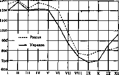
Figure 3 gives an idea of the morbidity from relapsing fever in the troops of the Red Army in 1919-22 by months of the year (per 1,000 of average strength). 3. Morbidity from relapsing fever by age and sex can be illustrated by the following data for the city of Moscow for 1920 and 1922 and for the Moscow province for 1906-10 (Table 10). Morbidity for all these periods (for the city of Moscow in 1920, 3,333 case cards of relapsing fever patients were processed, of which 1,934 were men and 1,399 were women; in 1922-13,497, of which 8,611 were men and 4,886 were women) among men is significantly higher than among women. Morbidity from relapsing fever increases with age; it was highest in 1920 and 1922 in the age group 15 to 19 years, in 1906-10 in the age group 15 to 39 years; subsequently, with increasing age, it gradually decreases. A significant excess of morbidity from relapsing fever among men was also noted in previous epidemics: in St. Petersburg in 1866 and 1867, 6,499 patients with relapsing fever were admitted to civilian hospitals, of which 5,700 were men (87.7%) and 799 were women (12.3%). Of all patients admitted during these two years, 1,701 patients, or 26.2%, were in the age group 15 to 20 years, 2,641, or 40.6%, in the age group 20 to 30 years, and a significantly smaller percentage falls on other age groups. In Nizhny Novgorod for the period from 1889 to 1896, 85.5% of relapsing fever patients were men and 14.5% were women; during these 8 years, of all relapsing fever patients, 30% were in the age group 16 to 25 years, 59.2% in the age group 26 to 55 years, and only 10.8% in all other age groups. 4. Morbidity in cities and rural areas. Relapsing fever is registered in cities significantly more often than typhus fever (see). Table 11 shows how many, according to the RSFSR, out of 100 patients with these two infections, were in cities and rural areas. Table 11a gives the number of cases of relapsing fever and typhus fever per 10,000 of the respective population. 5. Morbidity from relapsing fever by months of the year. Seasonal morbidity from relapsing fever differs
by its constancy, and its distribution by months of the year gives a characteristic curve, the maximum of which falls in the winter-spring period, and the minimum in the summer-autumn period. The rise of R. fever usually begins in November. Fig. 4 shows the monthly curves of Table 12. Monthly movement of R. fever in the USSR in 1920-22 and 1926 (absolute numbers). Months 1920 1921 1922 1926 January . . February . . March . . . . April . . May .... June . . . July . . August . . . September . . October . . November . . December . . movement of R. fever by months for the period from 1902 to 1914 in European Russia and Ukraine (average daily = 1,000). This typical course of the curve was, in general, preserved also during the pandemic of R. fever, as is evident from the figures in table 12. b. Mortality from R. fever is illustrated by table 13, which shows mortality rates (on average per year) per 100,000 population in the cities of Moscow and Leningrad for the period from 1881 to 1927, and in Odessa for 1901-1927. Table 13. Mortality from R. fever in Moscow, Leningrad and Odessa (per 100,000 population). Years Moscow Leningrad Odessa 1881-1885 . . 1886-1890 . . 1891-1895 . . 1896-1900 . . 1901-1905 . . 1906-1910 . . 1911-1915 . . 1916-1920 . . 1921-1925 . . 1926-1927 . . 75.6 15.2 3.3 4.8 2.6 11.8 0.6 21.2 49.4 0.08 46.2 6.4 4.8 3.6 2.0 4.6 0.04 71.4 57.6 0 2.1 8.8 2.8 39.8 68.4 0 Movement of mortality from R. fever by individual years is shown in Fig. 5. These curves, more accurately reflecting the course of relapsing fever than the corresponding morbidity curves (incompleteness of registration of patients), give a clear picture of the nature of the epidemic of R. fever. Almost completely disappearing in individual years, it manifests itself each time in the form of an epidemic. Its duration: for Moscow-7 years (1881-87), 5 years (1894-1898), 8 years (1903-10) and 7 years (1916-22); for Leningrad-7 years (1881-37), 3 years (1890-92), 2 years (1895-96), 6 years (1904-1909) and 8 years (1916-23). The longest period free from epidemic relapsing fever (in 48 years) was 5 years for Moscow and 6 years for Leningrad. Mortality by sex and age. Table 14 gives the mortality figures for Moscow for the period 1919-1922 per 100,000 people of the corresponding sex. Mortality among men during all these years was significantly higher than among women. Mortality by sex and age in 1920 and 1922 in the city of Moscow is presented in the figures given in Table 15 and in Fig. 6. \ '8 k \ ' / g' b 3_ /1 ch \ / . * | : - ' \ v /\ ! \ ; \ 3\ ' ' ; \ / \ ; \ ', / ' /IV ' .; ^ i-: / \ ! "* ch y ^ z 1881
1900 Figure 5. Mortality from R. fever in Moscow and Leningrad for the period 1881-1927 (per 100,000 population). Table 14. Mortality from R. fever in Moscow by sex. Absolute numbers Per 100,000 population Years men women men women 1919 . . . . 80.5 41.0 1920 .... 73.8 51.4 1921 .... 81.0 40.0 1922 .-.. 242.2 101.8
Mortality from R. fever, in general, increases with increasing age. In each age group, mortality among men is higher than among women. The highest morbidity, as was indicated above
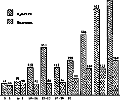
Figure 6. Mortality from R. t. in the city of Moscow in 1922 by age and sex (per 100,000 population of each group). (see table 10), falls on the young age groups. High mortality in old ages is explained by the high lethality in these ages (see below). Table 15. Mortality by age and sex in Moscow per 100,000 population of the corresponding group. year 1922. Age : MALE female sex male female sex 0- 4 years 12.9 6.4 9.6 66.4 56.0 61.1 5- 9 » 14.6 9.3 11.9 80.7 67.9 10-14 » 20.6 6.3 13.1 153.0 63.0 105.8 1 5-19 » 54.6 33.9 44.2 258.5 66.3 159.9 20-29 » 55.4 51.7 53.5 149.4 68.9 110.5 30-39 » 81.2! 61.7 71.8 190.2 90.3 143.3 40-49 » 138.7 78.9 105.5 332.9 148.7 24 5.3 5П-59 » 126.6 102.8 113.1 476.8 150.7 297.9 60 and over 147.6 63.9 88.4 520 , 4 190.0 290.9 7. Lethality (mortality). Data on registered cases and deaths from R. t. in Moscow over the last 27 years see in table 16. For Leningrad, the percentage of mortality from R. t. in hospitals for the period from 1886 to 1926 is indicated in table 17. Total for this period (without data for 1917 and 1918) in medical institutions of Leningrad there were 57,437 patients with R. t., of whom 3,201 died, i.e. 5.6%. Mortality from R. t., both in Moscow in relation to all registered patients and deaths, and in Leningrad in relation to those in medical institutions, for the same periods, roughly coincides. It varies depending on the epidemic. It was lowest during this period in 1910-14, highest in 1921-1925, with the maximum in 1922 (in Moscow - 14.2%), the year of the greatest rise of the epidemic. In the Russian army, mortality from R. t. for the period from August 1914 to September 1917 was 2.4%, on the Romanian front in 1917 - 1.9%. In Poland it was 2.9% in 1921, 3.5% in 1922, with fluctuations in individual districts in 1921 from 1.1% (Warsaw) to 15% (Krakow), and in 1922 reaching 7.4% (Warsaw) and 15.6% (Krakow). In Romania during the war it averaged 12%, with hospital mortality in individual districts ranging from 3 to 25%; for the period from 1920 to 1923 mortality ranged from 1.65 to 2.82 per 100 cases. In Serbia during the war mortality reached 30-40%. In Tonkin for the period from 1907 to 1918 mortality per 100 cases on average was 24.6%, with at the height of the epidemic (from 1907 to 1911) it fluctuated in individual years from 25.1% (1907) to 42.3% (1911); at the end of the epidemic it dropped to 4% (1918). In Punjab it was about 10% in 1920, 23% in 1923, 9% in 1924. In Egypt for the period from 1912 to 1921 it fluctuated from 6.6% to 18.3%. In western and central Africa in 1926 it averaged 20%, reaching 44% in some places. -Lethality by age groups (percentage of deaths per 100 cases of the corresponding age group) according to data from hospitals in Leningrad for the period from 1886 to 1909 is presented in table 18. The danger of mortality when contracting R. t. increases with age. It is highest in old age. Table 18. Lethality in R. t. in hospitals in Leningrad by age groups. Ages f4 «O oo eou й <o 1 CO sta from J, O co co co co Up to 1 5 years . . 16-20 » . . 21-30 » . . 31-40 » . . 41 - 50 » . -51g. and older 1.2 1.9 3.1 5.9 10.2 16.1 0.6 1.9 2.4 5.4 9.3 22.3 0.4 0.4 1.3 4.2 7.3 15.7 1.2 1.1 2.6 4.5 9.8 22.8 0.5 0.6 1.5 3.1 10.0 17.7 0.6 0.9 1.9 4.1 9.3 18.3 III. R. t. as a social disease. Soc. factors have the strongest influence on the spread of R. t. Wars, famine and popular disasters, accompanied by deterioration of sanitary conditions and mass development of lice, serve as the cause of the strongest outbreaks of epidemics, and sometimes pandemics of R. t., as observed during the imperialist war and after it in Serbia, Poland and USSR (see above). The first researchers of it in Russia pointed to the connection between epidemics of R. t. and famines, crop failures and poor grain harvests: Botkin, Lesch, Goryachev, Zorn and others. (the works of these authors belong to 1865-1868). English authors called R. t. "famine typhus". R. t. is observed mainly among the poorest strata of the population living in the worst sanitary conditions. Zorn (1865) writes: "In our country, as in the Irish and Scottish epidemics, relapsing fever almost exclusively affected the lower class of the people", "the people of the lower class contracted relapsing fever the most". Reitlinger (1874), as a result of his extensive review of epidemics of R. t. in Russia, concludes that "relapsing fever occurs predominantly where, for some reason, a concentration of people has formed under poor living conditions: in overcrowded barracks, seminaries, prison castles and shelters of the working class, among workers building railways, factories and factories, among the poor urban residents and among rural residents during famine after crop failure". "Relapsing fever occurs predominantly among manual workers, settlers, pilgrims, convicts and lower ranks of the military department". All later research confirms these data. -The study of the distribution of R. t. in individual parts of large cities indicates that R. t. is concentrated in certain areas of the city, among the least well-off strata of the population living in the worst housing conditions. Thus, in Petersburg during the epidemic of 1895-1896, the most affected (according to Likhachev) were the 3rd Spassky, 1st Narvsky districts and the Alexander Nevsky part; during the epidemic of 1906-1909, according to Kamanin, R. t. was concentrated in the same parts of the city, as well as in the Rozhdestvenskaya and Moskovskaya parts - "in districts abundant in lodging houses and inns, populated by poor people with their overcrowding and poor hygienic living conditions", while in the Admiralteyskaya, Kazanskaya, Liteinaya and other parts "with a wealthier population, living widely and freely, the epidemic was limited to a small number of cases". Binshtok, summarizing data on R. t. over 25 years, gives the following table of distribution of relapsing fever by individual parts of Petersburg for the period 1897-1911: Alexander Nevsky Narvsky . . . Spassky .... Rozhdestvenskaya Moskovskaya . . Kolomenskaya .

Petersburg Vasilievsky Vyborgsky . Liteinaya . . Kazanskaya . . Admiralty 4.1 3.7 2.7 2.4 1.3 0.4 The first 5 parts of the city, populated by the poorest and most crowded strata of the population, account for about 80% of all cases. In Moscow, during the epidemics of relapsing fever, the latter was concentrated, mainly, in those parts of the city where night lodgings, cubicle apartments, and inns were located. The highest number of cases (in particular, for almost all years) was registered in the 3rd Myasnitsky district (Khitrov market, night lodgings) and the 3rd Rogozhsky district (Morozov night lodging house). The number of cases in these parts of the city for some years is as follows: Years Total 3rd Myas- 3rd Rogozh- diseased nitsky dist. sky dist. 1896....... The 3rd Myasnitsky district (Khitrov market) in individual years accounted for 20 to 30% of all patients registered in Moscow. Grazianov, who surveyed relapsing fever in Nizhny Novgorod for 25 years (from 1873 to 1896), concludes that relapsing fever is observed predominantly in the Rozhdestvenskaya part of the city, where the largest number of lodging houses is concentrated and where 'the housing conditions of the working-class people are especially difficult.' For the epidemic of 1896-1897, he cites the following morbidity figures per every 10,000 population of the respective parts of the city: 1st Kremlin-19.7; 2nd Kremlin-24.0; Makaryevskaya-61.5; Rozhdestvenskaya-262.3. The distribution of relapsing fever patients by the nature of dwellings and apartments can be illustrated by the following data: in Petersburg during the epidemic of 1906-1909, relapsing fever patients were distributed as follows according to the nature of their apartments (see table on page 475). If we exclude from this table apartments 'without designation of character,' then lodging houses, inns, and corner apartments account for about 60% of all cases observed in the city. In Moscow for the period from 1894 to 1896, the distribution of patients by individual apartment types was as follows: Apartment types % Private apartments Cubicle-room apartments Artel apartments Craft establishments Lodging houses . 108 241 229 236 102 1,055 204 274 137 65 653 151 164 290 154 92 1,174 352 609 793 527 259 2,882 6.5 11.1 14.8 9.7 4.6 53.3 Total. . . 100.0 Lodging houses for these three years provided more than half of all relapsing fever cases registered in the listed apartment types. A large percentage of cases falls on cubicle-room and artel apartments. Private apartments provided a relatively very small percentage of cases—only 6.5%. An outbreak of relapsing fever in 1928 in Nakhichevan (North Caucasus) was also observed in an extremely overcrowded lodging house. The distribution of patients by professions gives the same picture—predominant morbidity of persons in the most destitute professions. According to Reitlinger, among relapsing fever patients admitted to Petersburg hospitals in 1867-68, there were: 1. Servants of all kinds, day laborers, and common laborers ................... 867 2. Persons engaged in trades and crafts (including carpenters-191, cabmen-137, shoemakers-129, tailors-102, tavern keepers-81, joiners-76, etc.)......
34 0 4. Persons engaged in factories and plants ...
7 According to Binshtok, in the 1908 epidemic, among the total number of 6,963 sick men, there were: common laborers-1,919, cabmen-506, merchants (peddlers)-361; among 932 women: servants-116, common laborers-111. Medical personnel (orderlies, nurses, feldshers, and feldsheritsas) accounted for 142 cases in this epidemic (in 1895, 101 cases). According to Kamanin, for the period from October 1906 to March 1909, in Petersburg out of 9,164 sick men there were: common laborers-3,110, cabmen-795, carpenters and painters-259, shoemakers-320, unemployed-777, etc.; among 1,241 women-common laborers-182, unemployed-110, laundresses-149, etc. Per 10,000 persons of the respective professions, illness occurred among men (with an average morbidity of 116 per 10,000 population): Common laborers.... Hospital attendants . . Masons .... Cooks, waiters Bakers . . . -. Factory workers . Domestic servants (swiss, maids, etc.) . . Wireworkers, etc. In Moscow for the period from 1894 to 1896, among 7,216 patients there were: day laborers-1,304, servants-596 (including waiters-228, hospital servants-72), common laborers-545, persons without definite occupations-396, cabmen-249, merchants-298, bakers-244, laundresses-139, janitors-90; the rest are almost exclusively artisans (shoemakers and bootmakers-231, joiners-103, etc.). Among persons of intellectual professions, cases were noted in isolated instances. Thus, teachers and students of Moscow (in all open and closed educational institutions) over three years produced a total of 5 relapsing fever cases. According to Grazianov, in Nizhny Novgorod from November 1896 to April 1897, among 770 relapsing fever patients there were: merchants-0; officials-19; servants-43; traders-48; artisans-181; common laborers-479. The groups of 'day laborers' and 'common laborers' who do not have permanent dwellings and live in the worst housing conditions produce the highest morbidity figures everywhere. The same picture is given by the distribution of patients by affluence. According to Binshtok, in the 1895 epidemic in Petersburg, not a single wealthy person and only a few well-to-do persons were noted among the relapsing fever patients. The distribution of patients in 1906-09 in Petersburg is as follows (per 100 patients): wealthy-0; well-to-do-1.6; poor-15.5; unknown-82.9. Kamanin refers the entire group of 'unknown' to the poor. Grazianov, based on an analysis of Nizhny Novgorod material, concludes that 'relapsing fever in Nizhny Novgorod, due to the extreme lack of hygiene in the housing conditions of the homeless people, spreads from year to year in winter among the working poorest part of the population.'
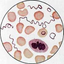
Figure I. Spleen in relapsing fever (longitudinal section, reduced by 2 times): abundance of miliary necroses (gray-yellow) in the pulp (dark red parenchyma). Upper left, under the capsule—large necrosis in the form of an infarct; in its center—disintegration (from the collection of the Museum of the Pathological Anatomy Institute of the 1st Moscow State University). Figure 2. Liver in relapsing fever (staining—sudan-hematoxylin, high magnification): abundance of lipoids (bright red droplets) in the reticulo-endothelium (a); in the parenchyma trabeculae (b)—lipoids in small quantities; c—lumens of liver capillaries (according to the preparation of S. S. Vail). Fig. 3. Treponema of European relapsing fever. To the art. Relapsing fever
RELAPSING:
dissertation, SPb, 1865; Goryachev P., On relapsing fever, diss., M., 1868; Reitlinger L., Studies on the history, geography, and statistics of relapsing fever in Russia, P., 1874; Iogikhes A., Statistical materials on relapsing fever, diss., SPb, 1886; Likhachev D. A., Epidemic of relapsing fever in St. Petersburg in 1895 and 1896, diss., SPb, 1897; Grazianov N. A., Relapsing fever in Nizhny Novgorod, 'Bulletin of Public Hygiene, Forensic and Practical Medicine', bk. 6, 1898; Kamanin V. I., Epidemic of relapsing fever in St. Petersburg from October 1906 to March 1909, SPb, 1909 (lit.); Reports of the Moscow City Administration on sanitary matters for 1894, 1895, and 1896; 'Statistical materials on the state of public health in the USSR for 1913-23', M., 1926; same for 1924-25, M., 1927; same for 1926, M., 1928; 'Proceedings of the commission on surveying the sanitary consequences of the war of 1914-20', M.-L., 1923; Hirsch A., Handbuch der historisch-geographischen Pathologie, B. I-II, Stuttgart, 1881-83; Gouzien P., La fievre recurrente a spirochete d'Obermeir, Bulletin de l'office international d'hygiene publique, t. XVI, 1924; Wasslewski T. u. Kersting C., Ruckfallfieber (Handbuch der arztlichen Erfahrungen im Weltkriege 1914-1918, hrsg. v. O. v. Schjerning, B. VII-Hygiene, Lpz., 1922); Nicolle Ch. et Blaisot L., Fievres recurrentes (Nouveau traite de medecine, publ. sous la direction de G. Roger, F. Widal et P. Teissier, fasc. 5, P., 1922); Dopter Ch. et de Lavergne, Epidemiologie, P., 1925; Abbatucci S., Les services d'hygiene publique dans les colonies francaises, Geneve, 1926; Rapports epidemiologiques de la Section d'hygiene de la Societe des Nations pour les annees 1924, 1925, 1926, Geneve.
I. Dobreitser. significant sizes, which are relatively rarely observed in other acute infections—reaching, for example, 5-6 times the size; the capsule of the organ is strongly tense, which, especially with simultaneous necrotic changes in the pulp, can easily cause tears and ruptures of the organ with subsequent bleeding. Often the capsule is covered with a delicate fibrinous coating (perisplenitis). Upon sectioning the organ, its dark red color is noted, indicating sharp blood engorgement; in addition, in approximately 40-50% (for various epidemics the figures vary greatly) necroses of the Malpighian corpuscles, and partly of the pulp itself, are observed; these necroses have the appearance of small FEVER
of the (miliary) gray or grayish-yellow spots (see color plate, fig. 1), but sometimes they reach significant sizes, often having the form of an infarct; the number of necroses varies greatly. Their origin is not fully clarified; most likely, and in relation to infarcts undoubtedly, is that they are based on severe circulatory disorders in the organ, in particular, in the small arteries; in the latter, degenerative and thrombotic processes, as well as clumps of spirochetes, have also been found; the possibility of the direct necrotizing effect on the tissue of the breakdown products of the parasites themselves, which can be abundantly detected in the spleen tissue by the silvering method, is not excluded. Generally, in the bodies of patients with relapsing fever, spirochetes quickly undergo autolysis, and for their detection, early autopsies are recommended as much as possible. Among the most frequent complications of R. t. are catarrhal, sometimes hemorrhagic pneumonias in the posterior-lower parts of the lungs. Some epidemics are accompanied by a particularly severe complication of a septic nature called N-paratyphobacillosis. During epidemics coinciding with malnutrition or one-sided nutrition of the population, sequential diseases of osteochondritis, mainly of the ribs, are noted, which leads to chronic purulent processes, the formation of fistula passages, and even greater exhaustion of patients.
I. Davydovsky. V. Clinical picture. The incubation period for R. t. is 7-10 days. The disease begins suddenly with a severe chill and headache, as well as repeated muscle (especially often in the calf muscles) or joint pains. At the beginning of the disease, nasal bleeding is often observed. From this moment on, a sharp increase in temperature and rapid pulse can be noted. Usually, patients remain fully conscious, sometimes there is delirium, insomnia. Appetite disappears, the tongue is slightly coated with a white coating, moist; sometimes nausea and vomiting are observed, and almost as a rule, constipation. Already during the first day, enlargement and tenderness of the liver, as well as the spleen, are noted; the latter reaches large sizes and is dense and painful on palpation. Also in the first days, a slight yellowish discoloration of the sclera is noted, in rare cases general jaundice can also be observed, which during some epidemics of relapsing fever occurs as a common phenomenon. Catarrhal phenomena are usually absent. Sometimes small, sparse roseolas appear on the skin of patients, which, having existed for only a few hours, disappear. The temperature during the paroxysm remains at high figures, giving slight decreases in the morning (see fig. 7). In this state, the patient remains for about 3-5-7 days, after which the paroxysm ends with profuse sweating and a sharp drop in temperature by several degrees (usually below 36°), with complete exhaustion of strength. In the following days, the patient's condition gradually improves, he begins to get out of bed and feels almost well, but with the approach of the next paroxysm, weakness, headache, and muscle pains return. The intervals between paroxysms are usually 5-9 days, but can be shorter or longer. The number of paroxysms varies from 1 to 15, averaging 5-6. In some cases, especially at the beginning of an epidemic and at its end, mild cases occur, expressed in small increases in temperature, often one-day, with only slight muscle pains. During individual epidemics, "bile typhoid" (bilious typhoid), first described by Griesinger in 1851, also occurs. It sometimes complicates the normal
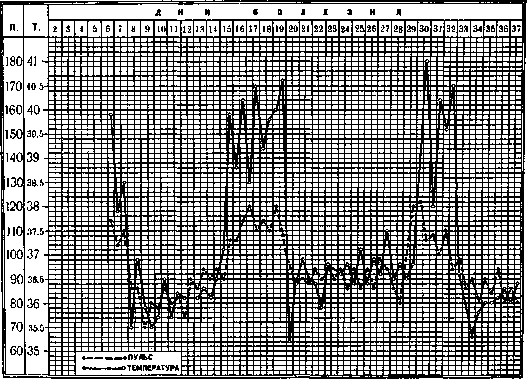
Figure 7. Temperature and pulse curve in European (louse-borne) R. t. (from the clinic of the Tropical
(of the institute in Moscow). healthy, but after 5-7 days with the same phenomena, a second attack occurs. Usually 2-4 such attacks are observed, rarely more, with subsequent attacks often being weaker. The duration of the afebrile period can vary considerably (in one case an attack was observed after 23 days). The liver and spleen decrease somewhat in volume during the apyretic period (rate en accordeon), but never return to normal. Perisplenitis is often observed. The pulse runs parallel with t° at all times. Nervous phenomena very often accompany R. f., and there are cases with typical symptoms of meningitis. In the kidneys, phenomena of nephrosis are not uncommon, and sometimes nephrosonephritis is also observed. From the blood side, a sharp decrease in the number of erythrocytes and Hb is noted; during an attack, a noticeable leukocytosis and polynucleosis with a slight shift in the leukocyte formula to the left are usually present, during the apyretic period the number of leukocytes decreases, and monocytosis is noted. The resistance of erythrocytes is increased. Along with sharply expressed cases of R. f., especially in the course of relapsing fever, but it can also develop independently from the very beginning. The usual symptoms of R. f. in typhoid fever are expressed more sharply, with jaundice, vomiting of bile, and a sharp increase and painfulness of the liver and spleen coming to the forefront. Persistent diarrhea is often observed; the tongue is usually heavily coated. In typhoid fever, brain phenomena (dizziness, loss of consciousness, drowsiness or delirium or excitement) are often observed. The temperature curve is usually irregular or of a remittent nature, as in septic diseases. Phenomena of hemorrhagic diathesis, abundant nosebleeds, hemorrhages in the skin and other organs, bloody vomiting and bloody diarrhea are not uncommon. Immunity in R. f. is extremely weak, and its duration is short. Repeated illness during the same epidemic is not a great rarity. The experiments of Nicolle and Conseil on monkeys confirm the stated position. Of 7 monkeys infected with R. f., 4 became ill with R. f. upon re-infection after 5-8 months, and 1 monkey even had two repeated illnesses. - Complications. One of the most severe complications in R. f. is rupture of the spleen and internal hemorrhage. Infarcts in the spleen, so characteristic of this disease, sometimes reach very large sizes and may suppurate, which in turn, upon opening the abscess into the peritoneum, can cause peritonitis (see Figure 8). Heavy nosebleeds and uterine hemorrhages are not uncommon. During pregnancy, abortion or premature delivery usually occurs; the child is usually born dead. Fairly frequent complications include iritis, then various neuralgias and neuritis, lesions from the gastrointestinal tract, colitis and enteritis, etc. Periostites and especially perichondrites, described as a complication in relapsing fever, probably depend on secondary infection (paratyphoid bacillosis) or are associated with impaired nutrition of patients (manifestations of avitaminosis). - Complications described in R. f. occur especially frequently in typhoid fever. The mortality rate in this disease reaches 60-70%. Whether typhoid fever is a special form of R. f. or represents a combined disease is a question that requires further study. The experience of Muchutkovsky speaks in favor of the first assumption, who transfused the blood of a patient with typhoid fever to a healthy person and thereby obtained ordinary relapsing fever (Kidney complications are described above.) - Diagnosis. The clinical picture of R. f. in a patient who has undergone several attacks is so characteristic that it presents no difficulties in diagnosis. From an epidemiological point of view, it is important to diagnose the disease at the very beginning, and it is especially important to be able to distinguish R. f. from typhus in the first days of the illness, as these diseases often occur as simultaneous epidemics. Four symptoms are characteristic of R. f.: a sharp rise in t°, frequent pulse, not diverging from t° (as is the case at the beginning of typhoid fever), slight jaundice of the sclera, and a large, dense, painful spleen (in typhus the spleen is usually barely palpable); it is especially important, in addition, the characteristic change in blood, which easily allows relapsing fever to be distinguished from malaria, typhoid fever, and paratyphoids. In epidemic jaundice, in contrast to relapsing fever, clearly expressed prolonged jaundice, renal phenomena, and hemorrhages are found. Treatment. The best method in the treatment of R. f. should be considered chemotherapy. For the first time, salvarsan (see Neo-salvarsan) was successfully used in relapsing fever by Iversen and gave brilliant results. Injections are made intravenously (in children in the v. jugularis or suprafascially, under the angle of the scapula). Injections are best done at the height of the attack in the first days of the illness or before the beginning of the next attack. If the dose is sufficient, then within approximately 6 hours t° drops to normal, and the disease is eliminated (see Figure 9). One might think that in this way complete sterilization of the blood is achieved (therapia sterilisans magna), however, as shown by the research of Krivchevsky on animals, this usually does not happen, and a small number of treponemes may remain in the body for some time without causing any pathological phenomena. According to Steiner and others., spirochetes can remain in the white matter of the brain for a long time even in immune animals. Salvarsan preparations, giving success.
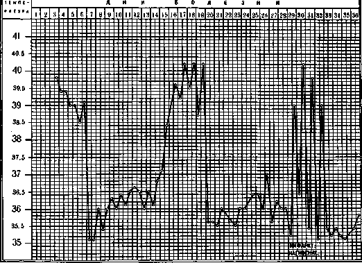
Figure 8.
Temperature curve in European R. f. Large infarct of the spleen with transition to suppuration on the 3rd attack (from the clinic of the Tropical Institute in Moscow). Injections are better done at the height of the attack in the first days of the illness or before the beginning of the next attack. If the dose is sufficient; then within approximately 6 hours t° falls to normal, and the disease is eliminated (see Figure 9). One might think that in this way complete sterilization of the blood is achieved (therapia sterilisans magna), however, as shown by the research of Krivchevsky on animals, this usually does not happen, and a small number of treponemes may remain in the body for some time without causing any pathological phenomena. According to Steiner and others., spirochetes can remain in the white matter of the brain for a long time even in immune animals. Salvarsan preparations, giving success.-The regimen of the patient and care measures during fever are usual, as in other infectious diseases.
VI. Prevention and control of R. f. Prevention and control of R. f. present great difficulties. Here, just as with some other infectious diseases, it is necessary to "break the epidemic chain" [patient with relapsing fever (carrier of spirochetes)-lice (transmitters of the disease)-healthy person], as well as to change the conditions that contribute to its formation. Early diagnosis and widely conducted salvarsan therapy of R. f. can themselves play a major role in the fight against the epidemic. n n i 12 34 56 7 8 9 10 11)2 13 14 15 16 17 18 19 20 2122 23 24 25 26 27 28 29 30 3132 33 34 36 36 41 - 40 39 - 38 - 37 - 36 -

sh
Figure Temperature curve in European (louse-borne) R. F. Treatment with neosalvarsan (from the clinic of the Tropical Institute in Moscow). powerful action on treponemes in the human body, in vitro they have almost no effect, and, as observations by some authors show, the blood of R. F. patients, containing treponemes, when mixed with neosalvarsan, remains virulent and in this form can, for example, be used to infect patients with progressive paralysis. The downside of chemotherapy for R. F. is that patients, due to the rapid elimination of the disease, do not have time to acquire long-term immunity and can easily be re-infected (examples of which have been observed). Gabrichevsky made attempts at serotherapy for relapsing fever, but without special success. Reducing and even reducing to zero parasitic carriage, one can achieve a "break" in the epidemiological chain, as a result of which an epidemic may cease. General measures to combat R. F. come down to expanding the medical network, improving laboratory work, and investigating and identifying foci of R. F. and its first cases. During epidemics, the first place is also occupied by the deployment of special departments and barracks for isolating the sick, as well as the concentration and proper distribution of neosalvarsan reserves in particularly threatened areas. Special measures to combat the disease should be directed against the destruction of lice. In the spread of relapsing fever, clothing lice play the main role, but head lice can also transmit the disease. As is known, the former lay their eggs on clothing, while the latter lay them on hair. The destruction of nits on clothing is achieved by ironing the seams, boiling the clothing in lye, or roasting it lightly in a Russian stove. Lice attack a person, guided mainly by smell. Weak, exhausted, sweaty and untidy people, as well as the obese, are more easily subject to this attack. On dirty clothing, impregnated with decomposing pus, in folds and seams, lice lay their eggs (nits), attaching them at the crossing of individual fibers. To protect oneself from lice attacks, some recipes can be recommended: sprinkle naphthalene on the sleeves and collar of a shirt; in this regard, the formula proposed by the English N. C. J. (in equal parts naphthalene, creosote, iodoform) works even better. For washing hair, the following composition is recommended instead of soap: Picis liquidi 5.0, Spir. sapon. kalin. 100.0. For washing hair, the following composition can also be recommended: 1 part of sublimate to 500 parts of water and 500 parts of vinegar. This composition has the good side that it allows for the removal of nits from the hair, as vinegar softens their chitinous shell. All these measures of protection against lice (personal prophylaxis) are to some extent only palliatives. It is generally necessary to create such sanitary conditions that the population does not have lice. Sanitary supervision over places where masses of people gather, inns, lodging houses should be strengthened both during the development of R. F. epidemics and in normal times to prevent them. Passage baths, barber shops, disinfection installations, etc., should be organized. In moments of large epidemics, the proper supply of the population with fuel, soap and baths should be especially ensured. Regarding baths, it should be noted that they sometimes, with improper construction, can themselves serve as a source of mass infection of the population and military units; therefore, their sanitary condition, proper construction, and systematic cleaning and disinsection should be given first place. Passage baths are the most expedient in this regard. For organized population groups, it is necessary to specially organize visits to baths (schoolchildren, troops, etc.). Hair cutting also has hygienic significance here; from this point of view, one can only welcome the custom of cutting hair short. Sanitary education should be aimed at promoting cleanliness of body and clothing.'
E. Marcinovsky.
VII. Tick-borne relapsing fever. Synonyms for tick-borne relapsing fever: Persian relapsing fever, Central Asian relapsing fever, tick fever, courdoud (Somalia), bobo (Uganda), miana (Persia). The first information about tick-borne relapsing fever dates back to Livingstone's journey to South Africa in 1857. He described it under the name human tick disease. This disease was more thoroughly studied by Dutton and Todd in the Congo; independently of them, R. Koch in East Africa discovered the causative agent of this disease—a treponema, and also clarified the conditions for its spread through tick bites. Since then, this disease has been described in many countries, and it has been established that the carriers are various species of Ornithodorus [see separate table (art. 579-580)]; thus, African relapsing fever is transmitted by ticks O. moubata (see table, fig. 5), O. Savignyi; American relapsing fever, first described by Bildow in 1865 as a small epidemic in the Andes, is transmitted by ticks O. talaje (see table, fig. 4) and the closely related O. vene-zuelensis, as well as O. turicata; Spanish relapsing fever—O. marocanus; Persian relapsing fever—O. lahorensis (see table, fig. 3 and 6); Asian relapsing fever—O. lahorensis, O. tholo-sani, and possibly O. talaje. Causative agents of the disease. Dutton and Todd in Africa in the blood of patients with tick-borne fever discovered treponemas, the same parasites were found in similar diseases in other countries and described under various names. Morphologically, these treponemas differ little from each other and from the causative agents of European relapsing fever. However, some biological features require them to be classified as separate species; for example, treponemas of African relapsing fever cannot develop in lice, and therefore lice cannot be carriers of the disease. Animals that have acquired immunity to African relapsing fever caused by Dutton's treponema are easily infected with American relapsing fever, the causative agent of which is Treponema venezuelensis. Similarly, there is some difference between these treponemas in terms of their pathogenicity for laboratory animals. In the blood of patients, treponemas are usually found in small numbers, but sometimes there are very many of them. Unlike European relapsing fever, in tick-borne fever (at least Central Asian), treponemas are easily found even during the afebrile period. The development of treponemas in the body of the tick Ornithodorus is of great interest. Dutton's treponemas, entering the tick's stomach, according to Koch's observations, are captured by special cells of macroph type and disappear from the stomach after 3 days. Part of the infected cells detach and, being excreted with feces, due to the special structure and location of the tick's reproductive apparatus, infect the ovaries along the way, on the surface of which treponemas are found in the form of whole coils. Dutton and Todd in the intestinal contents of ticks found rod-shaped formations 1-3 times the length with chromatin granulation; they associated them with the development of treponemas; they also found them in Malpighian tubules. Leishman describes treponemas with lateral and terminal swellings containing one or more chromatin grains in the stomach of ticks; similar grains are also found freely inside Malpighian tubules, as well as enclosed in cells. If infected ticks are kept for 10 days at t° 34°, short curved treponemas appear in them. Wittrock denies the connection between treponemas and the granules described by Leishman, since the latter are also found in uninfected ticks. Kleine and Eckard, examining ticks in native huts, found about 50% of them infected with treponemas. When dissecting these ticks, they found treponemas in the ovaries (very long, with signs of division into 2-3 individuals), in coxal glands, in Malpighian tubules, in head glands, in the stomach, and in salivary glands. They managed to infect monkeys with relapsing fever through the tick ovaries. Rocha-Lima found treponemas in the tick oviducts and in eggs. Schuberg and Manteufel note that ticks, once infected with treponemas, cannot be infected with them a second time; thus, they seem to develop immunity to treponemas. Ticks feed on blood many times during their lives and can thereby infect a large number of people with relapsing fever. Infection in ticks is transmitted to their offspring through eggs up to the 6th generation. As early as the 10th day after infection, treponemas are easily found in the fluid from the coxal glands, and they are also found in the tick excretions. The main carriers of infection are tick nymphs, not adult ticks. Infection with tick-borne relapsing fever occurs not through a tick bite, but through contamination of the bite wound with coxal fluid or excretions containing treponemas. Tick bites are painless, but on the second or third day, an itchy, copper-colored nodule appears; sometimes, apparently, with primary bites, a hemorrhannagic ring-shaped spot forms. The study of Central Asian relapsing fever has advanced significantly thanks to the research of Latyshev, Troitsky, and others. Latyshev, to prove that ticks Ornithodorus in Central Asia are carriers of the disease, conducted the following experiment: on April 25 and 26, 1926, he allowed himself to be bitten by several ticks, and on May 4, the first rise in temperature was noted in him, and on May 8, treponemas were discovered. Pikul infected several people with tick-borne relapsing fever outside the endemic focus of this disease (in Moscow). In addition, he inoculated relapsing fever into mice and guinea pigs, and with the blood of a guinea pig, in turn, infected a person. Thus, on the basis of all these experiments, it can be definitely stated that tick-borne relapsing fever exists in Central Asia. Tick-borne relapsing fever, at least Central Asian, gives DISEASES
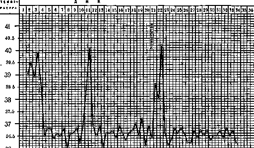
of tick-borne relapsing fever. Treatment with neosalvarsan at the clinic of the Tropical Institute in Moscow).
two peaks: spring and autumn (Latyshev), with the latter depending on a new generation of ticks.- Infection in laboratory conditions of mice, including Mus musculus, Mus silvaticus and other rodents, allows one to assume that in natural conditions small rodents can serve as a reservoir for the virus. In addition, it has been noted with respect to certain species of ticks that they usually parasitize other animals as well. Thus, Junkovsky often found O. tholosani (O. lahorensis) on sheep, and O. tarocaim usually parasitizes pigs (the latter, however, are not transmitters of the virus). Ticks live in animal barns and in human dwellings, where they hide in the cracks of clay buildings, in corners of earthen floors, behind baseboards and behind wallpaper; they usually attack humans at night; during the day they sit, hiding in their shelters. Ticks are afraid of high humidity, but during severe droughts they burrow deep into the ground, as a certain degree of humidity is still necessary for them. Ticks are apparently attracted by the smell of humans. To detect ticks in the ground, it is usually sifted through a sieve or stirred in water, after which the ticks float to the surface. Geography and distribution of tick-borne relapsing fever: Africa: Uganda, Congo, east coast, Abyssinia, Madagascar and Brazzaville; America: Colombia, Panama, Venezuela; Asia: Persia (Ardebil, Hamadan, Kermanshah, etc.), Central Asia: Fergana, Kulob, Guzar, etc., and probably the Transcaucasian republics; Europe: Spain.-Clinical picture. The incubation period is 7-10 days. The disease usually begins suddenly, without chills. Temperature rises, head hurts, general feeling of fatigue; in Europeans, vomiting, constipation or, conversely, a stool resembling dysentery are very common; mild jaundice of the sclera can often be noticed, pulse is rapid, up to 150 beats per minute. The attack lasts from 1 to 3-4 days, then temperature drops with slight perspiration, which occurs far from always. After some time, a new attack occurs, with intervals between individual attacks varying—from one day often up to two weeks, sometimes even a month (see figure 10). Among complications, keratitis are encountered. Along with sharply expressed cases of relapsing fever, abortive forms are often encountered, when the patient goes through the entire illness 'on the move', not paying attention to individual small temperature elevations.-Diagnosis of tick-borne relapsing fever presents great difficulties, as it is usually confused with other tropical febrile diseases and most often with malaria (see figure 11). The correct diagnosis can be established only on the basis of finding treponemes.-Mortality in tick-borne typhus, according to some authors, is higher than in European typhus; in Central Asia, fatal cases are a great rarity. - Treatment. The use of neosalvarsan preparations here does not give such results as in European relapsing fever, and recurrences of the disease are a common occurrence. Somewhat better results are obtained from treatment of orsanol, also an arsenical preparation, with simultaneous administration of methylene blue. - Prevention and control measures. The fight against tick-borne relapsing fever presents great difficulties. It should be based primarily on identifying the foci of this disease, determining the zone of distribution of Ornithodorus ticks, as well as informing the medical community and the population about this disease and the ways of its spread. In endemic foci of tick-borne relapsing fever, it is necessary to mark houses where patients and ticks have been found, and to place the residents of these houses under control and medical supervision. In some premises, relapsing fever exists for years, and anyone who settles in this house again is doomed to get sick. There are known cases, for example, in Kulob (Latyshev), when in one small house consisting of two halves, in one half there were many ticks and cases of relapsing fever were constantly observed, while the other half of the house remained healthy all the time. The usual methods of fighting ticks, i.e., disinsection (fumigating the room with sulfur, tobacco smoke and even disinsection with hydrocyanic acid), are little applicable in conditions of native dwellings and do not achieve the goal, as the ticks burrow deeper into the ground or go into cracks, remaining inaccessible to the action of the gas. In this respect, one can recommend good arrangement of human housing (plastering, painting walls, making good floors, changing wallpaper, etc.) and arranging animal dwellings away from human dwellings. For now, one has to resort to palliatives in terms of protection from ticks: since ticks do not tolerate moisture, it is useful to moisten the floor with water, even better with the addition of some resinous or aromatic liquids, especially before going to bed. It is also recommended that if there are earthen floors, a ditch should be dug along the inner walls and filled with water (to prevent mosquitoes from laying eggs in the water, the latter should be poured with kerosene). One can also fence off the room 0.2 m from the wall with a continuous tin box, extending 0.1 m into the ground and protruding approximately 0.2 m above the ground surface (for ease of attachment, the tin strip is attached to a wooden plank lying horizontally on the ground). Ticks can hardly crawl on tin, and it is impossible for them to cross such a barrier.
Related articles
Mentioned in
Cite this page
“Relapsing Fever.” Soviet Medical Encyclopedia. English translation of Bolshaya Meditsinskaya Entsiklopediya, 1st ed. (Moscow, 1928–1936), ed. N. A. Semashko. https://sovietmedicalencyclopedia.pages.dev/article/relapsing-fever/