Spirochetes
Historical document, translated for reference. It reflects medical knowledge of the 1920s–30s and is not medical advice.
Summary
This article from the 1928–1936 Soviet Great Medical Encyclopedia provides a detailed overview of spirochetes, describing their morphology, movement, reproduction, and biological classification. It discusses the historical debate regarding their relationship to bacteria and protozoa, as well as theories about their life cycles and potential transformations.
Encyclopedia article (1928–1936)
SPIROCHETES, SPIROCHETOSES. Spirochetes constitute a group of microbes characterized by a thin, spirally twisted body. They are very widely distributed as free-living forms in fresh and salt water, in soil, and even in hot springs (Cantacuzène), as well as in the form of parasites of the most diverse invertebrates and vertebrates (mollusks, starfish, worms, snails, insects, fish, amphibians, reptiles, birds, and mammals). Spirochetes have even been found in the milky sap of a plant—the spurge. In animals, they can live either on the surface of the body (ectoparasites) or in body cavities, the gastrointestinal tract, blood, and tissues (endoparasites). Intracellular parasitism of spirochetes has also been described, but it occurs very rarely and is denied by some altogether. Parasitic species of spirochetes are the causative agents of diseases—spirochetoses. The size of spirochetes varies within wide limits. Spirochaeta plicatilis has an average length of 100–200 μ and can even reach a length of 500 μ (Zuelzer). Some mollusk spirochetes also reach
a length of 100–150 μ, but the majority of
species are no more than 10–50 μ, and some
even 2.5–5.0 μ (Spirochaeta micro-
gyrata, Spirochaeta carnivorum). The length
can vary extremely among
representatives of one and the same species and depends (Zuelzer) on age, the medium in which the spirochetes develop, and other conditions. The thickness of spirochetes is very small; in the majority, it does not exceed 0.5 μ; in large mollusk spirochetes, it can reach 1.0–1.5 μ. The body of spirochetes consists of a layer of protoplasm spirally twisted around an axial elastic filament (Fig. 1) ("primary spirals"). Along with this, the body of the spirochetes forms bends of greater magnitude and less regular shape. The character and shape of the coils change under the influence of environmental conditions and are different for spirochetes in natural conditions, for example in the blood, and in cultures, and therefore cannot serve as a classification feature. Many authors believed that spirochetes, with the exception (according to Schaudinn) of Spirochaeta pallida, have a ribbon-like shape; however, subsequently, the wider distribution of a cylindrical shape was pointed out (Zuelzer for Spirochaeta plicatilis, Schellack for relapsing fever spirochetes, etc.). Spirochetes possess sharply defined motility, expressed by: 1) bending or wave-like movement of the entire body; 2) screw-like rotation around the longitudinal axis; and 3) translational movement with jerks forward and backward. Spirochetes, bending in different directions, form not only wavy but also more complex figures in the form of eights or tangles. Under the action of unfavorable external factors on spirochetes, e.g., low temperature and chemical substances, movements stop, which, however, does not always indicate the death of the spirochetes, since upon the removal of the harmful factors, movements are restored. Spirochetes that have lost their motility are apparently incapable of reproduction, and this explains the loss of virulence observed in such spirochetes. Some questions of the detailed structure of the spirochete body were studied with particular care to clarify the position of spirochetes in the system of microorganisms. To these belong questions about
the existence of a membrane, an undulating membrane, and flagella in spirochetes. These formations could be considered an indication of the belonging of spirochetes to bacteria (which have a membrane) or to protozoa (Protozoa, often lacking a membrane, or in particular to Flagellata, which have an undulating membrane). A membrane can be considered proven, apparently, only in one group of spirochetes—Cristispira. It is generally accepted that the surface of the spirochete body is covered by a thin layer of protoplasm, denser than the rest—the periplast. The presence of an undulating membrane has not been positively proven for any group of spirochetes. As flagella, some authors have described thin, mobile, thread-like outgrowths of the periplast at the ends of the body of some spirochetes. Investigations by other authors show that we are dealing here not with true flagella, but only with somewhat elongated and thinned ends of the spirochete body. Under the action of sodium taurocholate, distilled water, or carbolic acid on spirochetes, threads resembling peritrichous cilia sometimes form on the surface of the entire body of the spirochetes, but in reality, they are an artificial formation (artifact). As already mentioned, a thin elastic filament runs along the entire body inside the spirochetes (Fig. 1). Many authors denied its presence for the majority of spirochetes, but recently Zuelzer proved that it is present in many spirochetes. It was possible to detect it in relapsing fever spirochetes and bird spirochetes obtained from cultures, but not from animal blood. It can be detected by staining with hematoxylin or by the action of bile. Apparently, an axial filament is present in all spirochetes. A morphologically distinct nucleus has not been found in spirochetes, but some describe small chromatin granules and chromatin substance mixed with the protoplasm (Zettnow). In some species, the body is as if divided by septa (Cristispira) into chambers. Whether this is an expression of multi-chamberedness or simply a cellular structure of the protoplasm is a question not yet resolved. Reproduction of spirochetes occurs by transverse division. Spirochetes stretch in length, thin out in the middle, and finally break into two parts. Sometimes, during intense division and slow separation of daughter individuals, long spirochetes are formed, consisting of the union of many specimens. This phenomenon is often observed in cultures. Longitudinal division of spirochetes was described on the basis of microscopic images by many prominent researchers, such as Schaudinn, Prowazek, and others, but these data cannot be considered sufficiently convincing, and the majority of researchers at the present time hold the opinion that the vegetative reproduction of spirochetes occurs exclusively by transverse division. A very interesting, complex, and still unresolved question is the existence of sexual reproduction or a genetic cycle in spirochetes. A regular alternation of forms has not yet been established; however, many authors describe formations that are possibly links in a not-yet-known cycle of development. In many species of spirochetes, it has been repeatedly possible to observe two types—one thicker with wide coils, the other thinner with narrow coils. The question of whether these spirochetes are different (sexual?) forms of the same species or representatives of different species cannot be considered resolved. Levy observed in a dark field in the blood of a patient with relapsing fever the fusion and subsequent separation of spirochetes, which he takes for copulation. Much has been discussed regarding the genetic relationships between spirochetes and so-called "fusiform bacilli" (Martsinovsky, Tunnicliff, Wright, and others). Spirochetes are often found together with these microorganisms in the oral cavity, in feces, in ulcerating cancerous tumors, etc., and on the basis of such observations, conclusions are sometimes drawn about the origin of spirochetes from fusiform bacilli or that these forms are members of a complex developmental cycle. Tunnicliff and others observed in cultures the transition of fusiform bacilli into spirochetes. Recently, Sanarelli, in pure cultures of spirochetes from the intestine of a guinea pig, obtained fusiform rods, which subsequently turned back into spirochetes. However, Mühlens, during separate cultivation of spirochetes and fusiform bacilli, never observed the transformation of one form into the other. Of great interest is the far-from-resolved question of the existence of granular or filterable stages of development in spirochetes. Gross saw granules in large spirochetes, which he considers spores, although he did not observe their development into spirochetes. Collier confirmed these observations in relation to
Figure 1. Spirochaeta plicatilis (schematized). The axial filament is visible.
the spirochetes.
regarding Cristispira helgolandica, with which he succeeded in infecting starfish spores, in which they developed into spirochetes. In general, in the majority of spirochetes, both in the blood and organs of animals and in cultures, one observes, alongside ordinary forms, specimens that disintegrate entirely into granules or contain several granules. The significance of these formations is controversial. Some authors consider them products of the disintegration or degeneration of spirochetes, but the majority of researchers hold the view that these granules are developmental forms of spirochetes (one of the stages of their existence) (Leishman, Nicolle, Salimbeni, Meirowsky, and others). Leishman even describes the development of spirochetes from granules that he observed (Fig. 2). The existence of granular and invisible forms in the developmental cycle of spirochetes was proven by experiments involving the filtration of material containing spirochetes through bacterial filters, whereby the resulting filtrates, which did not contain spirochetes, proved to be infectious to animals. These experiments, however, cannot currently be considered convincing, as there are reports of whole spirochetes passing through bacterial filters. Meirowsky, in a whole series of reports, describes the formation of lateral or terminal outgrowths, or 'buds,' in spirochetes, from which new spirochetes are formed. These observations have been confirmed by a number of authors. The existence of a developmental cycle of spirochetes, which undoubtedly includes granular and invisible forms, has indirect confirmation in the cyclical nature of infectious processes caused by spirochetes and in certain peculiarities of the transmission of spirochetal infections by vectors (the disappearance and reappearance of spirochetes in vectors, the incubation period), and the infectivity of the vector; however, the question as a whole still requires further development. Cases have been described in which spirochetes are encountered in the form of coiled (annulated) forms. Some authors consider these forms to be 'resting forms' (Schaudinn, Prowazek, Mayer) or forms analogous to cysts (Marcinkowski), while others consider them products of degeneration (Levaditi and others), and Aristovsky and Geltzer encountered them in old cultures of relapsing fever spirochetes. In spirochetes of the genus Cristispira and Cristispirella, the formation of cysts has been described. In this process, the spirochetes coil into a ball and secrete a mucous mass, which, upon hardening, forms the cyst wall (Hollande). Spirochetes can live and multiply in artificial cultures, but this requires certain special conditions. Firstly, the presence of protein in the form of inactivated human, rabbit, or other serum, egg white, or pieces of organs is necessary. Secondly, the oxygen content in the nutrient medium is apparently of significance. Usually, media in which spirochetes are cultivated are covered with a layer of liquid paraffin. It was believed that layering paraffin in the presence of pieces of protein or organs created anaerobic conditions in the liquid medium; however, it has recently been proven that paraffin does not stop the access of oxygen, but only retards evaporation and changes in the reaction of the medium (the most favorable pH for the growth of spirochetes is 7.2–7.4). The majority of spirochetes apparently require anaerobic conditions (Spir. pallida, Spir. dentium, and others), however, the opinions of researchers regarding the significance of anaerobiosis for some other species of spirochetes differ extremely. Thus, Weil's spirochete (Leptospira icterohaemorrhagica), according to Dietrich, grows both aerobically and strictly anaerobically; according to Wolf, it grows aerobically; and according to Noguchi and Griffith, it is even an obligate aerobe. The same diversity of opinion exists regarding the relapsing fever spirochete. The ability to grow in artificial nutrient media is in close connection with the biological characteristics of spirochetes. Strictly parasitic forms, such as, for example, Spir. pallida, are cultivated with difficulty, require a greater amount of protein and strict anaerobiosis, whereas saprophytic forms of spirochetes are cultivated easily, are satisfied with a low protein content, and can grow in the presence of oxygen. Pure cultures of spirochetes on solid media have been obtained in the form of individual colonies. They are cultivated in hermetically sealed dishes in the presence of bacteria that strongly absorb oxygen. In view of the small thickness of the spirochete body and the weak light refraction of some of them, they are visible in the living state only when examined in a 'dark field.' There are special methods, such as the addition of India ink or other dyes, to reveal the presence of spirochetes (Burri's method and others). For the study of spirochetes in smears and sections, the silver impregnation method (see Levaditi's method) or staining of spirochetes with Giemsa is most commonly used. With Giemsa, relapsing fever spirochetes stain violet-blue, and the pale spirochete stains reddish-pink, which apparently depends on differences in the biochemical composition of the spirochetes. Among spirochetes, there are saprophytes living in the bodies of vertebrate and invertebrate animals, and parasites causing various infectious processes in humans and animals. Transitional forms are observed, representing various degrees of adaptation to the host organism, from saprophytism (Trep. bronchiale, Trep. intestinale) to the strictest parasitism (Trep. recurrentis, Trep. pallidum); the causative agent of infectious jaundice in this respect presents an interesting example of a relatively easy transition from parasitism to a free-living state and back. Parasitic spirochetes are of great importance as causative agents of infection. Some are transmitted to the host through the skin or mucous membranes upon direct contact (Trep. pallidum, Lept. icterohaemorrhagica), others through the skin by rubbing in a crushed louse (Trep. recurrentis, var. Obermeieri), and others by inoculation, i.e., through the bite of a vector—a tick (Trep. recurrentis, var. Duttoni). A peculiarity of the majority of pathogenic spirochetes is that they spend a certain period of life in the body of an intermediate host or vector, from which they are transmitted to humans. For different species of relapsing fever spirochetes and others, the vectors are lice and ticks; for sodoku, rodents; and for infectious jaundice spirochetes, rats. The infectious process caused by spirochetes has an acute character in some cases and a chronic one in others, but it is always distinguished by a relapsing course in the form of periodically alternating bouts of disease. It is noted that spirochetes causing an acute infectious process possess significant antigenic capacity and induce the formation of antibodies (Trep. gallinarum, Trep. recurrentis), while in the causative agents of chronic spirochetoses, these properties are expressed extremely weakly (Trep. pallidum, Trep. pertenue). In relation to chemotherapeutic effects, species of spirochetes are divided into two groups: salvarsan-susceptible, to which belong Trep. pallidum, Trep. pertenue, Trep. recurrentis, and salvarsan-resistant, such as, for example, Leptosp. icterohaemorrhagica. Among spirochetes of the same species, for example, relapsing fever spirochetes, which are generally salvarsan-susceptible, it has been possible to discover varieties (Krichevsky) possessing a significant degree of salvarsan resistance. Some spirochetes are characterized by ectodermotropism, i.e., the property of predominantly affecting organs of ectodermal origin—the skin and the central nervous system (Trep. pallidum, Trep. pertenue), while others are distinguished by endodermotropism, i.e., the property of affecting organs of endodermal and mesodermal origin—the intestine, liver, kidneys (Leptosp. icterohaemorrhagica, Trep. recurrentis). The position of spirochetes in the system of organisms is not entirely clear. They possess morphological and biological features inherent to both protozoan animals (Protozoa) and bacteria. The absence of a differentiated nucleus, reproduction by transverse division, and the absence of anterior and posterior ends of the body are signs that bring them closer to bacteria; on the other hand, the transmission of spirochetal infections through insects, the developmental cycle described by some authors, and the periodic, relapsing character of spirochetal infections speak for a kinship with Protozoa. Apparently, the most correct opinion is that according to which spirochetes are separated into a special, independent group of microorganisms (Spirochaetaceae, Fantham, 1908). As for the classification and subdivision of the group of spirochetes into genera, the simplest is the proposal of Zuelzer, who divides all spirochetes into two genera: 1) a small group lacking an elastic filament (Cristispira) and 2) all others, which possess an axial filament. Since the presence of an axial filament in smaller forms is not recognized by all researchers, the simplified classification of Noguchi (Wenyon, Epstein), adopted by many authors, should be considered the most correct, according to which spirochetes are divided into four genera: Genus I Cristispira Gross, 1910, 100 μ in length, with a wide membrane along the body. Typical species Sp. balbiani (Certes, 1882). Genus II Spirochaeta Ehrenberg, 1834, up to 500 μ in length. Lives in water. Typical species Sp. plicatilis Ehrb. 1834 (Fig. 1). Genus III Treponema Schaudinn, 1905, 15–20 μ in length.
Two types are distinguished: 1) forms with large coils—the typical species is Trep. recurrentis Lebert, 1874 (Fig. 3). This includes the causative agents of various forms of relapsing fever and avian spirochetosis; 2) forms with small coils, the typical species is Trep. pallidum Schaudinn, 1905—the causative agent of syphilis (see) (Fig. 4); other representatives: saprophytes of the oral cavity and intestine, Tr. pertenue Castellani, 1905—the causative agent of yaws.
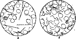
Figure 3. Treponema recurrentis: a—spirochetes in blood in the living state; b—the same spirochetes in a fixed and stained preparation.
and others. IV. Leptospira Noguchi, 1917, 15 microns in length. The coils are small, with hook-like curved ends; the typical species is Leptospira icterohaemorrhagica Inada, Ido, 1914, the causative agent of Weil's disease (Fig. 5). Further division of spirochetes into species cannot be performed on the basis of morphological characteristics, since within the genus, spirochetes are absolutely indistinguishable from one another by this method, e.g., Tr. pallidum, Tr. cuniculi, Tr. pertenue, and various species of relapsing fever spirochetes. Subdivision is performed on the basis of biological differences: susceptibility of animals, character of the infection, immunity reactions, and specificity of vectors. Spirochetoses, diseases caused by spirochetes, can be divided according to the morphological and biological properties of the causative agents and pathogenesis into three groups: I. Certain infections of the digestive and respiratory systems, in which spirochetes are usually encountered together with other microorganisms. This includes: 1) alveolar pyorrhea, 2) ulcerative angina (angina Plaut-Vincenti) and noma, 3) broncho-pulmonary spirochetosis. The role of spirochetes in these diseases is not entirely clear. In alveolar pyorrhea, they are found together with various microbes and possibly play the role of a concomitant factor or secondary infection. In ulcerative angina and broncho-pulmonary spirochetosis, in particular in gangrene of the lung, they are found in association with a fusiform bacillus (fuso-spirochetosis). To this same group of spirochetoses belong skin ulcerations of the thighs, known by the name "ulcus tropicum," in which spirochetes are also detected in association with fusiform bacilli (Fig. 6). Many species of saprophytic spirochetes have also been described in the respiratory and digestive tracts. II. A group of spirochetoses representing acute septicemias with fever, having a tendency to occur in the form of short, recurring attacks. In this
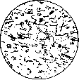
Figure 4. Treponema pallidum in a section from the liver of an infant with congenital syphilis.
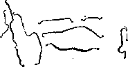
Figure 5. Leptospira icterohaemorrhagica.
the organs of entodermal and mesodermal origin (liver, kidneys, spleen) are primarily affected. This group includes: A. Infectious jaundice (see Botkin-Weil disease). B. Relapsing fever (see) in its two main forms: a) European (louse-borne) relapsing fever and b) tick-borne relapsing fever. To this group of spirochetoses, it is necessary to assign a disease of birds, manifesting in the form of acute
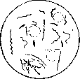
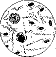
Figure 6.
Figure 7.
Figure 6. Spirochetes and fusiform bacilli in a smear from a "tropical ulcer." Figure 7. Spirochaeta anserina from the blood of birds. septicemia and first described by Sakharov in the Caucasus in 1891. He named the causative agent Spiroch. anserina (Fig. 7). Transmission occurs via the tick Argas persicus. A similar disease has been discovered by many authors in various birds. The spirochetes described under different names are apparently identical to Spir. anserina (Wenyon). Morphologically, they are almost indistinguishable from the spirochetes of human relapsing fever.—III. Spirochetoses of the syphilis group (see). Representatives of the spirochetoses of this group are, besides human syphilis, yaws (see) and rabbit syphilis, a synonym for spontaneous rabbit spirochetosis (see Pathology of laboratory animals). A somewhat special position is occupied by the rat-bite transmitted spirochetosis sodoku (see). This spirochetosis is interesting in that it combines in its manifestations the features of spirochetoses of the II and III groups—syphilis (primary lesion, lymphadenitis, etc.), relapsing fever (fever, relapses), and Weil's disease (carriage of the virus by rats).
Related articles
Mentioned in
Cite this page
“Spirochetes.” Soviet Medical Encyclopedia. English translation of Bolshaya Meditsinskaya Entsiklopediya, 1st ed. (Moscow, 1928–1936), ed. N. A. Semashko. https://sovietmedicalencyclopedia.pages.dev/article/spirochetes/