Flagellata
Historical document, translated for reference. It reflects medical knowledge of the 1920s–30s and is not medical advice.
Summary
This article from the 1928–1936 Soviet medical encyclopedia defines the class Flagellata (Mastigophora) within the phylum Protozoa. It details their morphology, reproductive cycles, nutritional modes, and ecological distribution, including both free-living and parasitic forms.
Encyclopedia article (1928–1936)
FLAGELLATA, Flagellata Conn 1851, Mastigophora Diesing 1865, a class of the phylum Protozoa, the representatives of which are characterized by the presence of flagella throughout the entire vegetative period of their life. Most Flagellata have an elongated body, usually pointed on one side. For the most part, they have a constant shape; only a few are metabolic, i.e., capable of slight changes in body shape. We distinguish: 1) Flagellata that do not have definite body outlines and, along with flagella, form pseudopodia in various directions like rhizopods, for example, the free-living Mastigella vitrea (Fig. 1); however, many doubt the classification of Mastigella and similar forms as Flagellata (see below—systematics); 2) Flagellata that are partially metabolic, i.e., capable of slight changes in shape (however, only in one direction, usually perpendicular to the main axis of the body), for example, Astasia tenax (Fig. 2); 3) finally, Flagellata (the majority), characterized by a constant and definite body shape, for example, Trichomonas (Fig. 3), Trypanosoma (Fig. 4). In the latter, the body shape is determined by the dense nature of their pellicle, and in many cases also by the presence of a so-called axial filament, axostyle, or axoneme. In these forms, the body is in some cases capable of bending along the main axis. The nucleus in most forms is single; in some (Distomatina) there are two, for example, in Giardia (Fig. 5). In the suborder Polymonadida, the number of nuclei reaches several dozen, for example, in Calonympha (Fig. 6). The number of flagella in different species ranges from 1 to 8, while in the group Hypermastigina it can reach several dozen or more with a single nucleus, for example, Lophomonas (Fig. 22). Flagella are usually connected to a central apparatus, and this connection is expressed differently in various Flagellata. In some, the flagellum originates from an intranuclear centriole; in others, from a centriole lying outside the nucleus. In some forms, for example, in trypanosomes, the extranuclear central apparatus is built in the form of a complex nucleus-like formation—a blepharoplast or kinetonucleus. Besides the blepharoplast, Flagellata may also have a so-called parabasal body, in some forms, for example in trypanosomes, connected to the blepharoplast into a so-called kinetoplast. The role of the parabasal body has not been finally clarified. It is possible that it is connected with the activity of the flagella. The number of flagella usually corresponds to the number of blepharoplasts. In some forms, for example, Trichomonas, Trypanosoma, the flagellum is connected to a special ciliary membrane, or undulating membrane. In others (Choanoflagellata), the base of the flagellum is surrounded by a special collar, for example, Codonosiga (Fig. 7). Many Flagellata form protective cysts. In others, the process of schizogony occurs in cysts. Most Flagellata are free-swimming; only some attach themselves to a substrate by special stalks. Besides solitary Flagellata, colonial ones are also encountered. Reproduction. Some Flagellata reproduce sexually and asexually; for others, only asexual reproduction is known (Trypanosoma). Asexual reproduction of Flagellata occurs by longitudinal division of vegetative forms or by schizogony, which in some Flagellata occurs in the free state, and in others—inside cysts. Sexual reproduction has the character of isogamous hologamy. In colonial forms, for example, Volvox (Fig. 8), a complex reproductive cycle is associated with strongly expressed polymorphism, manifesting in the formation of vegetative (trophic) and generative individuals, i.e., male and female elements. Nutrition and metabolism. In this respect, Flagellata are divided into holozoic forms, feeding by the animal type, i.e., on complex protein substances, with some representatives of this group having developed a special mouth opening—a cytostome. Holophytic (autotrophic) forms are equipped with photosynthetic pigments (chlorophyll, etc.) and feed by the plant type, assimilating CO2 (green Flagellata). However, a mixed type of nutrition is also known depending on environmental conditions. Finally, saprophytes and parasites feed exclusively osmotically at the expense of organic material, resp. products of protein decay, ranging from amine to fatty and simpler acids. In connection with metabolic processes in the body, various reserve deposits are found in many Flagellata—starch grains in green Flagellata, volutin in trypanosomes. In green Flagellata (Euglena), pigment spots are found. Some free-living forms have a contractile vacuole. Ecology. Flagellata are extremely widespread in nature and are found in the most diverse conditions. Free-living forms are found in all bodies of water, starting from freshwater puddles (Euglenoidea—e.g., Euglena, Fig. 9) and ending with seas [Dinoflagellata—e.g., Ceratium (Fig. 10), Cystoflagellata—e.g., Noctiluca (Fig. 11)]. As inhabitants of waters, Flagellata are organisms highly indicative for the biological assessment of waters. For clean bodies of water, Chrysomonadida are characteristic—e.g., Chrysamoeba (Fig. 12), and Craspedomonadida—e.g., Codonosiga (Fig. 7). Anthophysa (Fig. 13), Trachelomonas (Fig. 14) are found in ferruginous waters; Euglenaceae, Cryptomonadina—e.g., Cryptomonas (Fig. 15)—in ammoniacal ones. The group Bodo (Fig. 16) is characteristic of highly putrid bodies of water. Parasitic forms are found as ectoparasites [Costia necatrix (Fig. 17) on the skin of fish], endoparasites of the intestine [Trichomonas (Fig. 3), Giardia (Fig. 5), Trypanoplasma (Fig. 18)], blood (Trypanosoma, Fig. 4), or tissues and parenchymal organs (Leishmania, Fig. 19) in various animals. The phenomenon of blood parasitism in the class...
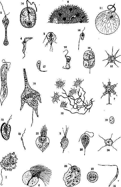
1-Mastigella vitrea; 2-Astasia tenax; 3-Trichomonas; 4-Trypanosoma; 5-Giardia; 6-Calonympha; 7-Codonosiga; 8-Volvox; 9-Euglena; 10-Ceratium; 101-Gymnodinium; 11-Noctiluca; 12-Chrysamoeba; 13-Anthophysa; 14-Trachelomonas; 15-Cryptomonas; 16-Bodo; 17-Costia; 18-Trypanoplasma; 19-Leishmania; 20-Cercomonas; 21-Phytomonas; 22-Lophomonas; 23-Mastigina hylae; 24-Multicilia. 769
MASTICATION
The origin of Flagellata is explained differently by various authors. Some believe that blood-parasitic Flagellata originated from intestinal forms by the latter penetrating through damaged intestinal walls into the bloodstream. Indeed, cases of finding intestinal forms (Giardia, Trichomonas) in peripheral blood have been described. Others consider blood parasitism of Flagellata in Metazoa to be the result of their inoculation into the blood by blood-sucking vectors, which are their primary hosts. Parasitic Flagellata have also been described in the tissue sap of plants (Euphorbiaceae). When examining intestinal material for the presence of parasitic Flagellata, it is necessary to account for frequent findings of so-called coprozoic saprophytic forms, which likely do not occur in the intestinal cavity at all, but easily develop from ingested cysts in excrement after it has been excreted from the organism (Bodo, Cercomonas - figure 20). Pathogenic significance. Many parasitic Flagellata are essentially harmless commensals, however, it is possible that under certain conditions they can manifest a pathogenic effect (Trichomonas, Giardia). Others are unconditionally pathogenic and even fatal forms (Trypanosoma gambiense - causative agent of sleeping sickness, Leishmania donovani - causative agent of visceral leishmaniasis), although other representatives of the same family may be little or not at all pathogenic to their hosts, such as, for example, rat trypanosomes (Trypanosoma lewisi), or those of fish, amphibians, reptiles, and birds. Methods of infection. The spread of Flagellata parasitizing in the intestine occurs through protective cysts by the ingestion of the latter by new hosts. Blood-parasitic Flagellata are spread either by direct contact through mucous membranes (Trypanosoma equiperdum - causative agent of dourine in horses) or through blood-sucking insects (Trypanosoma gambiense - causative agent of sleeping sickness via the fly Glossina palpalis; Leishmania tropica - causative agent of cutaneous leishmaniasis - via the sandfly Phlebotomus). Infection with blood-parasitic Flagellata in fish and amphibians is transmitted through leeches. Infection of plants with Flagellata occurs through insects - Phytomonas davidi (fig. 21) - the causative agent of cachexia in spurges - is transmitted through the bug Stenocephalus agilis. Phenomena of immunity. Infection with blood and tissue Flagellata entails in some cases the appearance in the serum of the infected animal of specific properties causing agglutination and dissolution of the corresponding pathogens, and in some cases also a complement fixation reaction. Methods of study. In addition to the usual methods of microscopic examination, culture methods are widely used in the study of Flagellata. Free-living Flagellata, especially chlorophyll-bearing forms, are usually cultivated in liquid or solid Knop's medium or similar mineral solutions. Intestinal Flagellata are cultivated for the most part in liquid protein media, while blood-parasitic Flagellata - trypanosomes, leishmanias - are well cultivated on blood media (NNN-agar). In the study of pathogenic flagellates, animal experimentation is widely used - infection of animals (rats, mice, guinea pigs, dogs, horses, monkeys). Systematics of Flagellata. The class Flagellata s. Mastigophora consists of 2 subclasses - Zoomastigina and Phytomastigina. A. Subclass Zoomastigina (s. Zoomastigophora) is characterized by an animal type of metabolism. The system of Zoomastigina is built on the basis of the number and ratio of nuclei and flagella. On this basis, they are divided into 3 groups. The 1st group - Monozoa (with one nucleus and a varying number of flagella and blepharoplasts) - includes the orders: I. Protomonadida - free-living, saprophytic, and parasitic forms. 1 nucleus; 1-6-8 flagella. Typical genera: 1) Trypanosoma (figure 4) - blood parasites of humans and other vertebrates, 2) Trichomonas (figure 3) - intestinal saprophytes in various vertebrates. II. Hypermastigida. 1 nucleus; many flagella. Parasitic forms, typical genus: Lophomonas (fig. 22) - parasite of the intestine of the black cockroach. III. Cystoflagellata. 1 nucleus; 1 flagellum. Free-living forms. Typical genus: Noctiluca (fig. 11) - sea sparkle. Causes bioluminescence of the sea. The 2nd group - Diplozoa (2 nuclei; 2 flagella; bilaterally symmetrical body) - includes the order: IV. Diplomonadida. 2 nuclei; 2-4 pairs of flagella; bilaterally symmetrical body. Parasites. Typical genus: Giardia (fig. 5) - intestinal parasite in various vertebrates. The 3rd group - Polyzoa (many nuclei, many flagella and blepharoplasts) - includes the order: V. Polymonadida. Many nuclei; many flagella and blepharoplasts. Parasites. Typical genus: Calonympha (fig. 6) - parasite of the intestine of termites. Some authors (Minchin) introduce into the system of Zoomastigina a 4th group - the order Pantastomina - encompassing free-living and parasitic forms with one or several nuclei and flagella, but also forming pseudopodia. Included here are the suborders: 1) Rhizomastigina (with 1-3 flagella extending from the anterior part of the body) with the genera Mastigamoeba, Mastigina, Mastigella (fig. 1). Typical genus: Mastigina hylae (fig. 23) - parasite of the intestine and skin of amphibian tadpoles. 2) Holomastigina. Free-living forms. Have several flagella extending without a definite orientation from various parts of the body. Example: Multicilia (fig. 24). B. Subclass Phytomastigina (s. Phytomastigophora) is characterized by a plant type of metabolism (presence of chlorophyll and other pigments, deposition of starch). Reproduction by division, as well as sexually. They form cysts. Representatives of this subclass are related to green algae and are for the most part free-living forms, with the exception of some inhabitants of the intestine of tadpoles. The system of Phytomastigina includes the orders: I. Chrysomonadida - carry brown pigment. Have 1-2 flagella. Typical genus: Chrysamoebe (fig. 12). II. Cryptomonadida - pigment of various colors. 2 flagella. Egg-shaped body. Typical genus: Cryptomonas (fig. 15). III. Dinoflagellata (s. Peridinea). Chromatophores are inconstant, 2 flagella, one of which lies in a transverse groove. Typical genus: Ceratium (fig. 10). IV. Euglenoidida. Large forms with 1-2 flagella and green pigment; equipped with a cytostome. Typical genus: Euglena (fig. 9), Astasia (fig. 2). V. Phytomonadida. Dense cellulose walls; 1-2 flagella; green pigment. Colonies are often formed. Complex life cycle. Typical genus - Volvox (figure 8).
Related articles
Mentioned in
Cite this page
“Flagellata.” Soviet Medical Encyclopedia. English translation of Bolshaya Meditsinskaya Entsiklopediya, 1st ed. (Moscow, 1928–1936), ed. N. A. Semashko. https://sovietmedicalencyclopedia.pages.dev/article/flagellata/