Trypanosomes
Historical document, translated for reference. It reflects medical knowledge of the 1920s–30s and is not medical advice.
Summary
Trypanosomes are parasitic protozoa of the genus Trypanosoma that infect the blood and tissues of mammals, birds, reptiles, amphibians, and fish. This article describes their morphology, classification within the family Trypanosomidae, and their characteristic appearance in the bloodstream.
Encyclopedia article (1928–1936)
Trypanosomes, trypanosomiasis. Trypanosomes, parasitic protozoa of the genus Trypanosoma Gruby, 1843, family Trypanosomidae, order Protomonadina, class Mastigophora. The family Trypanosomidae includes genera closely related to Trypanosoma: Leptomonas, Phyto-monas, Leishmania, Herpetomonas, Crithidia and Schizotrypanum (see figure). The latter genus is included by many in the genus Trypanosoma. Trypanosomes parasitize in the blood and tissues of mammals (including humans), birds, reptiles, amphibians and fish. In the blood, trypanosomes appear as highly mobile, elongated forms. Only
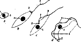
Diagram of morphological types of the family Trypanosomidae: a-leishmania; b-leptomonas (Leptomonas); c-crithidia; d-trypanosome; 1-nucleus; 2-blepharoplast; 3-flagellum; 4-undulating membrane.
some species, such as T. rotatorium of frogs, at certain stages of development have the form of flat structures, the width of which is slightly less than the length. The smallest trypanosomes are 12-15 μm to 25-30 μm in length (the average size of a number of pathogenic trypanosomes of humans and other mammals). Larger forms, for example the non-pathogenic trypanosome of cattle, T. theileri, reach 60-70 μm. There are also trypanosomes that reach 120-130-200 μm in length (trypanosomes of deer, fish). The body of trypanosomes is covered with a thin shell, the pellicle (periplast). The periplast has azurophilia and in preparations vigorously stained by Romanovsky's method, it takes on a reddish tint that masks the bluish tint of the basophilic plasma. In most trypanosomes, the periplast does not show special structures, but in some, for example in trypanosomes of amphibians, it has longitudinal striations. Along one edge of the trypanosome body, the periplast is raised by a flagellum that begins at the posterior end of the trypanosome and mostly extends beyond its anterior end. The duplication of the periplast raised above the plasma itself by the flagellum forms the so-called undulating membrane or undulating membrane. At the base of the flagellum, at the posterior end of the trypanosome, sits the so-called basal body, often merging with a larger granule, the so-called parabasal body. The parabasal body (or sometimes the combination of it with the basal granule) is called the blepharoplast, kinetonucleus, or kinetoplast. The blepharoplast stains like a nucleus and gives a positive Feulgen reaction. The nucleus is located centrally, it has a karyosome and peripherally located chromatin. Some trypanosomes in their natural state do not have a parabasal body (T. equinum). In others, normally a small percentage of specimens are found to be without it. Under experimental conditions, it is possible to obtain blepharoplast-free races of trypanosomes by the action of dyes (pyronin, etc.). At different stages of infection and in cultures, small round azurophilic grains, similar to volutin, are found in the plasma of trypanosomes. In the bodies of vectors and infected animals, trypanosomes can change their morphology, taking on forms characteristic of the closely related genera Leishmania, Leptomonas, Crithidia. The form closest to the proper trypanosome form is the crithidia form, which differs from the trypanosome in that its blepharoplast is located in front of the nucleus, accordingly the undulating membrane turns out to be shorter (see figure). Elongated forms with a free flagellum arising from the blepharoplast located at the anterior apex of the cell, lacking an undulating membrane, are designated as leptomonad forms (Leptomonas, see figure). Round or oval small forms with a nucleus and blepharoplast but without a flagellum are designated as leishmania-like forms (see figure). Reproduction of trypanosomes occurs by division. A sexual process for trypanosomes has not been established. Division of the nucleus is usually preceded by division of the blepharoplast, and the flagellum remains with one half of the basal granule with the adjoining half of the parabasal body, while from the other half of the blepharoplast a new flagellum begins to grow, running parallel to the old one and soon reaching the same length. Division of the nucleus occurs by mitosis. This is followed by longitudinal fission of the body. In some cases, a series of repeated divisions of the nucleus and blepharoplast with the formation of multiple individuals is observed. At different periods of infection, the reproduction of trypanosomes proceeds with different intensity. During periods of active reproduction of trypanosomes in the blood of an animal, specimens are found that are longer, with a free flagellum, and wider and shorter forms in which the flagellum ends at the anterior end of the body. At other times, forms that are uniform in their morphology are mainly encountered. A number of works have been devoted to the differentiation of different species of trypanosomes by their length. Later research showed that this length changes depending on the phase of infection and the species of animal to which the trypanosome has been inoculated. Due to the significant morphological similarity of pathogenic trypanosomes of many species of animals and the extremely pronounced ability of trypanosomes to change their biological properties depending on the duration of passage on one or different species of animals, the delimitation of individual species of pathogenic trypanosomes in many cases presents great difficulties. Laveran and Mesnil introduced a method for differentiating trypanosomes by so-called cross-infection, based on the fact that animals that have undergone infection with a certain species of trypanosome and have spontaneously recovered, are immune to this species but are susceptible to other species of trypanosomes. They also introduced a serological method for identifying trypanosomes, based on the fact that the serum of an animal immune to a certain species of trypanosome protects against infection with the homologous species but is not effective against heterologous species. In practice, however, both methods present a number of difficulties due to the fact that within each species of trypanosome there are numerous strains immunologically different from each other, as well as due to the fact that immunity after elimination of the infection does not last long. Differentiation of different species of trypanosomes is sometimes possible on the basis of their different relationship to chemotherapeutic substances and the specificity of the invertebrate host vector. The first attempts to cultivate trypanosomes belong to Danilevsky (1886-1889), who observed the reproduction of trypanosomes of birds and frogs in capillaries with blood. True cultures of trypanosomes with subcultures were first obtained by Novy and MacNeal on a medium of meat-peptone nutrient agar, to which at a temperature of 50°C was added 10% to 200-300% defibrinated blood of rats, dogs or rabbits. Growth occurred mainly in the condensation liquid at a temperature of 25-37°C. In cultures, the morphology of trypanosomes simplifies: they give crithidia and leptomonad forms. The latter can form rosettes completely similar to rosettes in cultures of Leishmania. Cultures of T. lewisi retained their virulence for rats after a series of passages in tubes at room temperature for a year or more. The same authors also obtained a culture of T. brucei, and after them Laveran and Mesnil grew T. evansi. The medium originally proposed by Novy and MacNeal was simplified by Noller (see Leishmaniasis). On this medium NNN it is possible to grow a number of trypanosomes (T. theileri, Schizotrypanum cruzi, etc.). T. theileri, T. melophagium and those close to them are also cultivated on broth with blood. Some species of trypanosomes require the addition of glucose to the medium. Noller cultivates trypanosomes on the surface of meat-peptone agar with 10% glucose, mixed half and half with defibrinated blood. On the surface of such agar, trypanosomes of birds, T. theileri, etc. give very characteristic forms of growth in the form of colonies with peculiar lateral outgrowths. In cultures at room temperature, trypanosomes give forms analogous to the stages of their development in the invertebrate host. When the temperature is raised to 37°C, they take on the form characteristic of them in the body of a warm-blooded animal. Cultivation of pathogenic forms of trypanosomes is possible only with great difficulty, and trypanosomes quickly lose their virulence. Behrens cultivated T. brucei on agar prepared from pea broth. Individual authors succeeded in obtaining cultures of T. congolense, T. brucei and T. gambiense [see Sleeping sickness and a separate table (t. XXXI, pp. 855-856, fig. 5)] on Ponsel's medium - defibrinated inactivated rabbit blood with the addition of distilled water and on other modifications of his medium containing different amounts of salt, peptone and gelatin. Cultivation of trypanosomes is often used as a method of enrichment for detecting trypanosomes when their content in peripheral blood is very small. Noller in 1916-28 applied this method to establish the vector of T. theileri: having obtained cultures of trypanosomes from the hindgut of tabanid flies, he infected calves with these cultures. The routes of spread of trypanosomes are specific to each species. The following mechanisms of transmission of trypanosomes are known: 1) contact route - direct transmission from animal to animal through contact of mucous membranes; 2) contaminative route - through the animal eating an intermediate host or the excrements of the latter containing virulent stages of trypanosomes; 3) inoculative route - through the bite of an intermediate host that introduces into the animal's blood infectious stages of trypanosomes. An intermediate position between the second and third method is infection through the introduction into a wound made by a bite of pathogenic stages of trypanosomes that have fallen on the animal's skin near the wound along with the excrements of an insect. T. equi-perdum is transmitted by the contact route (see Dourine). Trypanosomes of the lewisi group (T. melophagium, lewisi, etc.) are spread by the contaminative route. The vector for T. melophagium is the sheep ked (Melophagus ovinus), for T. lewisi - the rat flea (Ceratophyllus fasciatus). In the midgut of the insect, trypanosomes undergo division and then transform into crithidia forms.
The latter in the rectum of the insect after a series of repeated divisions again assume the form of trypanosomes. These so-called metacyclic trypanosomes exit outside along with the insect's excrement and infect the animal that eats the insect whole or licks its excrement from its fur. Thus, for trypanosomes with a contaminative method of spread, the development of infectious stages in the posterior part of the insect's intestine is characteristic. With infection by the inoculative method, the infectious stages of trypanosomes are ultimately localized in the anterior part of the digestive tract (in the proboscis) of the insect or leech. For different species of trypanosomes with the inoculative method of transmission, different types of localization and development of the parasites in the insect are characteristic. For some species of trypanosomes, the insect is merely a mechanical carrier, for example, T. equinum is transmitted through different species of horseflies carrying infectious blood from a sick animal to a healthy one on their proboscis. Other species of trypanosomes (T. congolense, gambiense) undergo a complex cycle of development in the anterior parts of the digestive tract and in the midgut of the insect (T. rotatorium in the leech), ending with the appearance of metacyclic trypanosomes that in some species penetrate into the salivary glands, while in others return to the proboscis, bypassing the salivary glands. In these cases, from the moment of sucking infectious blood to the moment when the insect becomes infectious, a certain period of time, usually several days, must pass, which is necessary for the trypanosomes to complete their development cycle in the intermediate host. Diseases caused by trypanosomes are designated as trypanosomoses (less correctly—as trypanosomiases or trypanoses). Of practical interest are primarily the trypanosomoses of humans and domestic animals. Human trypanosomoses are distributed only in tropical countries: sleeping sickness (see)—in Africa, Chagas disease (see)—in America. Animal trypanosomoses have a much wider distribution, but mainly, due to the conditions of spread, they rage particularly in tropical, subtropical, and steppe regions, where they often cause such great harm to livestock that they make the breeding and keeping of livestock completely impossible in a number of regions of Africa and India. Most important animal trypanosomoses. Dourine (see).—Surra, trypanosomosis of camels. In the USSR it is distributed in the Central Asian republics, Kazakhstan, and the Ural and Astrakhan steppes. The causative agent is T. ninae kohl-yakimov. The disease can have acute, subacute, and chronic courses. Acute forms lead to the death of the animal in one and a half to two months. Characteristic symptoms—lethargy, conjunctivitis, edema. Subacute and chronic forms lead to severe exhaustion, at the end of the disease paralysis of the hindquarters develop. A similar disease, caused by the same agent, is found in the Ural steppes in horses.—Surra, a disease widely distributed in India and North Africa, sporadically introduced to America, Australia, South Africa, and other countries. It affects mainly single-hoofed animals. Camels, elephants, and dogs also get sick, in which the disease usually has a fatal outcome. The causative agent is T. ovansi. The main carriers are two-winged insects of the genera Tabanus, Haematopota, and Stomoxys. In West Africa, a similar disease is known under the name Mbori.—Murrina, a disease of horses and mules in Central America. The causative agent T. hippicum is similar to T. evansi. It is transmitted mechanically through flies.—Mal de Caderas, a disease of horses and donkeys in the tropical zone of South America. It is accompanied by hematuria. The causative agent T. equinum differs by the absence of a parabasal body.—Nagana or ngana (syn. Baleri, Aino), a widespread fatal disease in Central Africa of horses, donkeys, dogs, and also affecting other domestic animals, in which however it has a milder course. The causative agent T. brucei is transmitted through the tsetse fly of the genus Glossina; it is very close to the causative agents of human trypanosomosis—T. gambiense and T. rhodesiense. Pathogenesis and symptomatology of trypanosomoses. The course and pathogenesis of different trypanosomoses have much in common. With acute infection, trypanosomes in many cases multiply uncontrollably in the animal's blood until its death. Death of the animal occurs in a short time: 2 weeks-2-3 months. With chronic course, it is often possible to establish initially a period of mass multiplication of trypanosomes in the blood with a pronounced febrile condition, swelling of the lymph glands, and edema. This is followed by a long period during which trypanosomes then appear in the peripheral blood (relapses) then disappear for a long time. In this period, rises in temperature also occur, anemia develops, general exhaustion, and accumulation of fluid in body cavities. Later, severe changes in the nervous system, paralysis, and mental disorders appear. Death occurs in a state of cachexia. In the pathogenesis of the disease, in addition to the toxic products of trypanosomes, the hypoglycemia that develops with the intensive multiplication of trypanosomes as a result of their consumption of blood carbohydrates probably also plays a certain role. In many species of animals, after the acute period or from the very beginning of the infection, it takes a very mild course. Animals whose blood remains infectious, which can be proven by transfusion of large amounts of blood to fresh animals, are practically healthy and capable of work. At the same time, such animals turn out to be resistant to reinfection with the same strain (non-sterile immunity, premunition). In other cases, complete recovery of the animal may occur, which after the complete elimination of the parasites retains immunity to homologous trypanosomes for a more or less long period.—The same species of trypanosome causes different pictures of the disease in different species of animals. Acute forms of trypanosomoses are more often observed in single-hoofed animals and camels; in cattle—more often subacute forms and chronic forms with a benign course. Pathological anatomy. Being primarily parasites of the blood, trypanosomes also affect certain tissues and organ systems. Significant development of trypanosomes takes place in the lymphatic pathways and glands, in which they sometimes develop in enormous numbers even before appearing in the blood. Some trypanosomes form significant accumulations in different organs: trypanosomes of frogs in the kidneys, trypanosomes of birds in the brain. In human sleeping sickness, the exit of trypanosomes from the vessels into the substance of the brain and the heart muscle and their multiplication in the cerebrospinal fluid is observed. A special position is occupied by Schizotrypanum cruzi, which forms leishmanial-like forms in the cells of the reticulo-histiocytic system of all organs and in the muscular elements of the heart. The picture on autopsy of animals that died from trypanosomosis is usually as follows: emaciation, edema, hemorrhages, enlargement of the spleen (especially in the acute period) and lymph glands. In the central nervous system, perivascular infiltrations from round cells and plasma elements are found, in the nerve trunks edema, degeneration, and small-cell infiltration. Immunological relationships between macro- and microorganisms in trypanosomoses to a large extent resemble the relationships that occur in spirochetoses. After infection, the first period of multiplication of the causative agent begins. By a certain time, a certain degree of immunity develops, accompanied by the appearance of specific antibodies in the blood; mass death of the parasites occurs. Part of the trypanosomes, however, survives, but turns out to be changed in its antigenic structure and insensitive to the antibodies of the first order. These seroresistant relapse trypanosomes, multiplying, lead to the formation of new antibodies. Again, most trypanosomes die, and then trypanosomes with changed antigenic properties appear again. Such a complex game continues throughout the infection. The antigenic properties of trypanosomes change not only in the formation of relapse strains in the same animal, but, as shown by Croo (Kroh), also when transferred from one species of animal to another. Anti-trypanosomal antibodies have lytic and agglutinating effects. These properties can be detected in vitro. The initial stage of the action of trypanolysins can be captured by the plaque load phenomenon of Bizzozero (Rickenberg). When blood containing trypanosomes is mixed with homologous trypanolytic serum, adsorption of antibodies by trypanosomes occurs, leading to a change in the physicochemical properties of the pellicle. The surface of the trypanosomes becomes as if sticky, and since the trypanosomes for some time still retain their mobility, they easily come into contact with the blood platelets floating in the blood and stick to these elements, which easily adhere to cellular elements with a damaged surface layer (cf. the role of blood platelets in thrombus formation). Under these conditions, trypanosomes are easily covered by other corpuscular elements and easily adhere to both erythrocytes and leukocytes (the phenomenon of attachment).
In later stages of infection, antibodies may be found that do not have direct trypanocidal action but retard their development (Taliaferro). Of particular interest are the trypanocidal properties of normal human blood serum and that of higher primates. These properties are found in relation to non-pathogenic to humans species of trypanosomes, such as T. brucei, evansi, equinum, congolense, as well as to the pathogenic form T. rhodesiense. Normal human serum, when injected into mice in certain amounts, has both preventive and therapeutic effects. Many researchers found that this action is inherent only in serum, but not in plasma, and that it manifests itself only in vivo, but not in vitro. Po-zental believed that the serum contains trypanocidogenic substances, from which trypanocidal substances are already formed in the mouse's body. Recent research by Yorke showed that human serum kills the mentioned trypanosomes in a test tube in high dilution and that plasma also has this effect. In relation to the pathogenic to humans T. gambiense, human serum does not contain trypanocidal substances. The trypanocidal properties of human serum depend on the normal function of the liver. Diseases leading to impairment of its function deprive the serum of trypanocidal properties. Thus, the determination of the trypanocidal strength of serum may have diagnostic value. Specific diagnosis of trypanosomiasis is based on the following methods: 1) Detection of trypanosomes in peripheral blood: smear, thick drop, enrichment methods (fractional centrifugation of large amounts of citrated blood, cultures). 2) Specific serum reactions: a) agglutination, b) precipitation, c) complement deviation, d) loading phenomenon-Rixnberg reaction. Among serum reactions, the complement fixation reaction in dourine has gained significant spread. 3) Specific allergic reactions: intrapalpebral or intradermal introduction of antigens from centrifuged trypanosomes treated with glycerin. These reactions do not give reliable results. 4) Non-specific serum reactions also play a certain role in the diagnosis of trypanosomiasis: sedimentation reactions and lipid-binding reactions; gelification reactions (adding formalin to serum in the ratio of 1:10 causes more or less pronounced coagulation). Immunization against trypanosomiasis practically fails: treatment with both killed trypanosomes and cultures that have lost virulence does not provide immunity. Even an infection that has been contracted but completely eradicated gives immunity for only a very short time. In chemotherapy of trypanosomiasis, arsenic derivatives with trivalent arsenic - arsenobenzols (neosalvarsan, silbersalvarsan) and with pentavalent-derivatives of phenylarsenic acid (atoxyl, triparsamide, etc.), antimony (mainly in the form of tartar emetic and trixidine=antimony trioxide), dyes (trypanrot, trypanblau, etc.). Great progress in chemotherapy of trypanosomiasis was made by the synthesis of Bayer 205 [germanin=naganol=moranil=furo, 309-naganin (Soviet drug)], which is the sodium salt of the sulfonic acid of a complex urea derivative containing 4 benzodiazepine and 2 naphthalene rings in its molecule. However, this compound is not universal and omnipotent. The best results are obtained by combining germanin with arsenic or antimony compounds. However, not all trypanosomiasis are amenable to such combined treatment. Of practical importance is the possibility of the emergence of so-called strains of trypanosomes resistant to this compound or to a whole group of compounds, occurring with insufficient dosage of the drug (see Chemotherapy). Some chemotherapeutic agents are also used for prophylactic purposes. Thus, naganol, due to its ability to circulate in the blood for a long time, is used for the prevention of surra, dourine and other trypanosomiasis. Neosalvarsan, added to the semen of horses suffering from dourine, under certain conditions sterilizes it with respect to trypanosomes without damaging the spermatozoa. Most important species of trypanosomes. Trypanosomes of fish: Trypanosoma giganteum, T. hemaki, T. carassii (transmitted through leeches). Trypanosomes of amphibians: T. rotatorium, T. inopinatum (frogs, intermediate host leech). Trypanosomes of reptiles: T. grayi (crocodile, transmitted through tsetse fly), T. erythrolampri (in snakes in South America). Trypanosomes of birds: Trypanosoma noctuae, Trypanosoma syrnii (in owls), Trypanosoma loxiae (crossbill)-intermediate host mosquito. Trypanosomes of mammals. Non-pathogenic trypanosomes: Lewis group; this includes T. lewisi of rats and a number of trypanosomes of small rodents, bats, insectivores, carnivores and monkeys (carrier-fleas); T. theileri, syn. T. transvaaliense, of cattle (carrier-tsetse flies); T. melophagium of sheep (carrier-ked, Melophagus ovinus).-Pathogenic trypanosomes. Brucei-gambiense group: trypanosomes of humans and cattle, transmitted through tsetse flies, in which they undergo a complex development cycle with localization of metacyclic trypanosomes in the salivary glands. T. gambiense (syn. ugandense)1, T. rhodesiense-see Sleeping sickness. T. brucei (syn. suis, pecaudi, togolense)-causative agent of nagana (see above). Congolense group: transmitted through tsetse flies, develop in the midgut and proboscis of the fly. T. congolense (dimorphon, nanum, pecorum, somaliense, Montgomery!)-causative agents of diseases of horses, pigs and other animals in tropical Africa; T. simiae (ignotum)-of monkeys, goats. Vivax group: transmitted through tsetse flies, develop only in the proboscis of the fly. T. vivax (cazalboui), T. caprae, T. uniforme-cause diseases of large and small ruminants in Central Africa. Evansi group-mechanical transmission through biting flies, tsetse flies. T. evansi-causative agent of surra (see above), T. soudanense-causative agent of a similar disease of horses chuapsini and camels in N. Africa, known under the name el-debab; T. elephantis was found by Bruce in an elephant; T. hippicum-causative agent of murine (see above); T. ninae kohl-yakimov (su-auru); T. equinum-causative agent of Mai de Caderas . T. equiperdum, causative agent of dourine (see), occupies a special position as being transmitted by contact without an intermediate host. The Schizotrypanum group differs by the absence of reproduction in peripheral blood and the development of leishmanialike forms in reticulo-histiocytic elements of various organs and in the heart muscle. Representatives of Schizotrypanum cruzi (see Chagas disease), Sch. pipistrelli of bats.
Related articles
Mentioned in
Cite this page
“Trypanosomes.” Soviet Medical Encyclopedia. English translation of Bolshaya Meditsinskaya Entsiklopediya, 1st ed. (Moscow, 1928–1936), ed. N. A. Semashko. https://sovietmedicalencyclopedia.pages.dev/article/trypanosomes/