Frog
Historical document, translated for reference. It reflects medical knowledge of the 1920s–30s and is not medical advice.
Summary
The article provides a detailed anatomical and physiological description of frogs, including their classification, external and internal structures, reproductive system, and life cycle. It also describes two specific species: Rana esculenta (edible frog) and Rana temporaria (common frog).
Encyclopedia article (1928–1936)
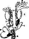
FROG, general name for representatives of the genus Rana, family Ranidae, order of tailless amphibians (Anura), class Amphibia. All Ranidae differ by the following characteristics: the toes on the lower side are provided with articular tubercles, the toes at the ends are not expanded; the tongue, attached by its anterior end, bears a deep incision on its free edge. The head of F. is generally triangular in shape; with its broad base it sits on the trunk; there is no neck; the body is short and wide; there are two pairs of legs, of terrestrial type; the front legs are short, the hind legs are long, jumping, and with swimming webbing between the toes. In the male, on the inner toe of the front leg, there is a wart-like pad from thickened skin glands (external sexual characteristic of the male). At the front end of the head opens a pair of nostrils; along the front edge of the head is a wide mouth slit; behind the eye on the side is a tightly stretched tympanic membrane; between the base of the thighs closer to the back is the cloacal opening. The body is covered with mucous skin, darker on the back than on the belly. The coloration depends on the presence of branched pigment cells. The epidermis is multilayered with flat surface cells. Under them lie simple spherical alveolar glands, the ducts of which pierce the epidermis. The glands secrete either serous (acidophilic) or mucous secretion. There are glands in which there are simultaneously cells producing both secretions. With age, the surface layer of flat epidermal cells cornifies and is shed in layers (molting). Under the skin lie extensive lymph spaces, thanks to which the skin is very movable. Due to the presence of terrestrial limbs, the vertebral column is divided into parts: cervical (one vertebra - atlas), dorsal (seven vertebrae), sacral (one vertebra) and caudal (the only coccygeal bone, fused from rudiments of caudal vertebrae). There are no ribs. The skull is narrow and long. The edge of the skull is bounded by a wide jaw arch; two occipital condyles. The anterior (pectoral) girdle consists of two scapulae, two coracoids, two clavicles and a large sternum. The pelvis - from fused together composite parts - has the shape of a tuning fork with a rudimentary handle. The skeleton of F. is only internal, built of bones and cartilage. In the oral cavity are vomerine teeth near the choanae (fig. 1); the upper jaw is provided with small conical teeth; the tongue is sticky; it is thrown outward for capturing prey (insects and other small animals). The esophagus is short, the stomach is elongated; near it lies a small spleen; in the duodenum opens the choledochus duct, formed by the fusion of the cystic duct and the hepatic duct and receiving the excretory ducts of the pancreas. The intestine is relatively short; the small intestine opens into the wide rectum, which opens into the cloaca, which in the male receives two ureters and the urinary bladder, and in the female additionally a pair of oviducts. The heart (fig. 2) is three-chambered, with a venous sinus, two atria, one ventricle; it is covered by the pericardium and is shifted far forward (lies above the sternum). From the ventricle goes the arterial trunk, dividing into two branches, each giving three arterial arches (carotid, aortic and cutaneous-pulmonary) (fig. 3). There is a portal system of the liver and kidneys. Among the veins, the abdominal vein in the thickness of the rectus muscle is of particular interest (convenient for injections). There are two circles of circulation; they are not separated, and in the ventricle both types of blood mix. The erythrocytes are oval, with nuclei. F. does not have its own body temperature. It breathes with a pair of lungs, skin and the mucous membrane of the oral cavity. The kidney is primary (Wolffian body). In the male, the ureters also serve for conducting sperm. The bean-shaped testes lie under the kidneys (fig. 4). The oviducts, long, twisted along the sides of the body, open into the body cavity with a funnel, not connected with the ovaries.
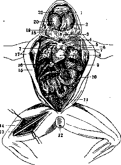
Figure 1. Internal organs of the frog: 1 - vomerine teeth; 2 - opening of the Eustachian tube; 3 - entrance to the larynx; 4 - tongue (cut off at the base); 5 - bulbus arteriosus; 6 - fragments of the sternum; 7 - atrium; 8 - ventricle; 9 - liver; 10 - stomach; 11 - ovary; 12 - cloacal opening; 13 - sciatic nerve; 14 - ischiadic nerve; 15 - pancreas; 16 - gall bladder; 17 - lungs; 18 - muscles; 19 - entrance to the esophagus; 20 - location of the eye; 21 - choanae.
The brain lies in one plane; all its parts are visible from above (fig. 5). There are 10 pairs of cranial nerves. There are internal and middle ears. Fertilization is external. Eggs are laid in a spherical, hard, transparent, glass-like shell; the mass covering them swells in water and connects the eggs into a mass ('frog spawn'). The eggs have a polar arrangement of abundant yolk. Cleavage is complete and uniform. From the egg hatches a legless tadpole with external feathery gills and a tail. Later these gills are replaced by internal ones. Metamorphosis consists in the sequential appearance of a pair of hind legs, then front legs, and the resorption of the tail; with the transformation is associated the transition to pulmonary respiration and the change from plant food to animal food, whereby F. changes from an aquatic animal to a terrestrial one, although still connected with water as a habitat. Rana esculena (edible F.) has obliquely lying between the internal nasal openings vomerine teeth in the form of oval strongly convex groups. The anterior and posterior edges of them go beyond the levels of the edges of the choanae. The head is weakly pointed. There is no dark spot on the temples. Length up to 17 cm. It goes north approximately to 59° north latitude; generally distributed in the European part of the USSR and Central Asia; absent in Western Siberia. Prefers to live in lakes and ponds, river bays overgrown with vines and sedges. Often stays in small ditches and stagnant puddles. Coming out on land, it usually does not move away from the body of water more than one jump. It winters by burying itself in silt. It wakes up in April. It spawns at the end of April, in May. It gives up to THOUSANDS OF EGGS. The transformation ends in late July, in August. Late tadpoles may go into winter hibernation. It feeds on insects, worms, mollusks, tadpoles and small frogs.
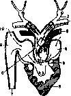
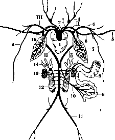
Figure 2. Opened heart of a tailless amphibian (diagram): 1 - carotid arch; 2 - aortic arch; 3 - pulmonary arch; 4 - left atrium; 5 - ventriculo-atrial valve; 6 - ventricle; 7 - posterior vena cava; 8 - arterial part of the arterial trunk; 9 - venous sinus; 10 - anterior vena cava. (Arrows show the course of blood.)
Figure 3. Diagram of the main arterial vessels: 1 - ventricle; 2 - right and left atria; 3 - common carotid art.; 4 - cutaneous art.; 5 - brachial art.; 6 - subclavian artery; 7 - lung; 8 - stomach; 9 - intestinal art.; 10 - mesenteric art.; 11 - iliac art.; 12 - kidney; 13 - testis; 14 - fat body; 15 - pulmonary art.; I, II and III - art. arches.
Rana temporaria (gray frog); vomerine teeth in the form of two m
Figure 4. Urinogenital organs of the male frog: 1 - fat body; 2 - testis; 3 - rectum; 4 - seminal vesicle; 5 - cloaca; 6 - urinary bladder; 7 - adrenal gland and below kidney; 8 - spermatic vessels. The transformation ends in late July, in August. Late tadpoles may go into winter hibernation. It feeds on insects, worms, mollusks, tadpoles and small frogs.
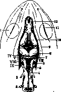
Figure 5. Brain of the frog: 1 - eyeball; 2 and 3 - II and III spinal nerves; 4 and 6 - medulla oblongata; 5 - recess of IV ventricle; 7 - cerebellum; 8 - visual lobes; 9 - intermediate brain; 10 - cerebral hemispheres; 11 - olfactory lobes; 12 - oculomotor nerve.
Bibliography: Pavlovsky E., Materialy po faune i ekologii amfibii i reptilii Kryma, Trudy Krymskoi ornitologicheskoi stantsii, 1928; Nussbaum M., Die Hautdrüsen des Frosches und ihre physiologische Bedeutung, Zeitschr. f. allg. Physiol., Bd. XX, 1921; Ballowitz E., Uber die Hautdrüsen der Batrachier, Anat. Anz., Bd. XLVII, 1919; D u d a B., Uber die Hautdrüsen der Batrachier, Anat. Hefte, Abt. I, Bd. LVII, 1919; H e r m a n n E., Der Frosch, Lpz., 1908; L e g o u y A., Rana temporaria possede-t-elle, comme la Bufo vulgaris, un poison cutane, Arch, intern, de physiologie, t. IX, 1910.
Related articles
Mentioned in
Cite this page
“Frog.” Soviet Medical Encyclopedia. English translation of Bolshaya Meditsinskaya Entsiklopediya, 1st ed. (Moscow, 1928–1936), ed. N. A. Semashko. https://sovietmedicalencyclopedia.pages.dev/article/frog/