Adenoid Tissue
Historical document, translated for reference. It reflects medical knowledge of the 1920s–30s and is not medical advice.
Summary
This article from the 1928–1936 Soviet medical encyclopedia describes adenoid tissue, a type of connective tissue that forms the basis of lymph nodes, spleen, bone marrow, tonsils, and the gastrointestinal mucosa. It details the tissue's structure, including reticular fibers and various cell types such as lymphocytes, macrophages, and fibroblasts.
Encyclopedia article (1928–1936)
ADENOID TISSUE (from Greek aden- gland and eidos- form), synonym reticular tissue, one type of connective tissue, forms the basis of lymph nodes, spleen, bone marrow, tonsils, and the mucous membrane of the gastrointestinal tract. In adenoid tissue one can distinguish a framework and the cellular elements lying within this framework. The framework of adenoid tissue appears different depending on the species of animal and age: in lower vertebrates (frog) and young individuals of higher species adenoid tissue consists of stellate cells anastomosing their processes with one another, thanks to which a delicate spongy tissue is obtained, in the nodes of which lie cell bodies with nuclei, and the crossbars of the network consist of the processes of nodular cells. With age in adenoid tissue of higher vertebrates special fibers appear, arising as a result of the formative activity of its cells—reticulin fibers. These fibers of different shape and thickness—sometimes in the form of threads, sometimes in the form of ribbons and plates—are distinct from ordinary fibers of connective tissue (collagenous and elastic): they are not digested by trypsin, do not swell from the action of diluted acids and alkalis, do not give a typical stain on elastin, and, unlike the elements of fibrous meshes, are otherwise impregnated with silver salts.
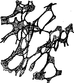
Adenoid (reticular) connective tissue from the substance of the lymph gland of a cat. Reticulin fibers lie inside the cells. With the development of reticulin fibers the cells remain on their surface, occupying the nodal points of the network, and with their protoplasmic processes cover the reticulin fibers—sometimes this sheath is well noticeable, sometimes, however, the protoplasmic covering is so thin that it remains completely unnoticeable. The mesh of adenoid tissue can be of different density in different places, for instance, in the sinuses of lymph nodes it is significantly looser than in lymphatic follicles and medullary cords. Cells of adenoid tissue often exhibit mobility and can completely free themselves from connection with the reticular framework and give free mobile elements exhibiting clearly expressed phagocytic activity (macrophages, necrophagocytes). According to data from tissue cultures in vitro, cells of adenoid tissue are pluripotent, i.e., they can develop in different directions, from them cells of the lymphoid series, macrophages, and fibroblasts can be formed. The latter, however, is not recognized by all. In the loops of adenoid tissue usually lie numerous lymphoid elements, the number of which can be so great that the reticular basis itself becomes completely unnoticeable, and for its detection it is necessary mechanically to remove the lymphoid elements by processing the section with a soft brush or by shaking the little compacted material in a tube with water (frozen sections or paraffin—after removal of paraffin from them). One can establish three types of cells lying in the loops of adenoid tissue: 1) large lymphocyte, or lymphoblast—a fairly large cell with a large pale nucleus and a narrow band of sharply basophilic protoplasm—the initial form for the other two types; in these cells figures of karyokinesis are often visible; it was previously supposed that in lymph nodes these cells usually accumulate in the center of the lymphatic follicle, which on stained preparations appears more pale—the center of multiplication of Flemming; however, such "centers of multiplication" are visible far not always, and sometimes figures of division can be seen scattered throughout the mass of the follicle or medullary cord; besides, there are views that the light centers of follicles consist not of lymphoblasts, but of rounded reticular cells due to some irritation; on the basis of this many propose to call these centers not "centers of multiplication" but "centers of irritation" or, better, "reactive centers"; 2) small lymphocyte, the most frequently encountered form—a small cell with a dense, well-staining nucleus, with a characteristic clotted arrangement of chromatin and a narrow band of basophilic protoplasm, obtained from the previous form; small lymphocytes do not contain figures of division, they are mobile and from the places of their formation migrate into the sinuses of lymph nodes, whence they are carried away with lymph, or enter the bloodstream directly through the wall of the blood vessel; 3) monocyte—a fairly large cell with a loose oval or bean-shaped nucleus and weakly basophilic protoplasm. All these three forms of cells filling the intervals of the reticular basis are connected by numerous transitions and can transform one into another, for instance, a small lymphocyte (both in adenoid tissue itself and in intertissue intervals in other places) can again take the form of a large lymphocyte and again can begin to divide and give rise to different cell forms; in tissues a small lymphocyte, stopping in place, can also transform into a monocyte or into a wandering cell (see.) in a state of rest and perform further transformations. All these three forms of cells can be considered free mesenchymal cells capable of being carried by the blood and lymph currents to other places of the organism and independently wandering through the tissues. As already mentioned above, besides these three types of lymphoid cells, the reticular basis of adenoid tissue also gives free cells separating from the mesh of adenoid tissue and phagocytizing in the cavity of the sinuses.-See also Reticulo-endothelial apparatus.
Adenoid (reticular) connective tissue from the substance of the lymph gland of a cat. Reticulin fibers lie inside the cells.
Related articles
Mentioned in
Cite this page
“Adenoid Tissue.” Soviet Medical Encyclopedia. English translation of Bolshaya Meditsinskaya Entsiklopediya, 1st ed. (Moscow, 1928–1936), ed. N. A. Semashko. https://sovietmedicalencyclopedia.pages.dev/article/adenoid-tissue/