Lymphatic System (a1605)
Historical document, translated for reference. It reflects medical knowledge of the 1920s–30s and is not medical advice.
Summary
The lymphatic system is a network of channels, vessels, and specialized formations that drain lymph from tissues. This article describes its phylogenetic development across different vertebrate classes and its structure in mammals.
Encyclopedia article (1928–1936)
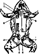
LYMPHATIC SYSTEM (GLANDS, VESSELS), a system of spaces, channels, vessels, and special formations (lymphatic glands) along their course, which drain from tissues the so-called lymph (see). The concept of the lymphatic system also includes certain formations from adenoid tissue (see). These include 1) individual lymphatic follicles (noduli lymphatici solitarii) and their conglomerates (noduli lymphatici aggregati), located in the walls of the digestive tube and in the respiratory tract, 2) the thymus gland (glandula thymus), 3) the spleen, and 4) bone marrow. The lymphatic system also includes the body's serous cavities: the abdominal, pleural, pericardial, and (in males) the cavity of the tunica vaginalis of the testes. (Regarding lymphatic circulation-see Lymph.) Phylogenesis of the lymphatic system. As a system distinct from the circulatory system, the lymphatic system is present in all vertebrates, starting from fish. But in most bony fish, lymphatic vessels cannot be detected in the injected state by ordinary research methods, with which these vessels can be easily found in other vertebrate animals standing at a higher stage of development. Usually, with this injection, only the veins fill with the injection fluid. To clarify the gradual development of lymphatic vessels, i.e., the replacement of lymphatic spaces by lymphatic tubes as the vertebrate organism develops from lower to higher forms, it is appropriate to give a brief description of the lymphatic system of some vertebrate animals by separate classes. The lymphatic system of frogs consists of extensive subcutaneous spaces formed by the skin and adjacent organs covered with fasciae, and of extensive milk sacs formed by two layers of the mesentery. Lymphatic vessels, easily detectable by the puncture method under the serous membrane of the digestive tract, flow into the milk sacs. Since the lymphatic vessels of the digestive tract are continuations of the spaces of the milk sacs along the course of blood vessels, the vessels detectable under the serous membrane of the digestive tract are the same perivascular spaces as the lymphatic pathways between the layers of the mesentery. By puncturing the muscles and skin, it was not possible (Iosifov) to detect lymphatic vessels of the body and limbs, although the organs are abundantly impregnated with a colorless fluid-lymph. From the subcutaneous lymphatic spaces and milk sinuses, lymph is pumped by the anterior and posterior lymph hearts. The two anterior hearts are located near the spine; they are covered by the shoulder blades. The posterior hearts consist of several bulbs and are located in the pelvic region, near the coccygeal bone. In a living frog, after an incision in the skin in the depression between the pelvis and coccygeal bone, one can see the posterior heart pulsing, which pumps the transparent fluid-lymph into the pelvic vein (figures 1, 2 and 3). The same can be observed from both the anterior and posterior lymphatic hearts in tadpoles after removing the skin.
Figure 1. Lymphatic system of the frog (on the left side, the lymph hearts are dissected, on the right they are covered by fasciae): 1- v. jugularis; 2- brachial plexus of nerves and subclavian artery; 3- left anterior heart and its connection with v. vertebralis, flowing into v. jugularis; 5 and 6- posterior heart, consisting of two bulbs, connected with the femoral vein (4); 7- partition between the subcutaneous lymphatic spaces of the back and thigh; 8~ right anterior heart visible under the raised edge of the shoulder blade.
FIG. 2.
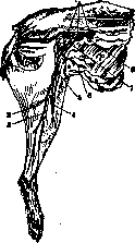
Figure 3. Lymphatic system of mammals. The most developed lymphatic system is in mammals. The main lymphatic vessels can be divided into Figure 2. Anterior lymphatic heart of the frog exposed: two semilunar valves are visible at the outlet into the vein; the inlet openings are marked by hairs inserted into them. Fig 3 Left anterior lymphatic heart of the frog, lying on the transverse process of the third cervical vertebra, with three inlet openings and a vein into which the heart pours lymph; the inlet openings are marked by hairs inserted into them (enlarged). subcutaneous and deep. These vessels are easily detected by the puncture method in tissues, with the exception of the lymphatic vessels of the spleen, brain, and placenta. In other areas of the body of mammals and humans, the lymphatic system consists of tubes running parallel to blood vessels and interrupted at certain points by lymph nodes. According to location, lymphatic vessels can be divided into subcutaneous and deep, main and secondary trunks, and capillary networks. The main subcutaneous trunks collect lymph from secondary trunks from the capillary networks of the skin and subcutaneous tissue and pass in the subcutaneous fat layer parallel to the main subcutaneous veins and nerves. They are easily detected by puncturing the skin and subcutaneous fat. The main deep trunks pass together with the main deep blood vessels and nerves and collect lymph from organs with the help of secondary trunks, which originate in the capillaries of organs located under the fascia (figure 4). The trunks are provided with valves and therefore have a distinctly tubular appearance. In the walls of the vessels and nodes, muscle tissue is embedded, the contraction of which moves lymph and propels it toward its origin in the veins. In addition, an auxiliary force for lymph movement is the suction effect of respiratory movements of the chest, the pulsation of arteries, and muscle contractions. The influence of diaphragm contraction on the initial expanded part of the thoracic duct (cisterna chyli) is so great that in both mammals and humans this cistern can be considered a passive lymph heart (figures 5 and 6). The rhythmic action of the muscle tissue embedded in the walls of the vessels between the valves is similar to the rhythmic action of active lymph hearts of lower animals; therefore
Figure 5.
Figure 6. Figure 5. Formation of the thoracic duct and its cistern (dilation) in a dog: 2-aorta; 2-v. azygos; 3-d. thoracicus; 4-trunci intestinalis; 5-cisterna chyli; 6-tr. lumbalis. Figure 6. Formation of the thoracic duct and its cistern (dilation) in humans: 1-d. thoracicus; 2-cisterna chyli at the initial end of the thoracic duct, formed by the fusion of the right and left lumbar trunks; 3-intestinal trunk; 4 and 5-lumbar glands. Ranvier considers each segment of lymphatic vessels between valves as a separate lymph heart. The lymphatic system of frogs, lizards, and eels (figure 7) represents a comparatively lower form of development compared to the lymphatic system of birds (figure 8) and Figure 7. Lymphatic system of an eel; 1- hair inserted through the cranial sinus cavity into the opening leading to the jugular vein; 2- hair inserted through the cranial sinus cavity into the opening through which the paravertebral trunk opens into the cranial sinus; 3- valve at the inlet opening of the cranial sinus; 4-right paravertebral lymphatic trunk; 5-jugular vein; 6-anastomosis of paravertebral lymphatic trunks; 7-aorta; 8-milk ducts; 9- kidney; 10- main intestinal sinus; 11-sinus of the sexual organ; 12-sexual organ; 13 and 14-caudal artery and vein; 15- continuation of paravertebral lymphatic trunks in the tail; 16-longitudinal intestinal sinus; 17-subserous lymphatic network of the stomach; 18-liver sinus; 19-cranial lymph sinus. mammals and consists of extensive lymphatic cavities, from which lymph is pumped by active and passive hearts. Milk sacs receive lymph from the wall of the digestive tract through channels that represent peri
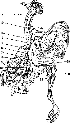
Figure 8. Lymphatic system of a pigeon: 1- lymphatic vessels accompanying the jugular vein with two trunks in the neck area; 2- lymphatic vessels of the forearm; 3- flow of lymphatic vessels of the anterior limb into the angle of fusion of the jugular and subclavian veins; 4- lymphatic vessels of the arm; 5- thoracic duct; 6- lymphatic vessels accompanying the abdominal aorta; 7- lymphatic vessels in the kidney area; 8- lymphatic vessels of the small intestine; 9- lymphatic vessels of the stomach; 10- lymphatic vessels accompanying the abdominal aorta; 11- lymphatic vessels accompanying the caudal vein; 12- lymphatic vessels of the posterior limb in the leg area with two trunks accompanying blood vessels; 13- lymphatic vessels of the thigh.



vascular spaces. Similar perivascular spaces are revealed by injection by the method of pricking into the thickness of the trunk muscles of eels, whereas in frogs and lizards they are absent, and it must be assumed that in the latter animals there are no preformed lymphatic capillaries and perivascular spaces in the area of the trunk and limbs; instead of them (capillaries and perivascular spaces) among the muscular tissue there exist simple lymphatic clefts. The existence of extensive lymphatic clefts in the aforementioned lower vertebrates fully corresponds to the function of these clefts as exclusively collectors of lymph, which is pumped into the blood by lymphatic hearts. In higher animals, the lymphatic clefts are replaced by tubes equipped with valves and muscles, serving to move lymph into the blood. Consequently, in higher vertebrates, lymphatic tubes serve not only as collectors but also as apparatuses for moving lymph. The gradual development of the L. s. corresponding to the complication of its function is indicated by the absence of lymphatic nodes (glands) in frogs, lizards, eels, and non-swimming birds. Lymphatic nodes first appear in a relatively small number compared to mammals in swimming birds (Fig. 9). In animals in which lymphatic glands are absent, the function of the latter is performed by adenoid organs (follicles) of the digestive tract, the thyroid gland, and bone marrow. Bartels, who studied the ontogenesis of the L. s., found that in pig embryos 67 cm long, the subcutaneous lymphatic vessels are already fully developed. Regarding the earlier stage of development of lymphatic vessels, Bartels says the following: "The development of lymphatic vessels and the thoracic duct is at least in the first stages even darker than the development of blood vessels. Indeed, some authors imagine that in the mesenchyme clefts form, which merge and the edges of which are covered with a sheath of endothelial cells, formed from the transformation of neighboring cellular elements into endothelium. But voices are also heard, which speak in favor of the development of lymphatic so
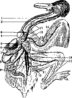
FIG '1 IlIMj) SPSTG(M1\ 11' 1 - SHMF.
Related articles
Cite this page
“Lymphatic System (a1605).” Soviet Medical Encyclopedia. English translation of Bolshaya Meditsinskaya Entsiklopediya, 1st ed. (Moscow, 1928–1936), ed. N. A. Semashko. https://sovietmedicalencyclopedia.pages.dev/article/lymphatic-system-2/