Placenta
Historical document, translated for reference. It reflects medical knowledge of the 1920s–30s and is not medical advice.
Summary
The placenta is a temporary organ that establishes contact between the maternal and fetal circulatory systems, facilitating nutrient and oxygen exchange. This article covers comparative anatomy, development, structure, physiology, and pathology of the placenta across various species.
Encyclopedia article (1928–1936)
Placenta. Contents: I. Comparative anatomy.............55.-1 II. Development of P. in humans.............. 556 III. Placenta of a full-term ovum..............5E8 IV. Physiology and biology 11.............55a V. Pathology of P. Pathological forms II........j..........563 Improper attachment И....... , . 564 Infarction, cysts, calcification and new growths of P....................5£5 Pathological changes II. in some diseases of mother and fetus.............. 567 «Placenlitis»..................... 568 Placental polyp..............569 Placenta accreta . .........-.......569 Premature separation of a normally situated P................... 573 Placenta previa............... . . 577 Placenta, placenta (from Greek placus- flat cake), child's place, the most important organ of a temporary character, in which a close contact is established between the circulatory systems of the mother and the developing embryo, so that the latter receives the necessary nutritional material and oxygen from the mother's blood and in turn returns to her the products of metabolism and carbon dioxide. I. Comparative anatomy. Among invertebrates, such relationships are rare, and only in tunicates and especially in salps does the placenta develop as a fairly constant formation abundantly supplied with blood. In this case, the fertilized egg is attached to the wall of the cloacal cavity and is overgrown with a circular fold of its epithelium. This fusion is associated with the loss of cellular boundaries and the transformation of both the follicular epithelium and the epithelium of the cloacal cavity lining into a continuous syncytium, which serves as an intermediary in the transfer of nutrient fluids from the mother's organism to the developing embryo. In some invertebrates, as for example in many bryozoans, the connection of the embryo with the mother's body becomes even closer, and sometimes the developing embryos feed at the expense of the mother's body itself and even completely destroy it. As for vertebrates, the placenta develops primarily in some sharks (Carcharias, Mustelus laevis), in which the embryo is attached to the wall of the oviduct ("uterus") by means of the yolk sac. At the junction, numerous folds and villi develop on the latter, growing into the corresponding depressions of the uterine wall walls. Similarly, in some viviparous lizards, the placenta develops at the expense of the yolk sac, however in some cases along with it the embryonic membranes (serosa with allantois) also participate in the formation of the placenta, and then (Seps ehalcides) this entire formation approaches what is found in most mammals. In mammals, the entire course of embryonic development is significantly changed due to the fact that the egg lacks the reserves of nutritional material (yolk) that are characteristic of all other vertebrates. After cleavage, the egg forms a rapidly expanding vesicle, the outer ectodermal wall of which plays the role of the "trophoblast"; through it, the absorption of nutrients secreted mainly by uterine glands (embryo-trophe) initially occurs. In many mammals, the trophoblast fuses with the uterine mucosa, forming a temporary, "ectoplacenta". The uterine mucosa at this site becomes more vascularized (but Strahl'io). J4f 'Jr. * *** 4V , FIG. 1 L G 111Й ДУЗЫрЬ ЛИСИЦЫ C n0.i_U_I] L.I.I- cent (no Strahl'io). rusts, in it develop b. or m. significant lacunae. Such a "preplacenta" later, with the formation of the chorion and the ingrowth of allantoic vessels, is replaced by the final P. (euplacenta). In marsupial mammals, the embryos usually still lie freely in the uterine cavity, surrounded by a nutrient fluid secreted by it. However, in some cases there is a connection of the yolk sac with the uterine wall (yolk or umbilical P.) (Phascolarctus, Halmaturus, Dasyurus), and in the bandicoot (PeramelPS) the P develops at the expense of embryonic membranes and specifically the chorion, in which the blood vessels of the allantois strongly proliferate and come into close contact with the vessels of the uterine mucosa. The epithelium of the latter transforms into a thin syncytium, so that the transfer of nutritional material and gas exchange between the embryo's blood and the mother's blood occur with the greatest ease. In "placental" mammals, the embryonic P. (pi. fetalis) always develops at the expense of the allantochorion, on which numerous villi develop, growing into the uterine mucosa. The epithelial lining transforms into a thin syncytium, and a close contact is established between the blood vessels of the allantois of the embryo and the vessels of the mother. The most primitive form in mammals is the "diffuse" P. (pi. diffusa) with simple villi scattered over the entire surface of the chorion (pigs, tapirs, horses,

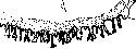
Figure 2. Cross-section through the fetal vesicle of the monkey Cercocebus cynomolgus with the primordia of two disc-shaped placentas (according to Strahl'io).
embryonic membranes and specifically the chorion, in which the blood vessels of the allantois strongly proliferate and come into close contact with the vessels of the uterine mucosa. The epithelium of the latter transforms into a thin syncytium, so that the transfer of nutritional material and gas exchange between the embryo's blood and the mother's blood occur with the greatest ease. In "placental" mammals, the embryonic P. (pi. fetalis) always develops at the expense of the allantochorion, on which numerous villi develop, growing into the uterine mucosa. The epithelial lining transforms into a thin syncytium, and a close contact is established between the blood vessels of the allantois of the embryo and the vessels of the mother. The most primitive form in mammals is the "diffuse" P. (pi. diffusa) with simple villi scattered over the entire surface of the chorion (pigs, tapirs, horses,
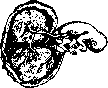
Figure 3. Embryo of the monkey Semnopithecus maurus with two leaf-shaped placentas (in the opened uterus) (according to StrahFio).
camels, semi-apes). In most ruminants there is a "multiple" placenta, consisting of many separate plaques, or "cotyledons" (see figure in t. III, st. 204), i.e., groups of complexly branched villi growing into the corresponding proliferations of the uterine mucosa (pi. multiplex, s. pi. cotyledonica). In such "semiplacentae" (scmiplacent'ae), during the birth of the offspring, the allantochorion with its villi separates from the uterine mucosa without special { violations of the integrity of the latter. In other mammals, the fusion of the system of complexly branched chorionic villi (pi. fetalis) with the uterine mucosa becomes more complete (pi. vera). The uterine mucosa proliferates (pi. uterina), its epithelium is scattered, the blood capillaries are stretched into wide sinuses, sometimes losing at the point of contact with the chorion even their endothelial walls, so that the villi, covered only by a thin syncytium, are freely washed by maternal blood. In these cases, during childbirth, along with the embryonic membranes, a significant part of the uterine mucosa is torn off (shed membrane, decidua), which is accompanied by noticeable bleeding. Such a more perfect placenta develops only in a certain area of the chorion, having the shape of a belt or more often a disk. Accordingly, a "belt-like" placenta (pi. zonaria, s. annularis) of carnivores (fig. 1) and elephants and a "disk-shaped" placenta (pi. discoidalis) of insectivores, rodents, bats, monkeys (fig. 2 and 3) and humans are distinguished.
I. Schmalhausen. The development and structure of the placenta in different animal species is so distinctive that these characteristics could seemingly be and were indeed used in zoological classification (thus, animals were divided into deciduata, adeciduata, placentalia, etc., based on the nature and features of placentation). However, it turned out that differences in the nature of placentation are observed even in animals that are very close in all other respects, and on the other hand, even in the same animal, placentation can proceed differently, i.e., according to different types on separate, isolated areas of the same egg. Thus, differences in placentation cannot serve for systematic purposes, but are instead very convincing characteristics and evidence of cenogenesis according to Haeckel's terminology (cenogenetic, as opposed to palingenetic, Haeckel calls such features and distinctions that develop in animals during embryonic development and serve as temporary adaptations in the struggle for existence). From what has been stated, it is already clear that placentation has undergone significant and numerous evolutionary, and perhaps also mutational, changes in phylogenesis, which is why existing animal species show such diversity in the development and structure of the placenta. In general, however, all this diversity can be reduced to four basic "types. 1. Placentation in which the villous membrane of the egg only comes into contact with the epithelium of the uterine mucosa, - villous-epithelial placentation (pl. epithelio-chorialis). In such placentation, the egg is nourished at the expense of the secretion excreted by the uterine mucosa and its glands (so-called uterine milk). 2. Placentation in which the villi of the egg destroy the epithelium of the uterine mucosa, grow into the connective tissue base of the mucosa, and are nourished, partly by dissolving it, - villous-connective tissue placentation (pl. syndesmo-chorialis). Here, the trophoblast of the egg already shows enzymatic, cytolytic ability with respect to maternal tissues. 3. Placentation in which the villi of the egg destroy both the epithelium of the uterine mucosa and its connective tissue base and lie directly against the wall of the maternal blood vessels, - against the endothelium of maternal capillaries. This is villous-endothelial placentation (pl. endothelio-chorialis, sive angiothelio-chorialis). In such placentation, nutrients are taken by the egg from the maternal blood, but must first pass through the thinnest wall of the maternal capillaries. 4. Placentation in which the villi of the egg destroy the endothelium of maternal capillaries, penetrating into the lumen of the vessels themselves and extracting nutrients directly from the maternal blood, - villous-blood placentation (pl. haemo-chorialis). II. Development of the placenta in humans. In humans, the placenta externally resembles a round flatbread, hence its Latin name, introduced into scientific terminology by Fallopius (16th century). In humans, placentation occurs according to the fourth type (see above), but in ontogeny, all four of the above-mentioned stages can be traced. Thus, initially, the human egg, after penetrating the uterus, merely lies against its altered mucous membrane and for a very short time is nourished at the expense of its secretion. However, very quickly, with its outer membrane - the trophoblast, in particular its syncytial layer, - the egg destroys the epithelium of the mucous membrane and lies against its connective tissue base, then destroys the connective tissue base, approaches the walls of blood vessels, but very soon penetrates through the capillary endothelium and thus its villi immerse directly into the maternal blood, which in turn flows out of the blood vessels and spreads throughout the intervillous space. This achieves very broad contact of maternal blood with the surface of the villi of the egg. The youngest human eggs described to date are barely two weeks old, and yet the villi of the trophoblast of these eggs are already immersed in maternal blood, and this blood fills the intervillous space. From this it is evident how quickly, in the process of implantation, the human egg passes through all the described stages of placentation. It must also be taken into account that of these two weeks, about a week is spent on the actual movement of the egg through the tube, and consequently the entire above-described process unfolds and concludes within the space of just one week. The further formation of the placenta - the formation of the placenta as a special organ - then proceeds no so quickly, occurs gradually and is completed by the end of the third month of pregnancy, and thereafter until the end of pregnancy the placenta only increases in size. The very formation of the placenta consists in the fact that in one place, namely where the egg lies directly against the uterine wall, its villi especially luxuriantly grow, forming the so-called branched chorion (chorion frondosum). Here the underlying uterine mucosa also grows particularly strongly, already transformed into a typical deciduous membrane (decidua basalis) (see Decidua). Throughout the third month of pregnancy, individual villi of the branched chorion densely fuse, better said adhere, with the deciduous membrane of the uterus. Such an adhesion also occurs along the entire periphery of the placenta directly between the chorionic plate itself (membrana chorii) and the deciduous membrane. Thus, the entire area of the egg surface covered by the branched chorion becomes completely delimited from the rest of the egg surface, where in the meantime the villi have died off, so that this entire part of the egg becomes as if balded (for which reason this area of the egg is called the "bare" chorion - chorion laeve). As stated, the fusion of maternal tissue with the chorion is completed by the end of the 3rd month, and then the placenta is already truly a delimited formation, a real organ. For this reason, if pregnancy is interrupted after the third month, the egg always emerges with a true placenta. Obviously, in such a placenta one can distinguish maternal elements - the entire layer of deciduous membrane covering the maternal surface of the placenta (placenta materna), and elements belonging to the egg, i.e., the entire mass of villi and the chorionic plate (placenta fetalis). Here it would be appropriate to mention that the changes occurring in the uterine mucosa during the formation of the placenta, i.e., the decidual metamorphosis, extend to a considerable degree to the underlying muscular wall of the uterus. Thus, here among the muscles one can find individual cellular inclusions that resemble in structure either decidual cells or cells of Langhans' layer of the villi, or epithelial islands; besides this, the muscle cells themselves and the connective tissue between them undergo certain changes resembling those that occur in the deciduous membrane at the point of its contact with the villi, namely - coagulation necrosis and the deposition of fibrinoid, in a word - the formation of the so-called Nitabuch layer. All this taken together gave the Belgian scientist Keiffer the opportunity to propose a special term for this area of the muscular uterine wall - placenta myometrial. This term has not yet come into general use, but the process described by Keiffer deserves attention in the further study of human placentation. From the end of the 3rd month of pregnancy, the placenta, as stated, no longer undergoes substantial changes, it only increases in size and at the same time as it were "ages." This aging is manifested, among other things, by the fact that the connective tissue base of the villi loses its embryonic character, and on the surface of the villi Langhans' layer gradually disappears, and they remain covered only by syncytium. As for the maternal part of the placenta, here necrotic changes intensify and in places even accumulations of calcium phosphate and calcium carbonate form. III. The placenta of a mature egg. By the end of pregnancy, the placenta acquires the well-known appearance of a round or oval fleshy flatbread 15-20 cm in diameter, 2-3; 4 cm thick and weighing 500-600 g. The surface area of the placenta is 100-150 cm², but the total surface area of all the villi forming the placenta is calculated at 6-10 thousand cm², and the total length of all the villi, if laid out end to end, is about 50 km. These figures give an idea of the enormous surface on which exchange between mother and egg takes place. The growth of the placenta is indicated to some extent by the change in its weight, which, starting from the end of the 3rd month, increases as follows (monthly): 30- 70-120-200-275-350-450 g and finally by the end of the 10th month - 600 g. The study of placental development makes the structure of the mature placenta understandable. Its maternal surface is covered with a compact (solid) layer of ashen-gray deciduous membrane; the entire surface is strewn with microscopic remnants of partitions of the spongy layer, since usually the placenta separates precisely within the thickness of the spongy layer of the deciduous membrane, with part of this layer remaining on the uterine wall, from which a new mucous membrane is regenerated, and another part passes onto the placenta.
The maternal surface of the placenta is somewhat rough and covered with a peculiar network of grooves that divide the surface of the placenta into a large number of sections of the most diverse shapes. From each groove, a more or less high projection of the tissue that will separate extends into the substance of the placenta, forming the so-called placental septa (septa placentae). These septa divide the entire placenta into a corresponding number of separate lobes, or cotyledons (cotyledones); it should however be kept in mind that the septa do not reach the chorionic plate, and consequently the individual lobes communicate widely with each other, ultimately forming a single common intervillous space. The placental septa initially consist only of decidual cells, but as the placenta grows, coagulation necrosis and the shedding of fibrinoid are also observed in them. Within the substance of each septa, branches of the maternal arteries - the terminal branches of the uterine arteries - penetrate deep into the placenta; they freely open into the intervillous space both on the surface of the septa and at their apexes. Naturally, the flow of maternal blood must slow down considerably at this point, since the channel for its flow is expanded many times over. The maternal blood drains through venous openings scattered directly in the separating tissue throughout the space between the grooves. These openings can sometimes be distinguished with a magnifying glass as blood points or the smallest depressions filled with blood. From this description it is evident that the intervillous space is bordered not by maternal endothelium, as was recently thought, but by the covering epithelium of the villi themselves, i.e., mainly by the syncytium. Despite the slow flow of maternal blood, individual syncytial masses, in the form of kidney-shaped coverings on the surface of the villi, may become detached from the villus and thus enter the maternal blood flow; usually they dissolve quickly in it, but as an exception they may be carried throughout the maternal organism. This is the so-called deportation of villi, to which Veit once attached great importance in the physiology and pathology of pregnancy. The possible harmfulness of such deportation of villi under normal conditions is greatly reduced by both the dissolution of the syncytium mentioned above and the fact that the syncytium is neutral in group relations, whatever group difference may exist between mother and embryo.
The entire thickness of the placenta consists of richly branched villi of the branched chorion. The total number of these main villi amounts to hundreds (from 400 to 1,000 units). Most of the villi float freely in the maternal blood, like the branches of aquatic plants in an aquarium. Individual branches firmly fuse with the maternal tissue on the surface of the separating membrane and on the surface of the septa described above. These villi - anchor villi according to Grusdev (Haftzotten) - firmly secure the fertilized egg to the maternal organism. In addition, such attachment takes place all around the periphery of the placenta, where here a marginal or venous sinus is formed - a very important additional drainage vessel for maternal blood (sinus terminalis placentae). At those places where the villi come into contact with maternal tissue, changes similar to coagulation necrosis occur in this tissue and in the covering epithelium of the villi themselves, with the shedding of fibrinoid. These peculiar changes were first described by the woman physician Nitabuch and are called the Nitabuch layer. In contrast to this, that layer of the separating membrane where such changes are absent or are weakly expressed is called the Winkler layer. As for the villi, they consist of delicate embryonic tissue penetrated by a wide-meshed network of thin-walled blood vessels (see separate table, figures 1-4). The villi are covered by a two-layer epithelium: the Langhans layer (otherwise - cellular trophoblast) and the syncytium (otherwise - plasmodial trophoblast). As indicated, the first layer [see separate table (art. 567-568), fig. 1] gradually disappears as the placenta grows.
IV. Physiology and biology of the placenta. The function of the placenta is twofold: 1) it serves for the purely mechanical attachment of the fertilized egg to the uterine wall, 2) it is an organ in which the extremely complex exchange between mother and fetus (sc. fertilized egg) takes place. The first function is simple and does not require additional clarification after the given anatomical description. The second is far from fully studied and consists of two categories of phenomena - physical and biochemical. The physical phenomena are determined by the character that the double circulation in the placenta has: on the one hand, the circulation of the mother in the intervillous space and parallel to it - the circulation of the fertilized egg in the villi (see Fetus, fetal circulation). Turning to the biochemical processes occurring in the placenta, it should be said that they are very complex, although until recently a simplified view prevailed regarding the placenta as a simple membranous partition between maternal blood and fetal blood. It was assumed that the exchange of substances through this partition occurs according to the simple laws of osmosis and diffusion. It turned out, however, that this partition possesses complex biological functions with a strictly differentiated selective relationship to the individual substances circulating both in the mother's blood and in the fetus's blood. Thus, the placenta at the same time is an organ assimilating from the maternal blood substances necessary for the development of the embryo, and on the other hand, it secretes into the maternal blood the waste products resulting from the metabolism of the same embryo. Even such a basic exchange as the gas exchange - the absorption of oxygen and the excretion of carbon dioxide - occurs in the placenta not according to the simple physical laws of osmosis and diffusion, but with the help of the complex biochemical activity of an enzymatic nature. And although essentially we have here the same exchange that occurs in the lungs during respiration, it is obvious that the exchange itself proceeds differently here, namely according to the type of intercellular exchange. Simple chemical compounds, including water, are exchanged between the fertilized egg and maternal blood not only according to the laws of diffusion and osmosis, but also through complex biochemical processes. This can be easily verified by comparing in the mother's blood and in the fetus's blood the level of such relatively simple chemical compounds as compounds of Ca, K, and Na - their level will be different. If we take iron, which, it is true, is in the mother's organism and in the fetus's organism in very complex compounds, then here the difference in level will be especially great: this is because the amount of iron stored by the fetus during intrauterine life considerably exceeds its current requirement; this is caused by the necessity of having a certain reserve of iron in the fetus's organism for the time of extrauterine life, when the newborn feeds exclusively on maternal milk, containing only negligible amounts of iron. Therefore, the iron content in the fetus's tissues and its level in the fetus's blood are considerably higher than in the mother's organism; the copper content in the liver is also considerably higher.
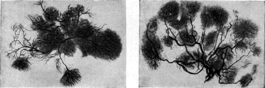
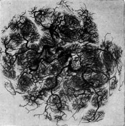
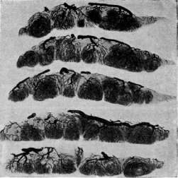
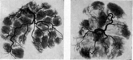
Hentgen photographs of the placental arterial network, injected with a contrast mixture. Figure 1. Normal placenta at the 3rd month of pregnancy. Figure 2. Normal placenta at the 6th month of pregnancy. Figure 3. Normal placenta at the end of pregnancy; the terminal capillaries are densely clustered, forming spherical clusters of uniform shape. Figure 4. Normal placenta at the end of pregnancy; the sections are perpendicular to the surface of the placenta; they show the direction of arteries penetrating into the thickness of the placenta, as well as the branching of the terminal capillaries. Figure 5. Placenta with two white infarcts (full-term pregnancy in a clinically healthy woman); at both sites corresponding to the infarcts, there is a complete absence of blood circulation. Figure 6. Syphilitic placenta (dead fetus). (After Fournier.) ft ft 2 of enzymes into the mother's blood. As for synthesis, it partially occurs in the organs of the fetus itself, but this synthesis also occurs already in the Placenta. This is at least obvious for proteins, since in the blood of the umbilical vein flowing to the fetus, it was not possible to prove the presence of those simpler compounds (albumoses) from which proteins are built, therefore the synthesis of proteins specific to the embryo has already occurred and has occurred in the Placenta.-From these general considerations, based however on numerous, diverse and complex experiments and research, it is obvious all the enormous significance of the Placenta in the assimilation of nutrients; in this respect the Placenta plays the role of both the lungs and the entire digestive tract together with its numerous glands. At the same time, the work of the fetus's own digestive glands as they are formed is of course not excluded. In particular, the liver of the fetus plays an enormous role in regulating and detoxifying the metabolism between mother and fetus. Turning to the question of the excretion of products of the fetus's vital activity, it can be said that here the matter is somewhat simpler. However, even the excretion of such simple chemical compounds as carbon dioxide, waste water and simple salts occurs by no means according to the simple laws of osmosis and diffusion, but by complex biochemical pathways. As for more complex chemical compounds (urea, uric acid, etc.), they are undoubtedly excreted in the Placenta through complex exchange reactions. In terms of excretion of metabolic products, the Placenta plays the role of the lungs, kidneys and skin. According to modern views, all the above-mentioned biochemical processes must occur with the help of various enzymes, and indeed, numerous enzymes necessary for all this complex and multifaceted chemical work have been found in the Placenta. However, the question arises whether these enzymes are formed in the Placenta itself or are brought into it with the mother's blood. This question cannot yet be considered finally resolved, and probably it is resolved differently for different enzymes, but there is every reason to assume that the cellular elements of the Placenta indeed possess the ability to form enzymes. This is clear, for example, for that proteolytic enzyme which is already present in the trophoblast of the youngest eggs and promotes their implantation (see above). In conclusion, it must be said that the Placenta, as a living, vigorously and diversely functioning organ, naturally also has its own metabolism, necessary for its own vital activity. Research conducted in this direction has shown that this own metabolism of the placenta proceeds with remarkable economy. Thus, the delivered placenta consumes 0.71 cm3 of oxygen per 1 kg/min., i.e., an amount significantly less than that established for the same own metabolism of other organs. The endocrine activity of the Placenta. Already at the beginning of the current century, individual indications appeared that the Placenta must possess and indeed possesses an endocrine function in relation to the mother's organism. This view received multifaceted confirmation, and at the present time it can be considered proven that in the Placenta there are-and in relatively large quantities-such hormones as the hormone of the ovarian follicle (folliculin), the hormone of the corpus luteum and finally prolane. With regard to hormones, the same question naturally arises as with regard to enzymes-are they produced in the Placenta itself or are they only adsorbed in it from the mother's blood, and are produced in the corresponding endocrine glands of the mother. This question also cannot be considered finally resolved, however, here too many facts speak in favor of the Placenta having the ability to produce these hormones independently or else hormones very similar to them in chemical structure and biological action. Thus, for example, with the onset of pregnancy, prolane circulates in the mother's blood and is excreted in her urine in such large quantities that it is difficult to assume its production exclusively in only the anterior lobe of the pituitary gland. In hydatidiform mole and chorioepithelioma'-so-called placental origin formations-prolane is produced in such enormous quantities compared to normal pregnancy that it becomes completely clear that it must be produced in the pathologically changed villi of the chorion. A hormone similar to the corpus luteum hormone can also be extracted from the Placenta in such large quantities that one has to assume that it is produced in the Placenta itself. In addition to the above-mentioned hormones, it is necessary to assume the possibility that the Placenta produces its own specific, and perhaps several specific, hormones still unknown to us. This has to be assumed, for example, when studying the effect which the Placenta has on the mammary gland and on the process of milk formation and milk secretion. Apparently it is in the Placenta that a hormone is produced that activates the development of the mammary gland during pregnancy, and perhaps at the same time another hormone that inhibits milk formation itself. The loss of the Placenta's function with the end of childbirth and consequently the disappearance (within three days) of this hormone from the mother's blood stops its inhibitory effect on milk formation and thereby causes the milk-secreting work of the mammary gland. There are some grounds to assume that in the Placenta, as it grows and matures, a hormone is produced and accumulates that causes the onset of uterine labor. Speaking of the endocrine activity of the Placenta during pregnancy, it is necessary to keep in mind that this activity manifests itself not only by direct action on the pregnant organism, but also by acting on the entire system of the mother's endocrine glands due to the correlation existing between the activity of individual glands. As to how complex this aspect of the endocrine effect of the Placenta is, one can to some extent judge by the schemes of Selitsky and Zeits (see vol. III, art. 215 and 219, figs. 5 and 7). In addition to the above-mentioned endocrine effects of the Placenta on the mother's organism, there is some reason to assume that the internal secretion of the Placenta also affects the intrauterine fetus, which it serves in all other respects, especially if this fetus is female. Thus, it is known that in the last weeks of intrauterine development, the reproductive system of a female fetus, in particular the uterus, grows strongly, and after childbirth, when the influence of the Placenta ceases, this enhanced development of the reproductive system stops, and the uterus of a newborn girl undergoes even involution, so that the uterus of a girl in the first years of life turns out to be smaller than that of a newborn. The loss of the Placenta's hormone also explains the appearance in newborn children of both sexes of swelling of the mammary glands (simultaneously with the same swelling in the mother) and the secretion of "milk" from them. In newborn girls, the same loss may explain the frequent appearance of bloody discharge from the uterus.-In recent years, the question has been raised about the formation of vitamins in the Placenta; it is premature to draw any conclusions, as the question has only begun to be studied. Finally, one can also mention the so-called "barrier" function of the Placenta (see Barrier function). It has already been said above that the Placenta has a clearly selective relationship to chemical substances circulating in the mother's blood and in the fetus's blood. It is important to clarify what is the relationship of the Placenta to those complex poisonous and protective substances which are produced or have already been produced in the mother's organism as a result of this organism's reaction to infectious and toxic agents entering this organism. A general answer to this question-naturally cannot be given, but still it can be said that a healthy Placenta also relates selectively to them and to some extent is able to serve as a barrier to those substances harmful to the well-being of the fertilized egg. The same must be said in particular with regard to the microorganisms themselves infecting the mother's organism-a healthy Placenta serves as a barrier to these organisms, and their penetration into the fetus's blood is as a rule possible only after the integrity of the epithelial covering of the villi is violated. The question is also generally resolved in the same way with regard to substances circulating in the fetus's blood-in terms of their transition to the mother's organism. Such in general outline are the complex and multifaceted biological functions of the Placenta.
Naturally, any pathological effects from the maternal organism can definitely influence both the morphology and the physiological function of the placenta; on the other hand, abnormal development of the placenta can be caused by factors inherent in the egg itself at the moment of conception, and can originate not only from the egg cell but also from the paternal element—the spermatozoon with its chromosomal apparatus. V. Pathology of the placenta. Pathological forms of the placenta. The usually round or oval shape of the placenta (placenta discoidalis) can vary considerably in different directions, but in general all these deviations are encountered far from often. Thus, the villi of the branched chorion may be preserved over a more extensive area than is normal, and then we will have a placenta that is very large in surface area but thin—pl. diffusa (such a placenta is common, for example, in pigs, horses). If at the same time the villi are very poorly developed, the placenta will be especially thin, almost membranous—pl. membranacea. Sometimes the placenta is as if divided into two or three parts (placenta bipartita, pl. tripartita), and this division may go so far that the placenta consists as if of two or more separate lobes (pl. bilobata, pl. multilobata). If the individual lobes are significantly smaller than the main placenta, one speaks of accessory lobes of the placenta (pl. succenturiata). This form often leads to serious complications in the postpartum period, as a small accessory lobe can easily detach from the "main placenta and remain in the uterus, which will not be noticed during examination of the placenta. In other cases, placental tissue is arranged on the ovum in the form of a belt or ring—pl. annularis, pl. zonaria (this form is common in carnivores). Underdevelopment or atrophy of the villi in one or another area of the placenta leads to the formation in the placental tissue of small membranous areas, like windows—pl. fenestrata (fenestrated placenta). A distinctive and not uncommon deviation in the form of the placenta is the formation around the placenta of a white margin (pl. marginata); sometimes this margin protrudes above the surface of the placenta in the form of a ridge (pl. circumvallata). Abnormalities in the formation of the placenta must also include improper attachment of the umbilical cord. As a rule, the umbilical cord attaches in the center of the placenta (insertio centralis), but it may attach eccentrically, at the edge of the placenta (insertio marginalis), or even outside the placental membranes (insertio velamentosa. Improper attachment of the placenta. The listed anomalies of the placenta's form can and do play their pathological role in the course of pregnancy, during labor, and in the postpartum period. However, greater significance is attached to those deviations from the norm that depend on improper attachment of the placenta or on the pathological nature of the attachment itself. As for the sites of attachment, as a rule the placenta develops at the site of egg implantation, i.e., on the posterior (more often) or anterior (less often) wall of the uterus. Sometimes implantation can occur in the tubal angle of the uterine cavity or along its lateral edge. In these cases, the placenta may acquire one or another abnormal form, and the very fact of its attachment not in the usual place can lead to one or another disturbances in its separation in the postpartum period. However, the listed forms of improper attachment of the placenta recede into the background in their practical significance compared to the serious complications caused by attachment of the placenta in the lower part of the uterus—in the so-called isthmus or lower segment (see below—placenta previa). The attachment of the placenta can be pathological not only in its location but, so to speak, in its essence. The point is that normally the chorionic villi penetrate the thickness of the compact layer of the decidua only to a certain depth. Their further growth is hindered, among other things, by the formation of the Nitabuch layer. However, in some cases, with insufficiency of the uterine mucosa or with its inadequate decidual metaplasia, the chorionic villi grow beyond the compact layer of the decidua, destroy its spongy layer, reach the muscular layer of the uterus, may pierce through this layer, reach the peritoneum, and finally destroy (eat through) the peritoneum itself. All these stages of what is essentially the same process are united under the general term "adherence of the placenta" (pl. accreta). During pregnancy this pathological attachment of the placenta usually gives no sign of itself, but during labor (in the postpartum period) the mechanism of separation of the placenta from the uterine wall is disrupted, and a series of very serious complications ensue, requiring one or another obstetric intervention up to and including hysterectomy (see below—adherence of the placenta). Among the numerous pathological changes observed in the placenta, there are some that occur quite often, which is why they represent a certain practical interest, although they do not have great importance in obstetric pathology. These include first of all the so-called "white infarct" of the placenta, "cysts" of the placenta, and petrification of the placenta (deposition of lime on the maternal surface of the placenta). The white infarct of the placenta is in essence nothing other than thrombosis of individual areas of the intervillous space. The origin of the white infarct is explained quite simply: maternal blood does not clot in the intervillous space because throughout its contact here with the epithelial covering of the villi, which like the endothelium of vessels protects it from clotting. However, if somewhere on the vast surface of the villi, under the influence of one or another harmful factor, the covering epithelium of the villi falls off or is pathologically altered, conditions for clotting of the blood immediately arise at this place, initially something like a "wall thrombus" forms, and then gradually the adjacent segment of the intervillous space of one or another length becomes thrombosed [see separate table (pp. 559-560), fig. 5]. It is clear that the mentioned harmful factors can originate from the mother, i.e., come from the side of the maternal blood; or they can initially be located in individual villi (for example, with obliteration of the blood vessels of the villus, there occurs sequential death of its covering epithelium and on this basis thrombosis of the adjacent part of the intervillous space). As a rule, white infarcts are of small size and usually do not exceed 1-2-3 cm in diameter. White infarcts are located both on the maternal surface of the placenta and, somewhat more often, in the very thickness of the placenta and near the chorionic plate. Under the microscope, a white infarct represents a collection of fibrin and fibrinoid, in which are embedded individual necrotized villi at various stages of the necrotic process, as well as groups of dying decidual cells, if the decidual septum of the placenta is partially included in the white infarct. Quite often white infarcts are located on the surface of the chorionic plate, covering large areas of it with a thin layer. Such formations have been described by Langhans under the name of "canalized fibrin." Besides the indicated, probably main cause of the formation of white infarcts, it is possible that they also occur as a result of excessive slowing of the blood current in one or another area of the intervillous space. Such slowing of the current of maternal blood can depend on disturbances of the circulation in the maternal organism (diseases of the heart), as well as on causes originating from the ovum (increase in intraovular pressure). As stated, thrombosis of the intervillous space usually is limited to small areas, but sometimes this process can involve very large areas of the placenta and then naturally the nutrition and development of the fetus can be severely affected, up to its intrauterine death. Such a widespread thrombosis is observed, for example, quite often in pregnant women suffering from chronic nephritis. On the other hand, after intrauterine death of the fetus, caused by an accidental cause, for example, tightening of the umbilical cord knot, if miscarriage does not occur soon, in the placenta of such a dead fetus sequential changes occur that are in essence a gradual, sometimes very slow thrombosis of the intervillous space. In particular, such changes are especially sharply expressed in the placenta in the so-called missed abortion (i.e., in very prolonged, sometimes multi-month retention of the dead ovum). Often in the central parts of white infarcts, liquefaction of the fibrin occurs, and then such an infarct turns into a "cyst." Sometimes the cavity of such a cyst is throughout or only in places lined with an "epithelial" covering. The origin of the cells of this covering is not quite clear: most likely they are cells of Langhans' layer of the villi, flattened under the pressure of the liquid contents of the cyst. In rare cases, the formation of white infarcts described here apparently does not proceed so slowly and gradually, but the clotting of maternal blood, i.e., the formation of a thrombus, occurs very quickly and moreover shortly before the birth of the fetus. In these cases, "infarcts" colored in various shades of red are observed in the placenta, depending on the time of their formation—these are the so-called red infarcts of the placenta.
One should not confuse such red infarcts with blood clots that are tightly seated on the maternal surface of the placenta and firmly attached to it. These clots form as a result of minor detachment of the placenta during pregnancy due to one or another trauma, if such trauma did not lead to an immediate termination of the pregnancy itself. If such a blood clot is removed from the placenta, a depression is found beneath it, covered throughout by a smooth and intact decidua. Cysts of the placenta. In addition to the above-described 'cysts' of the placenta forming from infarcts, other types of cysts are also observed on the placenta. These are the so-called subchorionic cysts. These cysts do not form in the thickness of the placenta or near its maternal surface, but directly beneath the chorionic plate or even within the thickness of this plate. Due to such a superficial position, these cysts are conspicuous upon examination of the fetal surface of the placenta and are therefore noted quite frequently. However, individual researchers give very different figures. Thus, there are indications that such cysts can be found in every second placenta, while in other statistics the ratio is 1 in 400 placentas. As for the origin of these cysts, it is not always clear. In a certain number of cases, these cysts undoubtedly originate from the formation, cavity, and liquefaction of a subchorionic infarct, in other cases it is obvious that the cyst formed within the thickness of the chorionic plate itself. In contrast to cysts that form in the thickness of the placenta, subchorionic cysts can reach quite a large size, containing several tens of cm3 of fluid (up to 250 cm3 have been described). This fluid is of almost neutral reaction, light yellow in color, specific gravity 1.010-1.015, contains a small amount of protein and a large amount of chlorides. Usually the fluid does not completely fill the cavity of the cyst, which is why the cyst has a 'flaccid' appearance. These cysts have no practical significance and it has not been possible to link their formation with any particular disease of the fetus or mother to this day—they are often found in completely normal placentas. Calcification of the placenta. The third pathological change of the placenta is the pronounced petrification of its maternal surface. It has already been said above that small deposits of calcium carbonate and calcium phosphate are often found on the surface of the placenta in its decidua capitis. Such a phenomenon can be considered normal. The very origin of these lime deposits is understandable if one takes into account that in the decidua capitis, as the placenta matures, significant degenerative and necrotic processes occur. Sometimes, however, the deposition of lime has a distinctly pathological character, and in these cases it affects large areas of the placenta—entire lobules, and in exceptional cases the entire maternal surface completely. If one runs a finger over the surface of such a placenta, it gives an impression almost like that of sandpaper. To this day, such petrification of the placenta has not been linked with any disease of the mother or fetus. It is observed in completely normal cases with the complete well-being of the mother and fetus. One must however think that some disturbance of calcium exchange between mother and fetus may play a role here. Clarification of this question belongs to the future. New growths of the placenta. Tumors of the placenta occur very rarely. However, as rare curiosities, all possible varieties have been described, including metastases in the presence of a malignant tumor in the mother. Angiomas are most frequently encountered, and sometimes they can involve such a large area of the placental villi that their mass already affects the development of the fetus. Among the new growths of the placenta must also be included that peculiar disease of the chorion, which is known under the name of hydatidiform mole (see Mole), as well as one of the most malignant tumors affecting women—the so-called choriocarcinoma. Pathological changes of the placenta in certain diseases of the mother and fetus. From all of the above, it is clear what important role the placenta plays during pregnancy, it is also clear how closely it is connected with the metabolism of both the mother and the fetus. It is therefore natural that certain diseases of the mother or fetus can cause specific changes in the macro- and microscopic structure of the placenta. Syphilis of the placenta. Characteristic changes in the placenta were first described in syphilis.1 For example, it was long noted that the placenta in syphilis differs in its color (the white placenta of syphilitic fetuses), its great size (thickness), and its great weight. Whereas in the normal ratio of the weight of the placenta to the weight of the fetus is 1:5-6, in syphilis this ratio changes in favor of the placenta and can reach a ratio of 1:3 (often), 1:2 and even in rare cases 1:1. It should however be borne in mind that even in the normal state, the indicated ratios undergo considerable fluctuations. As for the microscopic changes in the placenta, they consist in the edema of the villi, in the proliferation of their connective tissue, in the obliteration of blood vessels (endarteritis obliterans) [see separate table (art. 559-560), fig. 6]; in the epithelial covering of the villi all possible changes are described, up to the ingrowth of epithelium into the thickness of the villus itself. All these changes cannot however be considered absolutely diagnostic for syphilis, since they are also observed in other diseases with a definite absence of syphilis (e.g., in chronic nephritis of the mother). In addition to these changes, which are observed comparatively frequently, in individual cases of syphilis the formation of miliary abscesses (gummata) has been described. The syphilitic placenta is often characterized by the variety in size of the villi, especially by the abundance of small villi with hyperplasia of the villous epithelium. Truly diagnostic is the discovery of the pale spirochete in a suspicious placenta, although, on the other hand, even in definitely syphilitic placentas this is often achieved only with great difficulty. Tuberculosis of the placenta. Unlike syphilis, tuberculosis of the mother is apparently transmitted to the fetus very rarely. It is therefore natural that the placenta in pregnancy in tuberculous women does not bear changes characteristic of tuberculosis either macro- or microscopically. The formation of characteristic tubercular nodules has been described on the placenta in isolated cases, which, given the enormous prevalence of tuberculosis, clearly speaks to the rarity of the placenta being affected by this process. In the question of tuberculosis of the placenta, the mode of infection is also interesting—hematogenous or per continuitatem in the presence of a local tuberculous lesion of the endometrium. An answer to this question cannot be given on the basis of the available data. In the presence of tuberculous changes in the placenta, tuberculosis was found in the fetus in half of the cases. Changes in the placenta in other diseases. In chronic nephritis of pregnant women, changes in the placenta are always observed, provided only that the pregnancy goes to term. However, these changes cannot be called specific. It has already been said above that sometimes they coincide with those observed in syphilis; in other cases, abundant formation of white infarcts is observed. If certain changes are sharply expressed, the pregnancy is often interrupted, and if it is carried to term, the fetus is usually poorly developed and is characterized by congenital weakness. One must think that in nephroses of pregnancy and generally in toxicoses, certain pathological changes must occur in the placenta, but to date they have not been identified with definite certainty. In cardiac decompensation in the mother, in addition to the above-described changes, congestion of the intervillous space is quite often observed in the placenta. This phenomenon is of a stagnant nature and gives the placenta a distinctly bluish (cyanotic) coloration. It is observed however also with a healthy heart on the basis of acute and chronic poisonings, including in toxicoses (eclampsia). A peculiar change in the placenta is observed in congenital edema of the fetus; this edema extends to the placenta as well. The origin of this disease is unknown, and where the primary changes lie—in the placenta or in the fetus—is also unknown. "Placentitis". As for the question of isolated inflammation of the placenta, in the old obstetrical literature such a form of placental disease was widely recognized and bore the name placentitis. At present there are individual attempts to restore the right of citizenship of this term. However, it must be said that
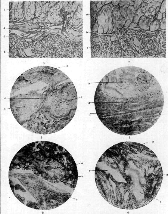
Fig. 1. Normal attachment of the placenta: a-well-developed spongy layer of the decidua (here in normal conditions the separation of the placenta from the uterine wall occurs); b-villi; c-muscle layer of the uterus; d-fibrous layer. Figure 2. Placenta accreta. The spongy layer of the decidua is completely absent, only a thin layer (b) of the fibrous layer (Nitabuch) is present; c-villus; o-muscle layer of the uterus. Figure 3. A fairly thick (compact) layer of the decidua (a) between the villi and the muscular (b). In thickness the layer is close to normal, but consists almost entirely of necrotized homogeneous mass, which explains the fact that the villus (c) grows through the decidua and branches directly on the muscular layer. Fig. 4. A very thin layer of the decidua (a) between the villi (b) and the muscular (c). Figure 5. Complete absence of the decidua between the muscles (c) and the villi (o), not even a layer of Nitabuch; a-island of dead decidual tissue. Figure 6. Structure of the decidua at the site of attachment of the membranes, i.e., in the area of dec. parietalis. In this preparation, unlike the previous three, the spaces of the spongy layer (a) of the decidua have been preserved; b-muscle layer of the uterus. (Figure 1 and 2 according to Seitz, figures 3-6 according to Kolosov.)
The conditions described in P. as characteristic for placentitis, according to modern views, do not indicate inflammation as such. In those cases where the inflammatory process, e.g., in acute infectious diseases, actually involves the placenta, the matter usually quickly ends in miscarriage or premature birth. On the placenta, inflammatory changes can then be found in the form of, e.g., small-cell infiltration of decidual tissue or in the form of numerous petechial hemorrhages into its thickness. Such changes were first described by Slavyansky in cholera. However, if acute infection does not disrupt the course of pregnancy and does not end in the death of the mother, then on the placenta, born at one time or another after the infection, usually no definite sharply expressed changes characteristic of the transferred 'inflammation' are found. Placental polyp. Among the pathological formations associated with the placenta are the so-called placental polyps. They form in the uterine cavity in those cases where, after a miscarriage or childbirth, a small particle of the placenta remains on the uterine wall. If infection of such a postpartum uterus occurs, the matter may end in the death of the mother or the remaining part of the placenta disintegrates and gradually is discharged with postpartum lochia or is finally removed by finger or curettage. In those cases where the uterine cavity with the remaining particle of the placenta does not become infected, this particle can remain in the uterus for weeks and months, sometimes causing more or less prolonged and profuse uterine bleeding. Eventually such a placental remnant takes the form of a polyp on a more or less thick stalk. It may finally detach, and the matter ends in recovery. More often, however, medical intervention in the form of curettage of the uterine cavity is required. Under the microscope such a polyp consists of layers of fibrin, in the thickness of which necrotized villi of the ovum, dying and detached decidual cells, and sometimes regenerating mucous membrane of the uterus are found along the periphery. The only conclusive proof of the placental origin of the polyp is the presence of chorionic villi, which is important to keep in mind in forensic medical examination when medical personnel is accused of leaving a part of the placenta. The fact is that formations, macroscopically quite similar to placental polyps, sometimes develop in cases of limited infection of the uterine cavity due to polypoid growth of granulation tissue or even of the mucous membrane itself without any participation of placental villi; such cases in no way should be interpreted as true placental polyps, and the presence of these growths in the uterus is not proof of leaving a part of the placenta after childbirth or miscarriage.
m- Kolosov. Placenta accreta. Placenta accreta denotes cases where the placenta does not separate spontaneously soon after the birth of the fetus, but is retained for a long time because its villi, adjoining directly to the muscular layer of the uterus (due to weak development of decidua basalis), fuse with it. Etiology. The etiological factors come down to more or less significant anatomical, and consequently functional, deficiency of the endometrium. Such inadequacy of it can develop 1) as a result of manual separation of the placenta in previous births, 2) after septic postpartum infection associated with significant rejection of necrotized mucous membrane of the uterus, 3) after other inflammatory diseases of it (e.g., gonorrhea), 4) after intrauterine application of cauterizing agents (e.g., T-rae Jodi), 5) after roughly performed curettage of the uterine cavity, especially in artificial abortion. In all these cases, further regeneration of the mucous membrane is difficult, and subsequently often prolonged amenorrhea, oligomenorrhea are observed, and in the very next birth placenta accreta may occur. Judging by the fact that placenta accreta occurs almost exclusively in multiparous women and in women who have had many abortions (especially after rapidly following births and abortions), it must be assumed that one or another inflammatory processes, often associated with both births and abortions (and even more so with operative procedures in this connection), indeed play an important etiological role here. However, in no way should the etiology of placenta accreta be reduced only to inflammatory diseases. Clinical experience shows that placenta accreta (in the absence in the anamnesis of any local inflammatory processes) is relatively often observed with its abnormal location in the lower segment of the uterus, in the cervix or even partly in its neck, i.e., in places generally unsuitable for implantation of the fertilized egg, because the so-called 'decidual reaction' of the endometrium to pregnancy is expressed here very weakly. In all precisely established, i.e., histologically studied cases of placenta accreta, the following changes were generally noted: complete absence of decidua basalis (where the placenta was attached), poorly expressed layer of Nitabuch, direct apposition of villi to the muscle, often their penetration into the very thickness of it and even into the lumen of uterine veins; further, cloudy swelling, hyaline degeneration and disintegration of uterine muscle fibers with extreme thinning (sometimes to several millimeters) of the uterine wall and consequently its increased vulnerability. Infiltration of the muscle by so-called chorionic wandering cells was also observed in an incomparably greater degree than is the case in normal conditions, and as if their splitting of muscle fibers, as well as formation of thrombi in uterine vessels, consisting of placental tissue. Kolosov in his dissertation (1910) makes the following main conclusion regarding the pathogenesis of placenta accreta: From the patho-anatomical side, placenta accreta is characterized by profound changes in the decidua, mainly of degenerative and atrophic nature, leading in milder cases to the destruction of the entire spongy and part of the compact layer of the decidua, in more severe cases-to the destruction of the entire decidua, with the growth of villi into the very substance of the uterine musculature (see separate table, figures 1-6). Later studies by Freund and Hitschmann give a new light to this question, pushing into the background the old 'inflammatory' theory of placenta accreta and bringing to the forefront apparently no less justified hypothesis about abnormally increased vital activity of the villous epithelium. These authors proved that the cause of placenta accreta should be sought not so much in changes of maternal tissues as in the growing egg itself. On their preparations they could convince themselves that sometimes trophoblast persists for a very long time (persistence of it); due to this the destructive, or rather - cytolytic, properties of the egg, which it normally possesses only in the very earliest stages of its development, are preserved for a long time. This peculiar 'excess of growth', this abnormal, excessive increase of the biologically inherent activity of the egg explains the abnormally strong connection subsequently between the placenta and the uterine wall and precisely due to the fact that trophoblast and villi first destroy the compact, and then the glandular layer of decidua, so that in the end the villi penetrate already into the thickness of the uterine musculature. It is possible that this is facilitated by the supposed local absence of antiferments, which in normal conditions counteract the enzymatic properties of trophoblast and villi, whereby the entire process of placentation takes on a sharply expressed destructive character (which partly resembles the development of destructive mole and choriocarcinoma).
The attachment of the placenta may be complete (placenta accreta totalis) or partial (placenta accreta partialis). Both of these occur in reality far from often, especially the typical complete attachment, which represents an extremely rare anomaly. It is very difficult to establish the exact frequency of these cases in general, since the indiscriminate use of the diagnosis 'attachment' is a common phenomenon in the practical activities of obstetricians and even in their scientific reports. Some authors indicate 1 case in 6,000 births, others 1:10,000 and even 1:40,000, but all these figures are very unreliable. If the criterion for a correct diagnosis is considered not only clinical data (which are often incorrectly interpreted here), but above all precise histological research, then in all the literature only several dozen perfectly correctly diagnosed cases will be found. The symptoms that could indicate impending disorders in the third stage of labor in connection with placental attachment are usually completely absent during pregnancy. Nevertheless, in some (very rare) cases, the abnormally deep penetration of the villi into the thickness of the uterine muscle tissue already manifests itself in the early months of pregnancy with bleeding, which apparently in no way differs from 'usual bleeding in abortion'; the true nature of these is revealed only when attempts are made to remove the ovum, and the obstetrician encounters very significant obstacles when he tries to remove all placental tissue completely; sometimes this proves to be completely impossible. In extremely rare cases, spontaneous rupture of the uterus may occur at the very beginning of labor or even long before the onset of uterine labor activity. Sometimes attention is drawn to the primary weakness of labor pains in both the first and second stages of labor, caused by the anatomical and functional insufficiency of the uterine muscle. In general, however, as a rule, any symptoms of placental attachment are completely absent during pregnancy and during the first two stages of labor, and disorders appear only in the third stage; strictly speaking, this stage also begins without complications at first; however, in most cases, very soon strong bleeding appears, and the cardinal symptom of placental attachment makes itself known - its persistent retention in the uterine cavity. One can only think of placental attachment when, with the condition of perfectly proper management of the third stage, the placenta is retained for a very long time. Therapy. If repeated, correctly applied manual expression of the placenta by the method of Credé (sometimes even under anesthesia) does not succeed, and at the same time the bleeding already takes on an obviously threatening character, then the operation of manual separation of it is quite indicated (see vol. 1, p. 320), and in the presence of truly complete true attachment, it is necessary to penetrate the hand deep into the tissues. However, with very firm and extensive attachment, this proves to be extremely difficult; it is usually not possible to remove the entire placenta at once, and therefore it has to be extracted in several stages, in pieces, which is always associated with the possibility of removing together with the placental tissue individual parts of the muscular wall of the uterus, which further complicates further orientation. If, despite careful and thorough efforts, it is not possible to separate all placental tissue at once, and at the same time the bleeding not only does not stop but even increases, then it is necessary to give up the idea of removing everything completely. Even for a very experienced specialist, it is very difficult to determine by touch some small piece of placenta in these cases, and yet to force the issue here is by no means safe, since one must always reckon with the danger of perforation of the uterus. In very difficult cases, the only way out is to tightly tamponate the entire uterus and vagina, and then apply a pressure bandage on top for the purpose of counter-pressure. This gives the possibility to stop or significantly weaken the threatening bleeding, thus gaining time and saving the woman from 'immediately threatening death' [in the third stage, by prescribing uterine-contracting agents, it is sometimes possible to achieve the expulsion of pieces of retained placental tissue from it (Stoeckel)]. The best thing after tamponade is to immediately transport the patient to a hospital institution in order to provide her, if necessary, with radical surgical help (immediate removal of the uterus by abdominal or vaginal route). Radical and moreover early surgical intervention in cases of truly complete placental attachment is the most rational method of therapy. When establishing indications for radical surgical intervention, one must always take into account the aforementioned danger of further septic infection. Removal of the uterus in the presence of true placental attachment is therefore not only urgent surgical help in a catastrophic situation, but also has a certain preventive significance. One must remember that in the vast majority of cases of placental attachment, we are dealing with multiparous women who already have children, and therefore the fear of removing the uterus, all the more in view of the often obvious danger to women, cannot and should not stop the surgeon. As for the recurrence of placental attachment, one should be somewhat skeptical about this, because at least true complete attachment of it either ends in a fatal outcome or is cured by timely radical surgery (removal of the uterus); therefore, a new pregnancy cannot be discussed here. In those cases where a recurrent placental attachment is reported, it is obviously a matter not of typical true attachment with its specific pathological-anatomical features and clinical symptoms, but of another, much less dangerous anomaly, i.e., of placenta adhaerens (adhered, firmly attached). (The latter is rather a purely clinical collective term; in contrast to placenta accreta, here no definite pathological-anatomical substrate has yet been found; nevertheless, even with placenta adhaerens, pathological changes in the separating membrane apparently also play an important etiological role.) Premature separation of a normally located placenta. Premature separation of the placenta includes cases when the placenta begins to separate in the first or second stage of labor (or even during pregnancy). Under normal conditions, due to a number of anatomical-physiological prerequisites (see Labor - third stage), the placenta cannot separate before the expulsion of the fetus. Therefore, premature separation of a normally located placenta (in general rather rare) is possible only in the presence of special etiological factors. The frequency of this complication (if we consider only cases with clearly expressed and more or less serious symptoms) according to Albeck - 0.6%, according to Prankl - 0.096%, according to Pankow - 0.41%. Premature separation of the placenta may be partial or throughout its entire extent, i.e., complete. Etiology. Among the etiological factors, the least significance is attached to direct mechanical (even if crude) trauma; taking this possibility into account, one should not overestimate it: from the rather extensive casuistry on this question (blows and strong jolts to the abdomen, falling directly on the abdomen, etc.) there is by no means any indication that such trauma would necessarily be accompanied by premature separation of the placenta. Much more often (already post factum) it is possible to establish a causal relationship between absolute or shortening of the umbilical cord and this anomaly. Similar mechanical conditions are also created with very delayed rupture of the fetal membranes, when the lower pole of the ovum produces tension on the lower edge of the placenta and ultimately detaches it from the uterine wall. Other mechanical factors may also have etiological significance. For example, the sudden departure of a very large amount of amniotic fluid in hydramnios, as well as the birth of the first fetus in twins, also create conditions favorable to premature separation of the placenta. With beginning overstretching of the lower uterine segment and threatening rupture of the uterus (in a narrow pelvis, in a neglected transverse position, etc.), premature separation of the placenta can also occur, since under these conditions the fetus, having no possibility to enter the pelvic cavity, due to gradually intensifying uterine contractions and increasing retraction of the uterine muscle tissue, moves more and more from the upper part of the uterus to the lower (as a result of this emptying of the hollow muscle of its contents, a decrease in pressure on the placenta from the inside occurs, which leads to its separation). However, all these purely mechanical factors by no means exhaust the etiology of premature separation of the placenta: some authors (Chautreuil, Winter, P'ehling, Henter, etc.) point to the frequent connection between this complication and kidney diseases of both inflammatory (chronic nephritis) and degenerative nature (pregnancy kidney, nephrosis). It has long been established that nephritis, extensive white infarcts in the placenta, degeneration of its vessel walls, and premature separation of villi from the deciduous tissue often coincide, forming one typical clinical picture.
Sometimes inflammatory changes in the deciduae (decidual endometritis), cell necrosis, thrombosis of vessels, and hemorrhages come to the forefront. Most frequently, a connection is observed between premature detachment of the Placenta and various toxicoses of pregnancy, and this usually manifests itself at the end of pregnancy. As is known, during this time, phenomena occur that prepare the ground for the onset of labor, namely, the gradual weakening of the connection between the ovum and the walls of the uterus due to degenerative and regressive changes in the decidua basalis. These purely physiological processes explain the fact of the greatest frequency of cases of premature detachment of the Placenta precisely at the end of pregnancy. As early as 1894, Hans Meyer first proved in two cases of premature detachment of the Placenta the presence of typical decidual endometritis (exactly at the site of its attachment) in the form of degenerative changes in the decidual cells and significant infiltration of the entire tissue by leukocytes. Very valuable are the microscopic studies of Albek on 1,015 placentas: 1) in 11 cases of clearly expressed premature detachment of the Placenta, hemorrhages were found in 10 (90.9%); 2) in 37 milder cases without clear clinical symptoms, such large and multiple hemorrhages were present in 27 cases (72.98%); 3) in 967 cases without any signs of premature detachment, small hemorrhages were observed 293 times (in 30.29%). Obviously, in premature detachment of the placenta, hemorrhages, which are generally not uncommon, represent a phenomenon typical of this complication. The following summary table by Albek is also of great interest from the point of view of pathological anatomy and etiology: Toxicoses of pregnancy Number of cases Hemorrhages in Placenta Premature detachment Percentage of cases with hemorrhages in Placenta Eclampsia . . . Threatening eclampsia . . . Albuminuria and edema..... Albuminuria . Edema...... Without symptoms . , k 7 11 76,72 64,51 44,26 44,85 40,24 23,3 From this it is clearly seen that placental hemorrhages and premature detachment of the Placenta often coincide with each other, and both occur the more frequently the more pronounced the phenomena of toxicosis of pregnancy. The etiological connection between these or other kidney diseases and premature detachment of a normally located Placenta has been established statistically on large numbers by many authors. Thus, for example, Brandt noted it in 73%, Hofmeier in 57%, Frankl in 56%, Albek in 51.3%, Pankov in 46.7%. And indeed, in premature detachment of the Placenta, a symptom such as albuminuria is very often observed, but on the other hand, there are also not a few cases where there is no protein in the urine; therefore, it is hardly possible to always consider kidney damage the primary cause of premature detachment of the Placenta. There are much more grounds to think that the same etiological factors that cause albuminuria, nephropathy of pregnancy, edema, and eclampsia also play a certain role simultaneously in the origin of premature detachment of the Placenta. The latter is sometimes also a result of toxicosis of pregnancy in the broad sense of this concept. It is precisely toxicosis that is often the common basic etiological factor and the true primary cause here. In the clinical picture of premature detachment of the Placenta, the dominant, often very severe symptom is bleeding as an inevitable result of the detachment of the chorion and the opening of the intervillous blood spaces in connection with this. The flowing blood accumulates behind the Placenta, forming the so-called retroplacental hematoma. If the lower edge of the Placenta has not detached, then the outflow of blood toward the exit from the genital canal is blocked, and therefore the bleeding remains internal and is recognized only by all the signs of increasing acute anemia. Otherwise, the blood easily finds its way between the fetal membranes and the walls of the uterus into the vagina, and then outward. It is clear that in connection with severe bleeding, both the mother and the fetus are in great danger, with the fetus being in greater danger. If the detachment of the Placenta has already occurred on approximately half of its entire surface, then the metabolism in the fetus is disrupted to such an extent that it inevitably dies; with less significant detachment, asphyxia threatens, often fatal if decisive and energetic help is not provided in time. Effective therapy here is ensured primarily by early and accurate diagnosis of only the beginning of premature detachment of the Placenta. Unfortunately, in most cases this is possible only in the presence of external bleeding. Usually, the differential diagnosis is between premature detachment of a normally located Placenta and its presentation. During internal examination, the question is resolved relatively simply, but not always, since there is often doubt as to whether there is really premature detachment of a normally located Placenta in this case, or rather its low attachment (when placental tissue cannot be palpated). In the absence of external bleeding, only very careful continuous observation can ensure timely and correct diagnosis. Local and general phenomena are of decisive importance. Due to the significant accumulation of blood, the uterus rapidly increases in volume, often takes on a spherical shape, becomes extremely tense and sensitive (stretching of its peritoneal covering). Sometimes the parturient complains of a sudden pain in the lower abdomen, which may be due to the sudden detachment of a large area of the Placenta, the rapid formation of a large retroplacental hematoma, and the sharp stretching of the uterine walls. Contractions usually cease because the uterus, already stretched beyond its elasticity, cannot contract well; in any case, they become weak. It is very difficult to palpate the parts of the fetus, determine its position, and listen to its heartbeat through the tense walls of the blood-filled uterus. The combination of these local symptoms with a clear picture of increasing acute anemia ensures the accuracy of the diagnosis and indicates that both the mother and the fetus are in great danger, with the latter possibly already dead. Nevertheless, even in very severe, apparently cases, decisively, quickly, and skillfully rendered help can still save the parturient from a life-threatening situation, and in rare cases, the fetus as well. In the choice of therapy for premature detachment of the Placenta, the following factors play an essential role: the severity of bleeding, its time of appearance, and then the circumstance that it can usually be stopped only under the condition of complete emptying of the uterus. If insignificant bleeding appeared only during labor, and with good contractions, one can temporarily wait, but be ready at any moment for active intervention: such cases can end favorably with spontaneous labor. If the fetal membranes have already ruptured and the uterine os is sufficiently dilated, then with increased bleeding, labor can be completed—depending on the conditions and position of the fetus—either with forceps or by version. With intact membranes, it is recommended to rupture them with the aim (sometimes) of accelerating labor and eliminating a possible causal factor, i.e., the tension produced by the membranes on the lower edge of the Placenta; however, it must be kept in mind that after rupture of the membranes, intrauterine pressure decreases, which can partially intensify the detachment of the Placenta and bleeding. In any case, rupture of the membranes is appropriate only with sufficient dilation of the os and with good contractions when a rapid end of labor can be expected. If with intact membranes there is primary weakness of labor pains, or if after rupture of the membranes the uterine os is still little dilated, and at the same time bleeding is clearly increasing, then to accelerate labor, version on the foot can be performed, and if dilation of the os does not yet allow this, to introduce a metreurinter (if necessary, preceding this with dilation of the cervix by Hegar dilators) or generally to apply one or another method of forced delivery (see 'Accouchement force'). If the fetus has already died, then perforation of the head is most appropriate. The task of the obstetrician is much more complicated in cases when bleeding manifests itself at the very beginning of labor activity, when the birth canal is not yet prepared at all; at this time, it is sometimes very difficult to make an accurate diagnosis of premature detachment of the Placenta, since such bleeding can also occur with placenta previa. With insignificant bleeding, one can assign bed rest and temporarily wait, but if it quickly increases or from the very beginning takes an obviously threatening character, immediate intervention is necessary. In the way of providing emergency surgical help here, the following operations are possible: cesarean section—vaginal or abdominal—or complete extirpation of the uterus, but preferably without opening its cavity.
Opening the uterus in severe cases is hardly rational, especially since the chances of saving the fetus here are more than questionable, yet the danger of sharply expressed uterine atony during the operation itself and immediately after it is very probable, since hemorrhage into the thickness of the uterine walls, fatty degeneration of muscle fibers, and abundant development of connective tissue between them (and especially in the presence of severe overstretching of the entire organ by the blood that has flowed into it) can cause such insufficiency of contractile ability even when it is emptied, that it is already difficult to expect a sustained tone here; rather, one must consider the possibility of its complete relaxation and consequently threatening sequential atonic hemorrhages. All these operations, if they are not undertaken too late, can still save the woman in cases where without this radical help she would certainly have died. If the fetus is obviously already dead, one can limit oneself to vaginal cesarean section with perforation of the presenting or subsequent head. If the fetus is still alive, abdominal cesarean section is best, which often gives good results here, saving both mother and fetus. In the presence of very severe anemia, when it is quite obvious that further blood loss, even if not very significant, may already be fatal in its consequences, supravaginal amputation of the uterus according to Porro or complete abdominal extirpation of it (without opening the cavity) is quite appropriate; both are fully justified in these very severe cases, always associated with the already mentioned danger of sequential atonic hemorrhages, which are often fatal in premature detachment of the placenta precisely because of the deep anatomical changes in the myometrium. If the placenta has not been expelled immediately after the birth of the fetus and bleeding continues, one must either express it by Credé's method or perform manual removal of it. The prognosis in premature detachment of the placenta is by no means always the same. In partial detachment with slight bleeding, it is relatively favorable for both mother and fetus. In more or less significant detachment of the placenta with strong external or internal hemorrhage, the outcome becomes doubtful and often fatal for both. Maternal mortality varies according to different authors; depending on many conditions, it fluctuates within wide limits, but still remains very high—up to 32%. Infant mortality is significantly greater—up to 80%. However, it should be borne in mind that these aggregate statistical figures only give a clear picture of reality when they are broken down into various groups corresponding to the severity4 of individual cases, which of course is by no means the same depending on the degree of premature detachment of the placenta and many other accompanying conditions. Placenta previa, placenta praevia (isthmica). PL praevia, i.e., standing in the path of the fetus's forward movement, is one of the most severe, life-threatening complications of the birth process for both the parturient and the fetus. Its frequency is approximately 1:1000 births, and it is observed mainly in multiparous women. According to large statistics covering hundreds of thousands of births, the frequency of placenta praevia according to Müller is 1:1,078, according to Schwarz is 1:1,564, according to Rick is 1:731, and according to Sicke is 1:1,300. The more births in the history, the greater the probability that in new births there may be placenta praevia. Interesting is a table illustrating material from Pankow's clinic. Out of 7,792 births, he observed 126 cases of placenta praevia, distributed as follows: Number of births Births Number of cases of placenta praevia Absolute % 4 567 1820 689 270 153 125 258 VI VII-XVI 17 - S5 19 8 8 0.37 1.9 8.2 2.9 5.2 8.0 11.2 If one does not start from the total number of births, but from the number of cases of the complication itself, it turns out that placenta praevia out of all 126 cases from Pankov's material occurred in primiparas only 13.5%. The same author presented a large comprehensive statistics compiled by him from data of many other authors, from which it is clear that out of 2,885 cases of placenta praeviae, it was observed in primiparas 341 times, i.e., in 11.8%, and in multiparas-2,544 times, i.e., in 88.2%. Etiology. The figures already presented above shed clear light on the etiology of placenta praevia. Apparently, here the pathological condition of the endometrium, which often occurs after complications of repeated pregnancies, frequently following each other abortions (the latter is possible but requires verification and proof), births, and postpartum periods, has very essential significance. The final result of all these puerperal processes can be very diverse both anatomical and functional changes in the entire sexual apparatus and first of all precisely in the endometrium. Various inflammatory and hyperplastic processes occurring in it often end in distortion of the normal histological structure of it and consequently in violation of its functions. The hypothesis of 'purely inflammatory' origin of placenta praevia, although it undoubtedly has a certain basis (see below), by no means yet exhausts the complex question of the pathogenesis of placenta praevia. Counterbalancing it is another hypothesis, putting in the first place not changes in the maternal soil, but biological peculiarities of the egg itself. For a fertilized egg to be able to implant, it must first reach a certain maturity, become capable of nidation; this 'nidational maturity' it acquires during its passage through the mucous membrane of the Fallopian tube. However, such maturity of the egg can apparently sometimes occur earlier, sometimes later. In the first case, it implants already in the tube itself, in the second (most often)—in the lower part of the uterus, which is called the isthmus.1 Both these anomalies of implantation, i.e., tubal pregnancy and placenta praevia isthmica, would find from this point of view a quite understandable explanation if the entire sexual canal, starting from the abdominal openings of the Fallopian tubes and up to the cervix, represented equally favorable soil for receiving the egg; however, the mucous membrane of the tube does not provide suitable conditions for this; therefore, it can be assumed that in those cases when the egg nevertheless implants in the tube, it really possesses some special biological properties, probably increased cytolytic ability of its trophoblast. In any case, such an egg, according to Poorten, should possess comparatively greater vital energy, greater potential growth force, the proof of which is the rapid deep embedding of it in the tube wall. A completely normal egg does not possess such properties and therefore does not remain in the tube. If this assumption is really correct, chat should be thought that in those cases when the egg implants only in the lower parts of the uterus, it must possess reduced growth energy, i.e., be partly defective in terms of its nidational ability. However, such an assumption is clearly contradicted by the very often observed in placenta praevia extremely deep (just as in ectopic pregnancy) embedding of the villi of the egg far beyond the limits of the mucous membrane. From this it is clear that the causes of implantation anomalies in general and in particular such as placenta praevia cannot be sought exclusively in the properties of the egg itself. The fact of significantly greater predisposition of multiparas compared to primiparas to placenta praevia clearly indicates that the greater wear and tear of the uterus and especially of the endometrium is really a factor causing abnormally low implantation of the egg. In addition to those already mentioned above, the following factors may contribute to this: 1) the movement of cilia of the ciliated epithelium in the mucous membrane of the body of the uterus towards the inside to the outside (as in the Fallopian tubes), proven by Hofmeyer, 2) the established fact by Gene (H61he) that in hypertrophic processes in the endometrium this movement becomes especially strong and continuous, 3) the progressive parallel with the increase in the number of births expansion of the uterine cavity. Undoubtedly, all these factors substantially facilitate the movement of the egg into its lower parts. In any case, to explain the pathogenesis of primary isthmic placenta, when the egg immediately attaches itself in the mucous membrane of the isthmus, one must assume an unusual width of its canal. The occurrence of secondary isthmic placenta (i.e., those cases when the egg initially attaches in the body of the uterus but still relatively low, and only in the process of its further development the placenta, gradually growing, turns out to be already a greater or lesser part of it in the area of the isthmus) can be caused by various factors. First of all, it should be noted that placenta praevia is observed much more often in multiple pregnancies (and namely—1:41 births;)
Strassmann); this indicates that the mucous membrane of the uterine body in these cases, with further growth of both placentas (or one large common one), may prove territorially insufficient and therefore a significant part of the placental tissue may capture the area of the isthmus. Qualitative changes in the maternal soil, however, have greater significance in the origin of placenta praeviae. For example, submucous uterine fibroids that stretch and thin its mucous membrane, and especially various intrauterine manipulations that in one way or another traumatize the endometrium, make it to a greater or lesser degree defective both morphologically and physiologically; such an etiological role is probably also played by gonorrheal endometritis and all other inflammations of the endometrium, even if not of a specific and not even of a bacterial character. A distant consequence of any trauma to the endometrium may be in the future an insufficiently expressed decidual reaction to pregnancy; the decidua formed under such unfavorable conditions is unable to fully provide the growing egg with the nutrition it needs. It is quite possible that partly for this reason the placenta grows significantly not so much in depth as on the surface, occupying more and more space not proper to it, gradually reaching the isthmus of the uterus and penetrating into its canal. Such an explanation of cases of secondary isthmic placenta praeviae is accepted by many authors as quite natural and understandable. In its favor, apparently, also speaks the circumstance that in these cases the placenta is often actually very large by its dimensions, sometimes extending from the fundus of the uterus to its very isthmus. At the same time, the os is often thinned and has an unusual shape (with separate processes at the periphery); often the placenta presents in the form of placenta circumvallata (this indicates that the growth of villi into the thickness of the decidua vera encountered an obstacle). The secondary os with a sharply expressed eccentric attachment of the umbilical cord, as well as a membranous attachment of it. The formation of additional lobes of the placenta is also frequently observed. On the secondarily formed isthmic placenta, an elongated tongue-like process can be noticed, obviously corresponding to that part of it (fig. 4) which developed in the isthmic canal. Pankow assumes that the origin of complete, i.e., central, placenta praeviae can also be explained in this way. To admit the possibility that the egg could be implanted immediately directly above the internal os of the uterus (as Bumm thought) is hardly conceivable. If such a deep, rather low, attachment of the egg is generally possible, this can only be explained by the fact that it falls on the mucous membrane of the isthmus, somewhere close to the internal os of the uterus, and only later, i.e., secondarily, when it has already developed more or less significantly, begins to gradually cover the os internum histologicum. To explain the pathogenesis of placenta praeviae in general, Hofmeier long ago proposed the hypothesis of the so-called 'reflex placenta' (placenta reflexa or capsularis). Its origin can easily be imagined, based on the following well-known embryological data. The fertilized egg is located on the mucous membrane of the uterine body, and then thanks to the cytolytic properties of its trophoblast, it dissolves the cells of the superficial epithelium and embeds itself in the decidua, which becomes increasingly thicker. With the progressive growth of the egg, the true (parietal) decidua is split in all directions into superficial and deep layers. As a result, the areas of parietal decidua surrounding the egg go partly to the formation of d. capsularis and partly to the formation of d. basalis, and the villi of the egg first spread in all directions, i.e., they go equally to d. capsularis and to d. basalis. The more the egg grows, the more d. capsularis is stretched, thinned and compressed; therefore, already starting from the second month of pregnancy, the growth of villi in d. capsularis gradually ceases, they here gradually atrophy, and their further development is limited exclusively to d. basalis, where the placenta begins to develop. However, it is quite possible that in some cases in the lower pole of the egg a part of this (original, primitive) 'capsular placenta' is preserved, if of course there were favorable conditions for this, i.e., if intervillos spaces remained in this part, thanks to which good circulation of blood and nutrition of the villi in this place was possible. On a series of preparations Pankow established with certainty the fact of this prolonged preservation of the 'capsular placenta' (placenta reflexa in the true sense of the word, as Hofmeier first proved). Once it is preserved throughout the entire pregnancy and precisely in the lower pole of the egg, the pathogenesis of placenta praeviae becomes completely understandable under such conditions.
In exceptionally rare cases, the location of the placenta is not only the isthmus, but even partly the cervix (placenta praevia cervicalis). Under this term should be understood such cases when the placenta lies in that part of the cervix which does not participate in the formation of the uterine cavity, is lined with a typical cervical mucous membrane and opens only during childbirth, therefore - the area between the external os of the uterus and os internum histologicum. A purely cervical placenta in the sense that the fertilized egg is entirely located only in the cervix (i.e., that its original implantation occurred here) has not yet been confirmed by anyone. In the described cases, the main part of the placenta was usually located in the isthmus, and only a relatively small part of the fertilized egg was attached directly to the wall of the cervix. In placenta praevia cervicalis, the villi usually far extend beyond the limits of the mucous membrane, deeply penetrate into the muscular layer of the cervix and destroy it so that it is split into two clearly distinguishable layers (outer - muscularis and serosa and inner - muscularis and mucosa). Such cases once again indicate that the reason for the deep penetration of villi into the underlying tissues lies not so much in the peculiarities of the egg itself, as Poorten thinks, but in the unfavorable conditions of the maternal soil. Most cases of placenta praevia cervicalis, after enormous hemorrhages, end in a fatal outcome, unless radical help in the form of immediate total extirpation of the uterus is not provided in time; this is understandable, since neither spontaneous separation of the placenta nor even artificial separation of it here is completely impossible (fig. 5). Implantation of the egg in the area of the isthmus (both primary and secondary) always causes a series of deep anatomical changes and the functional disorders caused by them: significant development of vessels and blood spaces, serous impregnation and loosening of the muscular layer, deep penetration into it of villi with destruction of the muscle and vessels.
From patho-anatomical and especially from a clinical point of view, it is important to distinguish between the various types of placenta praeviae. In order of increasing severity and danger to the life of the parturient and the fetus, they are arranged as follows: 1) low attachment of the placenta - its lower edge is near the os of the uterus, however not so much that it can be palpated even with complete opening of the uterus; 2) placenta praevia marginalis (marginal attachment)1 - the lower edge of the placenta
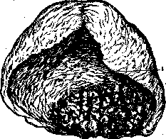
Figure 5. Placenta praevia isthmica primaria et cervicalis. The main part of the placenta was located in the isthmus (1) of the uterus. Continuous bleeding after manual separation of the placenta. Amputation of the uterus with recovery. (According to Pankow.)
can be palpated, following with a finger along the rim of the os of the uterus; 3) placenta praevia lateralis (lateral attachment) - the placenta is determined on one side of the os, and on the other side - the fetal membranes; 4) placenta praevia centralis, or totalis (central, complete attachment) - over the os of the uterus lies all around placental tissue (fig. 6); 5) placenta praevia cervicalis (cervical attachment) - an extremely rare, very dangerous anomaly. The clinical picture of placenta praeviae. The dominant symptom is bleeding. Extremely rarely cases are observed
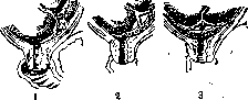
Figure 6. 1-placenta praevia marginalis; 2-placenta praevia lateralis; 3-placenta praevia centralis. (According to Stoekel.)
"bleeding without hemorrhage or with hemorrhage" only at a significantly more advanced stage of labor (Nurnberger). Usually bleeding appears already in the last months of pregnancy in connection with the impending stretching of the lower part of the uterus and, or more frequent, contractions of it; with the beginning of labor, i.e., with the beginning of the dilatation period, they clearly intensify. The cause of them is the premature separation of a normally low-lying placenta; with each uterine contraction there occurs a relative displacement of the two surfaces connected between them, one of which, namely the placenta, remains essentially almost immobile, while the other, i.e., the site of attachment of the placenta—the placental site—displaces during a contraction. Thus the contact that existed earlier between them, when the uterus had not yet begun to contract regularly, is disrupted. The result of this must be the gradual separation of the placenta from the underlying tissue, i.e., the placental villi must be torn away from the decidua basalis; in this case the intervillous blood spaces are opened up, and the blood in them, seeping between the separated part of the placenta and the uterine wall, gradually makes its way toward the exit from the birth canal. Any uterine bleeding in the second half of pregnancy or at the beginning of labor (before the fetal membranes have ruptured), unless there are some other obvious sources of it (for example, varicose dilatations of the vulva and vagina, cancer of the vaginal part of the uterus, etc.), should arouse suspicion regarding placenta praevia. Usually such bleeding at first does not have a threatening character; often appearing completely unexpectedly, sometimes at night, it soon ceases, probably due to thrombosis in the opened intervillous space. Much more rarely it is abundant from the very beginning. However, the most typical here are repeated bleedings, increasing more and more in connection with the increasing stretching of the lower part of the uterus; they cannot cease by themselves; on the contrary, they intensify as labor activity develops until the lower pole of the egg fixes the placenta at the uterine os, while the lower uterine segment stretches upward. After rupture of the fetal membranes the character of the bleeding usually, at least for some time, changes, i.e., the bleeding noticeably decreases, because from this moment on the fixation of the placenta ceases: it has the opportunity to follow the receding fetal membranes and again to press against the walls of the lower uterine segment. If after rupture of the fetal membranes, under the influence of good contractions, the presenting part (usually the firm head) descends downward, then it compresses the placenta against the uterine walls for quite a long time so strongly that bleeding ceases until the end of the expulsion period. But circumstances do not always develop so favorably; if the presenting part of the placenta is very large or there are not sufficiently strong contractions or the presenting part of the fetus cannot descend downward due to spatial discrepancy or abnormal position of the fetus, which disrupts the normal mechanism of engagement in the pelvis, then bleeding will continue even after rupture of the fetal membranes (due to the absent or at any rate insufficient compression of the separated part of the placenta). However, the danger of bleeding threatens the parturient also in the third stage. Despite the completed separation of more or less large parts of the placenta, this stage usually proceeds with great complications fraught with serious consequences; here incomplete separation of the placenta and retention of already separated parts of it are always possible; both often necessitate manual removal of the placenta. Meanwhile this operation, always responsible in terms of possible infection, is especially dangerous in this respect here. But even after the placenta has been expelled or after its manual removal, very strong bleeding of purely atonic character may immediately occur, since here usually there are neither those individual strong contractions of the uterus nor that persistent increase in its tone nor that good retraction which are absolutely necessary for the reliable closure of the vascular lumens at the placental site. Placenta praevia is always dangerous in terms of the immediately threatening severe anemia; while after other large blood losses during labor (1-17a) complete equilibrium is relatively quickly established, here blood regeneration proceeds very sluggishly; apparently the typical for placenta praevia repeated bleedings, summing up, quickly bring the organism to a threatening depletion of blood. In addition to this, placenta praevia is always associated with the danger of more or less severe puerperal diseases, since the abnormally low-lying placenta, with its large surface, with its large vessels, presents wide gates for infection. In contrast to normal conditions, when the placenta lies in the upper part of the uterus, which remains sterile both during labor and in the first days after it, placenta praevia is located close to that part of the birth canal which is always rich in microbes, and after effacement of the uterine os it almost directly borders the upper part of the vagina. Already with the first bleedings, often even before the beginning of labor, there begins here the unhindered invasion of microbes directly into the intervillous blood spaces. The possibility of infection is great also because cases of placenta praevia are usually associated with repeated internal examinations and various, often also repeated and prolonged operative manipulations, especially with (technically very difficult here) manual removal of the placenta. The third, though very rare, danger of placenta praevia—air embolism of the lungs due to the gaping venous sinuses at the site of attachment of the placenta and the possibility of air penetrating into them. This possibility must be especially taken into account during version, as well as during sudden sharp displacements of the parturient to the lateral position. The danger to the fetus in placenta praevia consists in the asphyxia threatening it due to more or less significant oxygen starvation in connection with the separation of the placenta. This threat, in itself very significant and serious, is intensified further because various operations, usually applied with the aim of stopping the bleeding and saving the mother from the threatening death, are ultimately based on strong compression of the bleeding surface of the placenta and the placental site. The diagnosis of placenta praevia is often possible already on the basis of anamnestic data alone, especially in those cases where all other possible and visible sources of bleeding can be completely excluded. Where bleeding from its very first appearance is very strong, one can sometimes suspect central placenta praevia. Sometimes the very first examination itself causes strong bleeding; therefore, when placenta praevia is suspected, it is always better to be prepared for the fact that immediately after examination the corresponding therapy for the diagnosis may be needed. One should in every way avoid frequent repeated examinations in the hope of necessarily palpating the placental tissue clearly, since any unnecessary manipulation can contribute both to bleeding and to infection, without giving anything particularly substantial in return. With low attachment of the placenta, where its lower edge cannot be reached with the examining finger, the diagnosis can sometimes be confirmed by the roughness of the fetal membranes. Concerning differential diagnosis between placenta praevia and premature separation of a normally situated placenta—see above. The prognosis in cases of placenta praevia is always very serious both for the mother and for the fetus; for both it represents a dangerous complication associated with the possibility of severe, often fatal consequences. If one considers the overall maternal mortality from large statistical summaries, i.e., regardless of the circumstances in which the births took place and the operative aids were applied, it averages 20%. The clinical material (although it usually consists of the most severe cases) does not present such a hopeless picture. For example, Pankov gives the following general summary: in 7,234 cases of placenta praevia, maternal mortality was in 504 cases, i.e., only 6.97%. More detailed information about the immediate causes of death is given only for 377 cases; it turns out that from bleedings caused by ruptures of the soft birth passages, 85 women died from this number (22.5%), from bleedings of purely atonic character—163 (43.2%), from sepsis—97 (25.7%), from various other causes—32 (8.5%). As for infant mortality, in the same material it appears as follows: in 3,489 conservatively treated cases of placenta praevia with a total of 3,511 newborns, 2,135 died (60.8%); in 1,989 also conservatively treated cases with a total of 1,887, but already viable newborns, 729 died, i.e., only 37%. As for the dependence of maternal mortality on the degree of placenta praevia, it is necessary to note significant and important practical fluctuations.
Thus, for example, according to Gichman's data, it is evident that with complete central placenta previa, the maternal mortality rate in clinics was on average 13%, while with partial placenta previa it was only 3%. Some authors present statistical data from which it is evident that the mortality rate was 0% (see below). Therapy for placenta previa. Placenta previa is the most typical and vivid example of those cases in obstetrics where a clear collision occurs between the interests of the mother and the interests of the fetus, where to save the mother from great danger it is necessary to consciously risk the life of the fetus. Such an action by the obstetrician, which goes against his basic and fundamentally correct striving to save both lives, unfortunately, is still quite often an inevitable, the only possible way out of a difficult situation, all the more so since the chances of obtaining a live, and above all viable, fetus here are generally relatively small, since, on the one hand, it is often premature, and on the other hand, it has already suffered significantly from oxygen starvation. Therefore, various attempts to accelerate labor (and even more so - forced labor) in the hope of obtaining a live fetus through a vaginal delivery operation (forceps, manual extraction of the fetus after a version has been performed, extraction by the breech in breech presentation), as being associated with the obvious danger of rupture of the extremely softened and exceptionally blood-filled tissues of the cervix, should here be considered fundamentally contraindicated. The obstetric aids and operations usually used for placenta previa are as follows: 1) amniotomy, 2) tamponade, 3) combined version according to Braxton-Hicks, 4) metreurisis, 5) cesarean section (abdominal). 1. Amniotomy. The beneficial, i.e., first and foremost hemostatic, effect of this method is based on the fact that usually after the rupture of the membranes, the presenting part (most often the head) descends, compresses the separating placenta and stops or at least temporarily reduces the bleeding. Such a simple, least dangerous in terms of infection, easily applicable under any conditions and often effective means should be considered first aid in these severe cases and therefore should be tried first, especially where it is difficult to apply other, more complex therapeutic methods. It is almost always effective, makes it possible to gain time and discuss further actions possible in the given situation. However, it cannot be considered absolutely reliable: sometimes it proves ineffective for the following reasons: 1) the contractions are not strong enough to quickly advance the presenting part; 2) this part is not voluminous and dense enough to firmly compress the placenta; 3) the placenta is located directly in the path of the forward movement of the presenting part and therefore hinders its engagement in the pelvic inlet. Therefore, amniotomy is most effective with marginal or lateral placenta previa, when the head is still above the inlet, and proper labor activity has already begun. The technical details of applying this aid in placenta previa are as follows: 1) the amniotic membranes should be grasped with some instrument (long forceps, bullet forceps, etc.) and manipulated downward; 2) under no circumstances should the membranes be ruptured by pressing on them with a finger, because simultaneously with the slightly upward-moving lower pole of the ovum, the placenta must inevitably shift upward and therefore separate even more, and as a result the bleeding will not only decrease but rather significantly increase. Döderlein in his report on the treatment of placenta previa at the International Congress in London (1913) presented a summary table of results from amniotomy obtained by various authors; the results of the table are as follows. Out of 309 cases, 306 mothers survived. Out of 260 fetuses, 192 survived, therefore the maternal mortality rate was 0.9%, and the infant mortality rate was about 25. From Pashkov's summary tables, it is evident that out of 4,607 conservatively treated cases of placenta previa, amniotomy was performed 546 times (11.8%). Out of 504 cases of this aid, there were 12 fatal outcomes (2.38%). In general, 35.6% of fetuses died, and in particular, 16.1% of viable ones. Such results must be considered quite favorable. It is interesting to compare them with the results of spontaneous labor in placenta previa (according to Pashkov's data). It turns out that out of 228 such cases, there were 7 fatal outcomes for mothers (3.1%), and 28.1% of viable fetuses died. This comparison would be more favorable if one takes into account the, presumably, unequal severity of individual cases, i.e., one must think that those cases of placenta previa that could be managed without any aid were relatively milder, while those where intervention was still required - even if only in the form of amniotomy - were already more severe. 2. Tamponade - undoubtedly the least reliable, and in terms of infection the most dangerous method. It is very often completely impossible to stop such severe bleeding, which usually occurs with placenta previa, by this means. The negative side of the method also lies in the fact that the gauze being pushed upward can easily, especially with significant cervical dilation, passing through it, further separate the placenta and thereby only intensify the bleeding. Another essentially important disadvantage of tamponade is the serious danger of infection. Nevertheless, tamponade sometimes turns out to be the only possible and unfortunately still inevitable means, serving as first (though risky) aid. This applies precisely to those cases where the surrounding situation does not make it possible to apply some other, more effective and less dangerous therapy, for example, primitive working conditions in remote areas where the midwife, and sometimes even the doctor, has to resort to such an imperfect method in order to transport the parturient to the nearest medical institution. However, even in such severe conditions, the doctor can still (especially if transportation is not feasible) find a way out by using methods such as amniotomy and combined version, which can still be relatively easily performed in any setting.

Figure 7. Combined version on the leg. Umbilical cord wrapped around the neck. (According to Stoeckel.)
one should in no way without need increase this tension with a weight and in general force it with the aim of accelerating labor; even with complete dilation of the cervix it is better if possible to wait for the spontaneous expulsion of the fetus. (The old classical saying by Schultze: 'Rupture the membranes, bring down a foot, but do not hasten with extraction.') From Dederlein's data it is seen that the combined version according to Braxton-Hicks in cases of placenta praevia (Fig. 7) gave the following results. Out of 1,434 cases of its application, 1,322 mothers remained alive, 112 died; 337 fetuses remained alive, 948 perished; consequently maternal mortality = 7.8%, and infant mortality = 73.7%. According to Pankov's summary tables, out of 5,083 cases of placenta praevia, version according to Braxton-Hicks was performed 2,298 times (45.2%). Out of 2,247 cases of this operation, 156 mothers died (6.94%). Out of 1,468 cases of this same operation, there were 1,113 fatal outcomes for fetuses in total, i.e. 75.7%, and out of 197 cases of viable fetuses, 128 died (65%).
4. Metreurynter. The metreurynter in cases of placenta praevia acts similarly to the fetal buttocks after combined version, but one can fully count on such an effect only when it is introduced intra-ovularly, i.e. into the cavity of the ovum (see Metreurynter). If the metreurynter is introduced extra-ovularly, i.e. with the membranes intact, then as it advances it may sometimes push only the lower pole of the membranes upward, bypassing the placenta, which will first remain unaffected, but later (when the balloon expands after being filled with fluid) it will be more or less compressed, and bleeding will nevertheless stop. However, when using this method, a serious complication is always possible, namely: the metreurynter, introduced between the uterine wall and the membranes, can easily, by pushing the lower pole of the latter upward, at the same time shift the presenting portion of the placenta, i.e. cause further detachment of it over a much larger area and as a final result not stop the bleeding but on the contrary intensify it. Proceeding from these considerations, the majority of obstetricians believe that intra-ovular metreurynter deserves decisive preference. One must also take into account a number of other negative factors inherent in the metreurynter, and therefore in practical activity one can be guided by the following principle: the metreurynter is appropriate only where version is still impossible, where the os is impassable for two fingers. It is precisely in these cases that one should first think of the metreurynter, for the introduction of which dilation of the os to at least one finger is sufficient (which is usually always ensured; in extreme cases, insufficient dilation can be quickly increased with the help of metal dilators). It should be remembered that in cases of placenta praevia, metreurynter gives a better prognosis for the fetus compared to combined version. According to Dederlein's summary tables, metreurynter gave 726 favorable outcomes and 51 fatal outcomes for mothers out of 777 cases, and for fetuses - 386 favorable and 322 fatal. Total maternal mortality = 6.5%, and infant mortality = 45.4% (i.e. although greater, but still much less than after version according to Braxton-Hicks). According to Pankov's summary tables, out of 1,332 cases of application of intra-ovular metreurynter in cases of placenta praevia, there were 68 (5.1%) fatal outcomes for mothers. Out of 1,148 cases of the same therapy, fetal mortality in general was 564 (49.1%). Out of 471 cases of intra-ovular metreurynter where fetuses were already viable, there were 214 (45.4%) fatal outcomes. Extra-ovular metreurynter gave about 10% mortality in mothers and again about 45% in fetuses. Both combined version and metreurynter, besides their direct hemostatic effect, have one more very significant positive side, namely both these methods to a greater or lesser degree (especially when hanging a weight) always enhance labor activity. The short but nevertheless very strong mechanical irritation associated with the performance of these manipulations itself, as well as the more or less prolonged mechanical irritation obtained as a result of their application (pressure of the fetal buttocks or rubber balloon as a foreign body on nerve endings and the uterine musculature itself), almost always proves to be a strong stimulant of uterine contractions. Such an enhancement of the contractile, resp. labor activity of the uterus is as it were a side (but at the same time also useful) result of applying these operative-therapeutic methods, since the main goal here is first of all to achieve a hemostatic effect. One must and can be quite satisfied with this acceleration of the labor process, since it occurs depending on mechanical irritations inextricably linked with these methods themselves. Any other types of operative assistance provided through the natural routes (whether manual or instrumental), if they are specifically directed at accelerating labor in cases of placenta praevia, are rather already 'a deviation from the art of obstetrics'; such actions, especially little justified and at the same time forced, performed through the vaginal route, are completely inappropriate here due to the danger of traumatizing the cervix and the possible threatening bleeding associated with this. Both combined version and metreurynter in most cases reliably stop bleeding for the entire duration of the expulsion period. If applied cautiously, they almost always prove effective both in the sense of hemostasis and in the sense of prevention of cervical ruptures. (In spontaneous labor there is no special reason to fear these ruptures.) Interesting is Gichman's table, drawing a parallel between the results of combined version and metreurynter: Fatal cases Combined version - 3,035 Metreurynter - 1,095 Total number of fatal cases ..... 191 (6.3%) 68 (6.2%) Death from bleeding ..... 39 (3.5%) Death from sepsis .....
14 (1.3%) From this table it is evident that both methods, in terms of outcomes for mothers, are almost completely equivalent. In the treatment of placenta praevia, skillful management of the third stage of labor is very important. From the very beginning and until the end of this stage (and even immediately after the completion of labor), the menacing specter of bleeding usually reappears; it can even become stronger than before. The reasons for this are as follows: 1) in the third stage of labor, there can be no question of any tamponade effect or any compression of the placenta, with the result that the placenta, since it had already separated earlier, again departs unimpeded from the uterine walls to which it had been temporarily attached in one way or another; 2) the anatomical and physiological prerequisites necessary for the rapid and complete separation of the entire placenta from the underlying tissues are not present here: in contrast to normal conditions, in these cases it does not lie entirely in the area of action of the most active, i.e., upper part of the uterus. In placenta praevia (which is usually isthmic placenta praevia), only a small part of the placenta is connected with the walls of the hollow muscle capable of powerful contractions, while a much larger part of it lies in the area of the very weak, inert—both in terms of contractile work and in terms of retractile ability—lower part of the uterus, namely the isthmus, which is capable rather of passive stretching than of active contraction; 3) the complete separation of the entire placenta, which is necessary at this time, occurs slowly and unevenly, which is all the more noticeable the larger part of it lies in the lower part of the uterus, the more it is stretched, and the more its (in itself very slight) contractile ability is weakened. Uneven separation of the placenta is always accompanied by severe bleeding. Therefore, in the third stage of labor in cases of placenta praevia, a very watchful obstetrical eye and the ability to quickly orient oneself are always required. For a physician who does not yet have sufficient experience in these critical cases, it is useful, as far as possible, to adhere to a known therapeutic general plan that includes a series of different measures, so that, starting with the simplest manipulations, one can proceed as necessary to more serious actions—up to the inevitable radical surgical intervention in very severe cases (see Labor, third stage and its pathology). (Incidentally, it should be borne in mind that severe bleeding in the third stage of labor may be caused by partial atony of only the isthmus while the body of the uterus has contracted well.) Even spontaneous, and certainly operative deliveries in cases of placenta praevia always cause certain concerns; it is almost impossible to avoid any risk with full guarantee in this matter. But since a physician, especially one working in unfavorable conditions, usually has no other way out of this difficult situation, it is understandable that he often has to be content with the above-described part of expectant and part purely symptomatic therapy in cases of placenta praevia. 5. Cesarean section in placenta praevia. The entire situation is quite different in cases where the pregnant woman or parturient enters the clinic or generally a hospital setting at the very beginning of labor activity. Under these favorable conditions, it is always possible to perform the operation of abdominal cesarean section with a favorable outcome not only for the mother but often also for the fetus, if it is more or less full-term and has not yet suffered from oxygen starvation. [Vaginal cesarean section in placenta praevia is now almost entirely abandoned. This operation was recommended by Dederlein; according to statistical data (Pankov), maternal mortality with it is 11.3%—Dederlein himself observed in 125 cases of anterior vaginal section 9.6% mortality, of which—7.6% from blood loss!] Krenig and Sellheim were the first to decisively and persistently begin to advocate the idea of applying abdominal cesarean section in placenta praevia, believing that only in this way, by immediate delivery at the very beginning of labor activity, can the stretching of the lower uterine segment (which is otherwise completely irreparable and dangerous in terms of bleeding) be avoided. In our country, G. E. Reinhart was the first to apply this operation a long time ago in cases of placenta praevia. Statistical data obtained in recent years from a large number of various clinics, maternity homes, and hospitals eloquently speak in favor of cesarean section in placenta praevia and testify to very good (compared to former times) results of this operation for both mother and fetus. For example, Dederlein in his summary table, compiled from data received from reports of many authors, gives the following cumulative result: out of 146 cases of abdominal cesarean section in placenta praevia (up to 1913), 133 mothers remained alive and 13 died; out of 141 newborns (for not all of them there is information in individual reports), 98 remained alive and 43 died (these figures are cumulative, without indication of the degree of maturity). Thus, maternal mortality up to 1913 was 8.9%, and infant mortality was 30%. According to Gichman, who compared literary data (up to 1921) on cesarean section in placenta praevia, the total number of cases of this operation was 191, with fatal outcomes in mothers only 7, i.e., only 3.6%. Jaschke, who presents data relating to a later time, gives for 125 cases of this operation in placenta praevia, collected from German literature, maternal mortality in even smaller amounts, namely—2.4%, and for 262 cases collected by him from foreign literature, it is 4.0%. Pankov presented at a later time the following table: Authors Total number of cases Mortality (in parentheses-%); mothers—total infant viable fetuses Ganssle-Vogt Panitow Walthard Stoeckel Schulte 58 52 31 25 6(8.4) 2(3.4) 2(3.8) 1(3.3) 0(0) 6(6.4) 6(10.0) 18(34.6) 14(14) 1(4) ? 4(7.5) 4(10.5) ? Total 11(4.3) 45(17.4) 15(6.4) Of course, cesarean section is indicated by no means in every case of placenta praevia. The indications for it must be carefully weighed. The main indication is the presence of severe bleeding (regardless of the degree of presentation) with insufficient dilation of the cervix (or especially with its absence in primiparas) and with signs of acute anemia. When there are indications, the duration of pregnancy must also be taken into account: the fetus must be alive, full-term, or at least viable. In equal measure, certain contraindications must also be taken into account. For example, the presence of signs of obvious infection or even strong suspicion of it contraindicates the use of cesarean section. Cesarean section is also contraindicated when there has already been significant stretching of the isthmus, which can be assumed when there is significant dilation of the os; only in cases of very severe bleeding, especially with complete presentation of the placenta, when it completely fills from all sides the already widely dilated cervical os, can one decide on this operation in order to avoid greater danger, i.e., further blood loss and ruptures during delivery by natural routes. Abdominal operation is hardly appropriate with a definitely non-viable or dead fetus: metritis, combined version, if necessary with subsequent perforation of the head—here is a much more correct way out of the situation. However, if from the very beginning of labor or generally in the presence of only very weak labor activity bleeding immediately takes on an obviously threatening character (sometimes even during pregnancy), generally when the cervix is not yet fully effaced and the cervical os is almost completely closed or only very slightly open, one has to decide on the operation of cesarean section without thinking about the viability of the fetus, in order to save the mother from inevitable death at times. The interests of the mother may also urgently require cesarean section in the presence of a full-term fetus, even if dead, if under the condition of the woman's already severe blood loss, one can reasonably fear that a sad outcome is very possible during delivery by natural routes due to inevitable new blood loss, while abdominal operation is after all a more conservative method of delivery in such dangerous cases. A strictly individualized approach to individual cases is necessary.
But in any case, purely surgical treatment in placenta praevia, namely abdominal cesarean section, is as a rule the only effective and reliable means for simultaneously saving both lives—that of the mother and that of the fetus—and thereby fulfilling the main task of all obstetrical therapy. [Classical cesarean section (in terms of the speed of its performance) is at least in threatening severe cases the most suitable method here.] Of course, for a further (even more significant than that already achieved) reduction in maternal and child mortality, it is necessary that every case of placenta praevia, recognized in a timely manner, should be conducted in the setting of a maternity ward or hospital department with the provision of sufficiently qualified surgical assistance, rendered on the principle of emergency surgery in unfortunate catastrophic cases.
Related articles
Mentioned in
Cite this page
“Placenta.” Soviet Medical Encyclopedia. English translation of Bolshaya Meditsinskaya Entsiklopediya, 1st ed. (Moscow, 1928–1936), ed. N. A. Semashko. https://sovietmedicalencyclopedia.pages.dev/article/placenta/