Pregnancy
Historical document, translated for reference. It reflects medical knowledge of the 1920s–30s and is not medical advice.
Summary
This article from the 1928-1936 Soviet Medical Encyclopedia covers physiological changes during pregnancy in humans and animals, embryonic development, implantation processes, and species-specific characteristics of reproduction.
Encyclopedia article (1928–1936)
212 Changes in organs and systems during P. ...
233 Diseases of the endocrine glands . .
257 Pregnancy in animals. Pregnancy is the physiological state of the female animal during the development of the embryo in her uterus. In invertebrates, fertilized eggs often develop without establishing a closer connection with the uterine wall; however, even in invertebrates, adaptations for nourishing the embryo with maternal juices appear—either through the formation of a closer connection with the uterine wall (scorpions, peripatus, etc.), or through the secretion of nutrients into the uterine cavity. Among vertebrates, embryonic nourishment from the mother is already found in cartilaginous fish—rays and sharks. In some rays (Trygon Bleekeri), the uterine walls form papillae that secrete a thick mucus containing protein; this mucus is absorbed by the embryo through the so-called spiracle (a modification of the first gill slit); in another species (Trygon Walge), these papillae form long threads (trophonemes) that secrete nutritional material directly into the spiracle. In sharks (Mustelus laevis), a similar nourishment system to that of mammals is established: the yolk sac of the embryo forms villi, which, penetrating into the uterine wall, terminate in blood lacunae, from which the necessary nutritional material is obtained.—In mammals, fertilization of the egg and the necessary copulation occur during periods called estrus, analogous to human menstruation. These periods, associated with egg maturation, are accompanied by profound changes in the uterine walls and at the same time in the animal's behavior.--The number of maturing (and correspondently fertilizable) eggs is characteristic of each species, and although not all matured eggs are fertilized or develop in the uterus, the number of developing embryos can generally be considered constant and fluctuates within certain limits (rabbit 4-12, hamster 6-18, opossum 12-16, mole 3-5, pig 6-14, goat 2, bats 1-2, dogs 4-16, cow, horse, whale—one each). The number of pregnancies per year may vary, depending on the number of estrus periods. However, the number of offspring born does not always correspond to the number of fertilized eggs; in some cases, for example, in armadillos, the following process is normal: the fertilized egg divides during development into several parts, from which several embryos are formed (a phenomenon analogous to human identical twins). This method of embryonic fragmentation into several embryos is also found among invertebrates (bryozoans, Hymenoptera insects).--True P., i.e., the connection of the embryo with the uterine wall, does not occur immediately after copulation or fertilization. The time interval between copulation and fertilization varies in different animals: in bats it stretches over an entire winter (copulation in autumn, P. in spring), but most often it equals several days.--The fertilized egg reaches the uterus mostly within 5-10 days (white mouse 5-6 days, guinea pig 7 days, dog, human 8-10 days), and quickly implants into the uterine wall (in the guinea pig within 8 hours). In many animals, it remains in the uterine cavity (marsupials, pigs, horses, wolves, carnivores, camels, rabbits), in hedgehogs and mice—in the uterine groove, while in the guinea pig, pocket gophers (Geomys), it embeds into the uterine wall itself, which here becomes overgrown with the embryo (the so-called interstitial implantation). With implantation, profound changes begin both in the uterus and in the embryo itself. The uterus greatly increases in volume and weight (in the horse—from 500 g to 7,000 g,
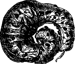
Amniotic sac of a sheep (according to O. Schultze). Greatly reduced.
in sheep-from 60 g to 750 g), and in the embryo, embryonic membranes and the so-called child's place, placenta (see.), are formed, through which the embryo is nourished. The form of the placenta (different in different orders of mammals) and the method of fusion with the uterine wall determines the greater or lesser ease of the act of birth.-With the onset of P. in female mammals, sexual desire disappears, and in some mammals the vagina after copulation is sealed by a special, so-called vaginal plug-a hardened secretion of the glands of the vaginal wall. The significance of the vaginal plug is not entirely clear, but in any case this plug prevents the outflow of the penetrated semen and makes a second copulation impossible. The method of embryo nourishment and the duration of pregnancy vary greatly. The shortest P. is found in marsupials (8-11 days) and is explained by the fact that they do not yet have a child's place, and the completely smooth chorion closely adheres to the uterus, the wall of which is greatly thickened and secretes together with the blood a large amount of protein mucus; the liquid part of these secretions is directly absorbed by the wall of the chorion, and the solid particles are assimilated by phagocytosis; in higher (placental) mammals, the shortest duration of P. is found in mice (21 days), the longest-in elephants (22 months); in general, the duration of P. varies within the following limits: mouse-3 weeks, rabbit-30 days, hare-4 weeks, rat-3 weeks, guinea pig-62 days, marmot-5 weeks, hedgehog-7 weeks, cat, marten-7-8 weeks, fox, lynx-9 weeks, wolf-9 weeks, badger-10 weeks, dog-9 weeks, lion-15-16 weeks, tiger-22 weeks, puma-15 weeks, musk ox-9 months, yak-9 months, bison-9 months, dugong-12 months, macaques-0 months, pigs, beavers-16-17 weeks, sheep-20-21 weeks, goat, roe deer-21-22 weeks, bears and monkeys-6-7 months, deer and horned cattle-30-40 weeks, human-40 weeks, horse-48 weeks, deer, zebra-51 weeks, camel-11-13 months, giraffe-14 months, rhinoceros-72 weeks, dolphin-10 months, llama and roe deer-10-11 months. The duration of pregnancy depends on many factors: the degree of development of the embryo being born, the number of embryos developing simultaneously, the biological conditions of life of the female, etc. According to Heinroth, P. of larger animals is longer than that of small ones; thus, in the small opossum-12 days, in the giant kangaroo-39. According to observations of livestock breeders, the duration of P. even in the same species of animals can vary depending on: a) the sex of the embryo (male offspring is carried somewhat longer than female), b) the season (P. of horses covered in early autumn is 4-5 weeks shorter than those covered in late autumn), c) the breed (so-called "early-maturing" ones carry less than ordinary ones), d) the number of embryos (twins are born sooner). The connection of the embryo with the uterine wall in mammals and nourishment at the mother's expense occur quite differently: either by assimilating various products secreted by the uterine glands, so-called uterine milk, or by assimilating nutritional material carried by the blood vessels of the uterus; accordingly, mammals are divided into two large groups-with non-shedding uterine mucosa (indeciduata) and with shedding (deciduata). The first group includes pigs, horses, elephants, camels, ruminants, whales, sirens, toothless (edentata) and some semi-apes. In these forms, the walls of the fetal sac connect with the uterine wall only by means of small papillae, while in the uterine wall itself a large number of glands develop. Nourishment occurs by osmotic and phagocytic means. The location of these papillae can be uniform over the entire surface (so-called diffuse placenta in pigs, horses, donkeys, semi-apes), or the papillae merge into special groups, so-called cotyledons (from 5 in roe deer to 100 in sheep). Nourishment here occurs mainly by absorbing disintegrating maternal cells and so-called uterine milk, which in sheep is rich in protein, salts, fat, disintegrating leukocytes, and protein crystals (Bonne). In these forms, maternal blood is directly processed by the chorionic wall. During birth, the exit of the fetus is associated with only a very slight separation of a layer of the uterus, from which the name indeciduata originated.-In the second group, characterized by the shedding membrane, the connection of the fetus with the uterine wall is very close, and nourishment by uterine milk takes a secondary place; nourishment occurs mainly at the expense of the mother's blood. In this group, only a certain part of the fetal sac becomes the placenta, and a belt-shaped placenta (pi. zonaria-in carnivores, seals) and a disk-shaped one (pi. discoidea-in insectivores, bats, rodents, monkeys, and humans) are distinguished. In the first case (pi. zonaria), the zone of papillae is located in a band, encompassing the middle part of the elongated fetal sac; in the second case (pi. discoidea), the papillae are located only in a certain area, and at the point of attachment the fetal sac papillae are very elongated and branched at the end, while in the uterine wall itself, destruction of cells, growth of connective tissue, and formation of large lacunae occur, into which the mother's arterial blood enters. The placental papillae are immersed in these lacunae, and here the embryo receives oxygen brought by the mother's blood and nutritional material-partly in liquid form, partly in the form of solid particles assimilated by phagocytosis. The duration of labor and contractions varies greatly and, apparently, depends both on the structure of the placenta and its greater or lesser fusion with the uterus, and on the number of embryos (in horses-15 min., in pigs-from 2 to 6 hours, in dogs-6-8 hours, depending on the number of puppies, in cows-11/»-5 h.).-The degree of development of the newborn young is also in a certain correspondence with the ecological conditions of life of the female. In ungulates and some rodents, the young are born already sighted, hearing, and with ready milk teeth, covered with a true hair coat (horses, cows, sheep, goats); some can nibble grass while simultaneously feeding on milk. In animals whose mothers raise the young in protected places or nests (rabbits, rats, mice, beavers, cats, lions, tigers, dogs, and wolves), they are born blind (in dogs puppies become sighted on the 9th day), toothless (first teeth-on the 5th week); the P. period of these forms is somewhat shorter than the first (rodents and insectivores-3 weeks, carnivores-2-5 months, pigs-4-5 months). In some species, the young are born in fetal membranes, which are then torn by the mother (dog); in humans and domestic animals these cases occur as exceptions (birth in a "shirt"). The umbilical cord tears off from the placenta by itself and gradually dries up (ungulates), sometimes it remains, and in these cases the animal drags the placenta along the ground for several days (e.g., seals). Carnivores, rodents, and monkeys bite it off themselves. Carnivores and some herbivores (goats, mice) eat their own placenta.
H. Bogoyavlensky.
Normal pregnancy. Normal pregnancy is pregnancy that proceeds in a healthy uterus of a healthy woman, causing no pathological changes in the organism, and ending in timely normal birth with a living, normally developed child. The beginning of pregnancy is considered to be the first day of the last menstruation. This is not entirely accurate, since the actual beginning of pregnancy is biologically timed to the moment of conception, i.e., the penetration of the sperm into the egg cell (impraegnatio), but this moment is not known with precision. The end of pregnancy is timed to the end, not the beginning of labor, since it is not always easy to accurately detect the onset of labor, because pregnancy can gradually and completely imperceptibly transition into the act of labor. On the contrary, the end of labor is precisely determined by the time of the expulsion of the placenta. The duration of normal pregnancy is taken as 280 days = 40 weeks = 10 lunar months. However, it can fluctuate within rather wide limits, not only in humans but also in animals. In humans, within limits of 40 days in either direction, i.e., it can be considered from 240 to 320 days. Knowledge of these facts is very important, as it forces the physician, especially in insurance and forensic medical examination, to determine the duration of pregnancy and labor with great caution. The site of conception should be in the nearest vicinity of the ovary, more precisely in the infundibulum of the fallopian tube, since conception occurs soon after the egg cell leaves the follicle. The possibility of conception in the abdominal cavity is proven by very rare cases of ovarian and primary abdominal ectopic pregnancy; however, here, apparently, favorable conditions for the vital activity of spermatozoa are absent: they quickly undergo spermolysis and are phagocytized by white blood cells. After conception, the egg moves along the fallopian tube for 7-10 days and only in the second week enters the uterine cavity, where it implants into its mucous membrane (implantation). Even during the movement of the egg along the fallopian tube, the uterine mucous membrane undergoes the so-called decidual metamorphosis. It proliferates, swells, a pronounced blood filling (hyperemia) and edematous impregnation of the stroma are noted in it, and its cells take on a special structure, transforming into decidual cells, very rich in glycogen and in appearance resembling squamous epithelial cells (epithelioid). The thickness of the mucous membrane in this case reaches up to 1 cm, while in a state of rest it is 1-2 mm. All these changes, however, do not capture the entire thickness of the mucous membrane, but only part of it, facing the uterine cavity, i.e., the functional layer; the part of the mucous membrane adjacent to the muscular wall of the uterus—the basal layer—changes little. For this reason, the shedding membrane divides into two layers, distinguishable under a microscope by their morphological structure: in the functional layer, the glands are compressed and then gradually completely disappear, so it appears to consist entirely of decidual cells; this layer, in addition, loses the covering epithelium and is called the compact layer. On the contrary, in the basal layer, morphologically and functionally unchanged, the lumens of the glands not only do not disappear but are strongly stretched by the contents, which now has no outlet into the uterine cavity. As a result, it acquires a cellular spongy structure, and therefore is called spongy or spongiotic (spongia-sponge). Thanks to this decidual metamorphosis, when the egg enters the uterine cavity, it finds its mucous membrane prepared for the process of implantation. Development of the ovum. While moving along the tube, the egg undergoes a series of cleavages and thus, from a single-celled organism, transforms into a multicellular one. In size, it does not change and still fits in its amorphous shell—the zona pellucida. Its size, therefore, is equal to that of a mature egg cell (0.2-0.3 mm). In the process of cleavage, the egg passes through the morula stage and already transforms into an egg vesicle (blastula, blastocyst) with a clear differentiation of cells into those located on the periphery and those occupying the interior of the vesicle. The former—'trophoblast'—form a new, already not amorphous but cellular, egg shell and take upon themselves the task of nourishing the entire egg; the latter cells give rise to the embryo itself—the 'embryonic pole of the egg'. Nutrition of the egg during this period occurs at the expense of the reserves of yolk laid down in the protoplasm of the egg cell itself. It is also possible that there is an exchange of substances not only within the egg itself but also between the egg and the maternal organism. It can occur by means of cells carried out by the egg cell from the Graafian follicle during ovulation and surrounding it in the form of the so-called corona radiata, as well as directly through the zona pellucida. In the process of such exchange, on the one hand, the egg receives various nutrients from the fluid filling the lumen of the tube, on the other hand—it secretes into this fluid, i.e., into the maternal organism, products of its own metabolism. Having entered the uterine cavity, the egg (larva) as if hatches from its shell and begins to embed into the thickness of the compact layer of the shedding membrane. This embedding is possible because the cells of the trophoblast possess a clearly expressed ability to dissolve the covering epithelium and the subepithelial tissue of the uterine mucous membrane. From this moment, the egg begins to feed at the expense of the lysed maternal tissue, i.e., already definitely at the expense of the mother. The egg in this early stage of its development already manifests its hormonal function and acts either through the corpus luteum or directly on the entire maternal organism and, especially, on the uterine mucous membrane, causing and enhancing its decidual metamorphosis. Thus, on the one hand, the life processes in the egg in the first time after conception are maintained by the trophic influence of the hormones of the developing corpus luteum, and on the other hand, the egg itself (and even the unfertilized but maturing egg cell) is that self-sufficient organism which subordinates the maternal organism to its interests and causes in it the changes described above. From this it follows that pregnancy must be viewed as a process of extremely great biological importance for the well-being of the female organism and that it by no means can be considered as a manifestation of the parasitism of the egg in the maternal organism (and such a view was still quite recently quite widespread among physicians and naturalists).—Of the series of human eggs described to the present time, the youngest is recognized as the egg recently described by Mollendorf. The age of this egg is determined as 13 days from the moment of conception. Thus, the first two weeks of development of the human egg (i.e., the entire tubal period and the first days of the uterine period) are still completely unknown to us, and we can judge about them only by analogy with the development of eggs in other animals, which must be done with the greatest caution, since in the development of eggs of different species very large differences are possible. By the end of the third week, the egg has a diameter of about 20 mm. In such an egg, already having completed implantation, the trophoblast has undergone further differentiation and split into two layers: the outer layer loses its cellular partitions and appears as a continuous protoplasmic mass with a large number of intensely staining nuclei (plasma trophoblast, or syncytium); the inner layer retains its cellular structure and consists of well-expressed cylindrical cells (cellular trophoblast, or Langhans' layer). The trophoblast not only differentiates into two layers but also actively proliferates along its periphery, forming a large number of root-like outgrowths in the form of lobes and thinner threads—the 'villi of the egg'. Initially, they consist of only the syncytium and have the ability to dissolve not only the basic tissue of the shedding membrane but also the walls of the blood vessels passing here, mainly capillaries. The erosion of the vascular walls leads to the fact that maternal blood flows into the space between the individual villi, filling the entire egg bed (egg capsule) and forming an intervillous space within it. Thus, the egg is immersed in maternal blood, from which it begins to absorb the necessary nutrients for its further development and at the same time secretes into it products of its own metabolism. Now the egg even more comes under the influence of the maternal organism, as well as itself more strongly affects it by hormonal products; it becomes as if a new gland of internal secretion in the maternal organism. Such a deep penetration of the egg into the very essence of the maternal organism is a special and very important feature of humans and some anthropoid apes; in other animals with intrauterine development of the egg—the latter enters only into more or less intimate contact with the changed uterine mucous membrane.—The interior of the three-week-old egg—its embryonic pole—also undergoes by this time considerable differentiation: in it two vesicles are formed, surrounded by mesoblastic tissue.
One of them - the ectoblastic - gives rise to the amnion (water sac) and the ectoderm of the embryo; the other - entoblastic: part of it forms the primary gut of the embryo (entoderm), while part goes to form the yolk sac. The mesoblastic tissue surrounding both sacs partially liquefies, forming the 'primary' cavity of the egg (exocoelom), and partially retains its reticular-fibrous structure, forming magma reticulare. This same mesoblastic tissue begins to compact on the surface of the yolk sac and under the layer of trophoblast. Distinct layers are formed, which play a very important role in the development of the egg,
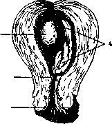
Figure 1. Uterus with egg at the end of the 1st month of pregnancy (l/i natural size): 1-egg covered by decidua capsularis; 2-orificium int.; 3-orificium ext.; 4-decidua parietalis (according to Bumm).
because in them the formation of blood and the circulatory system of the embryo first begins. That layer of mesoblastic tissue which is adjacent to the trophoblast from the inside soon begins to grow into its thickness and penetrates into its villi, forming their main connective tissue core. Thus, the structure of the villi is complicated; they consist of a connective tissue base covered by two layers of trophoblast - cellular and syncytial. The egg shell receives a new name - chorion (chorion), or villous membrane. In the connective tissue base of the villi, rudiments of the circulatory system begin to appear, which gradually merge into a common circulatory network with vessels forming in the wall of the yolk sac and in the mesoblastic tissue of the embryonic area itself. With the formation of the circulatory system and blood, the intra-egg circulation between the embryo and the yolk sac is established. This is the so-called yolk-sac circulatory system. It has a transient significance in humans, as the yolk sac quickly decreases in size, is almost completely resorbed, and ceases to nourish the embryonic area of the egg. Its functions are fully taken over by the circulatory system connecting the embryonic area of the egg with the chorion (chorion-umbilical circulatory system). With its formation, nutrients from the maternal blood pass through the cover
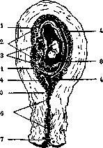
Figure 2. Uterus with egg at the end of the 2nd month of pregnancy (sagittal section, '/, natural size): 1-transition of decid. parietalis into decid. capsularis; 2-decidua basalis; 3-chorion frondosum; 4-decidua parietalis; 6-orificium Int.; c-cervical canal; 7 - orificium ext.; 8 - cavity of the fetal egg (according to Bumm).
of the villi into its base and from there into the fetal circulatory system. Subsequently, the circulatory system of the chorion concentrates
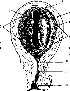
Figure 3. Uterus with egg at the end of the 3rd month of pregnancy (sagittal section, Va natural size): 1-chorion frondosum; 2-decidua basalis; 3-dilated arteries of decid. basalis; 4-site of origin of decid. capsularis; 5-uterine cavity; 6-cavity of the fetal egg; 7-decidua parietalis; 8-dec. capsularis and chorion laeve; 9-lower part of the uterine cavity; 10-orificium int.; 11-cervical canal; 12-orificium ext. (according to Bumm).
almost entirely in a separate area directly adjacent to the uterine wall (see fig. 1-3). Here the chorionic villi develop luxuriantly and give off strong branches, while on the rest of the surface of the egg they die off. The chorion thus breaks down into two parts, of which the first receives the name chorion frondosum (branched chorion), and the second chorion laeve (bare chorion). The first villi come into especially close contact with the maternal decidua, and here gradually develops the placenta—an organ in which all subsequent exchange of substances between mother and fetus is concentrated. In it, according to the latest data, also occurs that hormonal activity of the egg, which was mentioned above. The external form of the embryo itself initially resembles a simple cellular plate, in shape resembling an English biscuit. It is part of the wall of the ectoblastic vesicle. Subsequently, on its dorsal surface appears the neural, or primary, groove. Later it closes into a tube and gives rise to the nervous system. The embryonic plate itself rolls into the abdominal side, forming the embryonic body, at the same time drawing into itself the adjacent part of the entoblastic vesicle (the primary intestine). By the end of the second month and, even more so, in the third, the embryo is formed in its main parts; in the following 7 months of intrauterine development, growth occurs, mainly. The intensity of this growth can be judged by the length and weight of the embryo in individual months of pregnancy, as seen in the following tables: Fetal length by months of pregnancy: 1x1= 1 cm 2x2= 4 i> 3x3= 9 » 4x4 = 16 >v 5 X 5 = 25" » 6 X 5 = 30 » 7x5 = 35 » 8x5 = 40 9 X 5 = 45 )> 10 X 5 = 50 » Approximate weight of individual elements of the egg by months of pregnancy in grams (according to Zangemeister). Duration of pregnancy its o I V i o V i o V i o I a i i i I Metabolism in normal pregnancy is established in the direction of a plus. The pregnant woman continuously gains weight, with the greatest increase occurring in the second half of pregnancy and, especially, in its last weeks, when it reaches 50-60 g per day, i.e. about 400 a per week. The total gain for the entire pregnancy can reach 10 kg. If from this figure we subtract the weight of the fertilized egg—5,000 g—then it turns out that the pregnant woman's own weight increases by 5 kg. Zangemeister showed that the weight gain stops several days before childbirth; in the last 3 days a decrease is even observed, reaching 1 kg. This phenomenon (Gewichtssturz) was attempted to be used as a symptom indicating the approach of labor. However, it is far from constant, difficult to detect, and this symptom has not found application. The continuous increase in weight during pregnancy is mainly due to hypertrophy of individual organs, deposition of fat, and retention of large amounts of water in the body. Protein (nitrogenous) metabolism proceeds in the direction of protein accumulation. According to observations by Hoffstrom, the pregnant woman assimilated 310 g of nitrogen in 167 days, of which 101 g went to the fetus and 209 g to the maternal organism (quoted from Kehrer). Such increased protein deposition is caused by both increased assimilation and more economical expenditure. During intense protein exchange between mother and fetus, whole molecules as well as large fragments of fetal protein enter the mother's blood. Since the fetus's protein is foreign to the mother, the maternal organism responds to this phenomenon by the formation of 'protective' enzymes, with the help of which this protein should be neutralized. Abderhalden expressed this hypothesis and developed a special reaction (see Abderhalden's reaction), with the help of which one can determine the presence of these specific enzymes in a woman's blood and thereby diagnose pregnancy in her. This reaction and its modifications, due to their complexity, proved impractical for clinical use. Fat metabolism also increases, affecting both neutral fats and lipoids and cholesterol. Besides fat deposition in certain areas of the body (mammary glands, buttocks, upper parts of thighs), there is a characteristic increased content of fat in the blood, which explains the turbidity of the blood serum of pregnant women, resembling a fat emulsion. This also explains the fact that pregnant women are very susceptible to chloroform anesthesia and tolerate it easily. Carbohydrate metabolism in pregnant women changes in the sense that they show a decreased ability to assimilate carbohydrates absorbed from food, which is why sugar appears in their urine much more easily than in the non-pregnant state. This is the so-called physiological glycosuria of pregnancy. Nevertheless, the sugar content in the urine of pregnant women fluctuates within normal limits even with an increased intake of carbohydrates in food. Recently, attempts have been made to use this feature of carbohydrate metabolism in pregnant women for the recognition of early pregnancy. Inorganic metabolism in pregnancy has been little studied to date. The fertilized egg, to cover its need for mineral salts, absorbs them from the maternal organism, but how this affects the metabolism of these substances in the maternal organism is unknown. The egg's need for these substances (except for calcium, the amount of which in the mature fetus exceeds 20 g) is small and can be easily covered by their excess in the maternal organism. The deficiency in calcium is covered by reserves in the maternal organism, mainly from the blood and bone system. The nature of water metabolism in pregnancy has been extremely inadequately clarified. Pregnant women intensively retain water both in the blood and in the tissues, which explains their tendency to edema. This is the so-called physiological hydremia of pregnancy, which often stands on the border of pathology. Physiological edema at the end of pregnancy of the pelvic organs, cervix, vagina, as well as the pelvic and perineal muscles has a favorable effect on the course of labor, as it increases the elasticity and distensibility of those tissues that form the soft birth canal, which helps preserve their integrity during labor. Basal metabolism in pregnant women increases. Already from the 3rd month, an increase in oxygen assimilation can be noted, which increases with the progress of pregnancy and by its end rises by 25% compared to normal. Changes in organs and systems during pregnancyIn accordance with the above-mentioned changes in metabolism, all organs of the woman undergo certain changes in connection with pregnancy, both in morphological and functional aspects. The reproductive system, and in particular the uterus, undergoes the greatest changes during pregnancy. The weight of the uterus increases from 50 g in the non-pregnant state to 1,000 g by the end of pregnancy. This increase is mainly due to hypertrophy of the smooth muscle fibers, the length of which increases tenfold (reaching 500 ц), and the width quadruples. Along with hypertrophy of the muscular elements, there is also their hyperplasia. The thickness of the uterine wall reaches its greatest (2.5-3 cm) in the 4th-5th month of pregnancy. Until the 3rd month of pregnancy, the uterus also enlarges in ectopic pregnancy, i.e., its hypertrophy depends on endocrine influences, and not on the stretching of the uterus by the growing egg, which at this time does not yet fill its entire cavity. From the 4th-5th month, the egg increases more rapidly than the uterine cavity; therefore, from this time on, there is not thickening, but thinning of the uterine walls; by the end of pregnancy, their thickness decreases to 0.5-1 cm, and the uterine cavity increases more than 500 times, reaching a capacity of almost 2,500 cubic cm; its length (see figure 4) increases by months as follows: non-pregnant uterus—7 cm, in the 2nd-3rd month—11-13 cm, in the 4th—13.5 cm, in the 5th—17 cm, in the 6th—21.5-24 cm, in the 7th—24-30 cm, in the 8th—30-32.5 cm, in the 9th—32.5-37 cm, and finally, by the end of the 10th month—37-38 cm. All other elements making up the uterine wall,
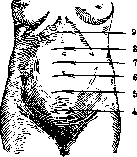
Figure 4. Level of the uterine fundus in different months of pregnancy (4-9 months).
also undergo the influence of pregnancy and hypertrophy. This is especially true of the vascular system, as the growth of the uterus and, especially, of the egg contained in it requires an increased supply of nutritional materials. Hypertrophy of uterine vessels proceeds so intensively that, becoming excessively elongated, they begin to spiral. In contrast to the body of the uterus, its cervix remains almost unchanged and only at the end of pregnancy undergoes that edematous infiltration which to a large extent contributes to its proper stretching during labor. The mucous membrane of the cervical canal does not undergo decidual metamorphosis. However, quite often in separate areas of the cervical mucosa, islands can be found that in their structure resemble Placenta '-,. 01 Thyreoidea
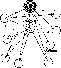
Uterus Hypophysis Parathyroid Mamma ChromaJ. syst. stimulation Thymus --------- inhibition Figure 5. Hypothetical scheme of the action of placental hormones on individual endocrine glands. . decidual cells. They were also found in other places, e.g., in the mucous membrane of the Fallopian tubes, ovaries, peritoneum, lymph glands, mucous membrane of the vermiform appendix; no satisfactory explanation has yet been found for the appearance of such an "ectopic" decidual reaction.-The ovaries during P. undergo important changes, which mainly concern the corpus luteum. It fully performs its internal secretory function only in the first half or even in the first 3-4 months of P.; later it begins to gradually, though slowly, wither and finally shrivels in the first weeks after childbirth. Among other functions, it is also attributed an inhibitory influence on the ovulatory activity of the ovary, which is why ovulation does not occur during P. Graafian follicles, having reached some development by the beginning of P., undergo atresia and transform into formations somewhat resembling yellow bodies, however, with the difference that in them lutein cells are formed not from cells of the membrana granulosa, but from cells of the theca interna, and they never reach such development as in the corpus luteum. The entire complex of such imperfect "yellow bodies" formed during P. is called the "interstitial gland" of the ovary, to which an endocrine function is attributed. However, in the second half of P. and even from the 4th month, the internal secretory activity of the ovaries, although it does not cease completely, decreases significantly. From this time, the functions of the ovary are taken over by the placenta, from the tissue of which it was possible to obtain a substance, hormonally identical to the corpus luteum hormone. Thus, the placenta in the second half of P. is a gland of internal secretion, which controls not only the exchange between mother and egg, but also the exchange in the maternal organism by a correlating effect on the entire chain of maternal endocrine glands (see Figure 5).-The Fallopian tubes undergo little change during P., their mucous membrane does not undergo decidual metamorphosis.-The round ligaments somewhat hypertrophy, but this hypertrophy reaches a significant degree only in individual cases.-The vagina in the last months of P. undergoes loosening and edematous infiltration. This increases its distensibility so much that by the end of P. it can pass the head of a mature fetus with a circumference of 34 cm without damage. The discharge from the vagina during P., especially in its second half, increases greatly; in connection with this, pregnant women often complain of the appearance of "leukorrhea." If such secretions do not differ in external appearance and acidic reaction from normal ones, their appearance is regarded as a physiological preparation of the birth canal for the upcoming childbirth: these secretions make the walls of the vagina more moist and slippery, which facilitates the passage of the fetus. The flora of the vagina of a healthy pregnant woman should correspond to the 1st degree of purity. This is due to the increased acidity of the vaginal contents during P. and its ability to physiological self-cleansing. The mammary glands under the influence of P. undergo significant changes, eloquently speaking for the endocrine function of P. itself. Their hypertrophy begins to manifest very early, starting from the second month of P. At this time, a small amount of clear or slightly turbid fluid can be expressed from the mammary glands. The nipple shows increased erectile ability; the areola becomes pigmented; Montgomery's glands in its area hypertrophy. All these changes in the mammary glands are only probable signs of pregnancy, since sometimes they are observed without any connection with pregnancy. The blood during pregnancy is enriched with water (hydremia) and fat-like substances. Its total amount increases by the end of P., approximately, by 15%. This increase is both absolute and relative to the weight of the pregnant woman: in the non-pregnant state, the amount of blood is 1/16-1/20 of the weight, at the end of P.-1/11-1/12. The specific gravity of the blood during P. decreases; the maximum decrease is observed at the end of it. The number of red and white blood cells increases. The amount of Hb increases. Thus, the morphological composition of the blood during normal P. improves. In view of the intense metabolism observed in the ovum, the acidity of the mother's blood increases (acidosis). The organism of a healthy pregnant woman reacts very perfectly to this physiological acidosis and maintains the normal concentration of hydrogen ions in the blood. This is achieved by enhanced pulmonary exchange, increased formation of ammonia compounds from disintegrating protein molecules, and mobilization of the so-called reserve alkalinity of the blood and tissue fluids. The question of acidosis during P. has extremely great importance, since apparently in the study of this phenomenon lies the key to solving many of the pathological phenomena that are now united under the general term- toxicoses of P. The sedimentation of red blood cells during P. accelerates. This phenomenon was first noticed Fåhræus when studying the blood of pregnant women, and according to his initial assumption, it could serve as a differential sign for recognizing pregnancy, more precise than the Abderhalden reaction. This assumption did not fully justify itself. The blood vessels and heart during pregnancy undergo significant changes. The vessels in the immediate vicinity of the uterus and especially in the uterus itself hypertrophy, and the venous system, in addition, gives expansion of the bed in the lower half of the body. All this, together with the increase in the total mass of blood, leads to an increase in cardiac work and hypertrophy of the heart, especially at the end of pregnancy. However, the reason for the expansion of cardiac dullness on percussion or cardiac shadow on fluoroscopy is not only hypertrophy of the heart, but also its change in position. At the end of P., when the growing uterus, occupying most of the abdominal cavity, rises to the diaphragm, the latter is displaced upward, which entails a displacement of the heart. At this time, the heart rises less and approaches the anterior chest wall more, and at the same time somewhat rotates around its axis. This causes twisting of the vessels bringing and carrying away blood, and thereby some insufficiency of cardiac activity, which is expressed by stagnation phenomena in the lower half of the body and shortness of breath. These phenomena reach their highest development by the end of the 9th month of P. and noticeably decrease in the 10th, when the bottom of the uterus descends (see Figure 6), and after it the diaphragm descends, which allows the heart to partially return to its usual position. The data obtained from autopsies of pregnant and parturient women are very uncertain. Thus, some believe that the weight of the heart by the end of P. increases, on average, not more than 3 g, while others accept the usual increase in heart weight by 25 and even 60 g. In any case, with a normally proceeding P., a healthy heart copes quite well with the increased work and the inconvenience of its changed position. The walls of the capillaries during P. become more permeable to the elements of the blood and more permeable to its liquid part, which partly explains the tendency of pregnant women
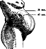
Figure 6. Level of the bottom of the uterus at the end of the 9th and 10th months of pregnancy.
to edema. - Blood pressure during normal P., as a rule, does not increase, with the exception of the time of labor, when it increases in connection with contractions and with the expulsive activity. Such stability of blood pressure within normal limits, despite a whole series of causes that could cause its change, is extremely convincing evidence of the adaptability of the pregnant organism, and also forces one to pay special attention to blood pressure (and even to a small but persistent increase in blood pressure, one should always be suspicious). The kidneys and liver as organs carrying the function of neutralizing and eliminating products of incomplete metabolism undergo a great test during P., when metabolism, on the one hand, is altered, and on the other, is increased due to the developing ovum with its exceptionally intense life processes. In the urine of pregnant women, one can quite often find the presence of small amounts (up to 0.5%) of protein. This so-called 'physiological albuminuria' of pregnant women is not accompanied by a persistent increase in blood pressure and easily yields to the most innocent hygienic and dietary measures. But in some cases, it can easily cross a certain boundary and then becomes a symptom of a pathological nature, which, in connection with other symptoms (edema, increased blood pressure), indicates the beginning of poisoning of the pregnant organism by products of metabolism (toxemia). The normal composition of urine in P. does not change; only rarely with a completely normal course of P. is there observed a saturation of urine with urates and chlorides and an increase in specific gravity. In such cases, a decreased excretion of urine is usually found, which can be explained both by the retention of water in the tissues and by the increased excretion of water by the skin through transpiratio insensibilis. In recent years, in studying the function of the kidneys in normal P. by studying the composition of blood and functional tests, no sharp difference in their work compared to the non-pregnant state has been found. But even minor disorders in kidney function are now interpreted as nephrosis, as nephropathy of pregnancy, in contrast to previous views, when the 'pregnant kidney' was also considered a physiological phenomenon of P. The liver of pregnant women, when studying its function by means of functional tests and microscopic examination, shows no pathological changes. All this taken together speaks in favor of the fact that a healthy organism copes quite successfully with the increased demands which are made on it in connection with P. The endocrine system undergoes particularly strong changes during P. The developing ovum is a new peculiar gland of internal secretion, with
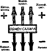
Chromaff. syst. Brain, append.,
Ovary Parathyroid glands Figure 7. Diagrams of S. A. Selitsky's (sugar exchange) and Seitz's (protein exchange). which, with the formation of the placenta, this endocrine function of the ovum as if concentrates in the placenta (see Figure 5). In the ovary, the corpus luteum and the interstitial gland begin to function. From the basic principle of endocrinology, according to which all glands of internal secretion are in constant and close correlation with each other, it follows that during P. certain changes must occur in the entire endocrine system. Unfortunately, not all these changes are sufficiently known. The appendage of the brain hypertrophies, increasing its weight 2 and even 3 times, and this hypertrophy mainly affects its anterior lobe. Here appear quite unique 'pregnancy cells,' which increase in number with the progressive course of P. The incretory activity of the appendage explains some of the peculiarities of metabolism during P.: retention of water in the tissues (pituitary edema), deposition of fat and change in mineral metabolism, as well as changes in the external appearance of pregnant women: acromegalic face with enlargement of the nose, lips, and sometimes the chin, and acromegalic changes in the extremities, lengthening and thickening of the fingers on the hands and feet. The thyroid gland during P. noticeably increases. This was explained by its increased function. Now many speak in favor of a decrease in its function and explain the increase of the gland by the retention of colloidal production. From this point of view, the edema of the face of pregnant women (mask of pregnancy) is as if myxedematous. The adrenal glands during pregnancy increase due to their cortical layer, which is greatly enriched with lipoid substances. It is unknown whether these lipoids are products of the activity of the adrenal gland itself or are only deposited here. However, apparently, in connection with the activity of the adrenal glands stands the characteristic for P. enrichment of the blood with lipoids, and in particular with cholesterol. The function of the adrenal glands is attributed to the deposition of pigment in the skin of pregnant women: on the face (chloasma gravidarum), in the areolas and along the white line. In the medulla of the adrenal glands, hypertrophy is observed, and in connection with this, increased secretion of adrenaline. The other glands of the internal secretory system undoubtedly change in connection with pregnancy. However, complete clarity in this complex question has not yet been achieved. The endocrine glands also take direct part in the metabolism of pregnancy (see Figure 7). The nervous system in general reacts to P. in the sense of increased excitability of reflex and neuromuscular activity, especially in its second half. - As for the autonomic nervous system, it is still difficult to say whether there is vagotonia or sympathicotonia during P. - The psyche of pregnant women changes less in connection with P. itself than from the woman's attitude to the pregnancy that has occurred. In those cases where P. is undesirable, the mental and nervous state of pregnant women undergoes a severe test, which is expressed by a whole series of clearly pathological manifestations. When P. is desired, manifestations of the opposite order are observed; sometimes they clearly do not harmonize with the general somatic state of the pregnant woman. Thus, one has to see pregnant women with a progressive deterioration of the general condition on the basis, for example, of toxemia, who show no depression of either nervous or mental activity and often even get rid of a whole series of functional nervous disorders observed in them in the non-pregnant state. This can be particularly sharply expressed in women of an hysterical nature, who sometimes literally transform with the onset of a desired P. The emotional side of the psyche of a pregnant woman decreases in the second half of P. and, especially, toward its end. The most 'frivolous' woman with the onset of P. obviously transforms, acquiring calmness and equilibration, becomes 'important' and 'serious'. Such a change in the psyche is, of course, a highly valuable biological factor, as it makes the pregnant woman less mobile, more cautious, and gives her the mental calm so necessary in anticipation of the final stage of P., which is labor. Among the mental changes associated with P., one can include the appearance or intensified manifestation of the maternal instinct.'-Twin pregnancy see Twins.'
abdominal dropsy (rapid weight gain according to W. Stoeckei, dropsy, cardiac disorders, visual impairment, headaches, etc.), i. e., intoxication of the organism (see Figure 1). With the termination of labor, all these phenomena rapidly disappear, and renal function is restored. Cases of recurrence of the disease in subsequent pregnancies are observed; transition to chronic forms upon the termination of pregnancy is rarely observed. The prognosis is almost always favorable; the pregnant kidney does not pose a danger, but sometimes more severe forms of it may develop; eclampsia as a complication is observed. Pregnancy in milder cases proceeds to term, and the disease does not affect the fetus; in severe cases, however, pregnancy may be interrupted prematurely, children are born weak and of low weight (intrauterine fetal death may also occur). The main therapy is the most careful prophylaxis throughout pregnancy; upon the appearance of kidney disease, even in the mildest degree, the usual therapy for kidney diseases is indicated. In addition, along with the restriction of proteins and salts and the prescription of a milk diet, absolute rest (bed rest) is recommended; the frequent use of baths is inadvisable, as they can often cause harm, especially when cardiac activity is already impaired. If therapeutic measures are unsuccessful, to avoid the progression of the process and the onset of eclampsia, artificial termination of pregnancy is indicated. Eclamptism, a term introduced by Bar, is a sharply defined intoxication of pregnant women, consisting of a series of pathological symptoms. It is regarded as a prodromal stage of eclampsia or even as one of the phases of eclampsia itself. Eclampsia (see), one of the most severe manifestations of intoxication during pregnancy, is a disease of the organism in toto; its prevailing symptom is convulsions (cases without convulsions have also been described). It is observed during pregnancy, labor, and the postpartum period. Chorea of pregnant women (see Chorea), or St. Vitus' dance, is very rare. Only 17 cases have been described in the USSR. The etiology of the disease is not precisely known. Past infections, rheumatism, and heart diseases as etiological factors are not always confirmed by clinical observations. Chorea of pregnant women can also be classified among toxicoses, and its emergence can be regarded as an intoxication depending on various physiological functions (mainly pregnancy), on any dysfunctions and diatheses existing in childhood. The prognosis of chorea of pregnant women is always serious; statistics show a high mortality rate (from 5% and even up to 50–60% in severe cases). Chorea is an indication for the termination of pregnancy if it does not respond to treatment and if, with the intensification of choreic convulsions, other pathological symptoms are observed (weight loss, general exhaustion, mental disorders, decrease in urine volume, albuminuria, elevated temperature, etc.). In a milder clinical course, strengthening treatment (arsenic, phosphorus) and the administration of solutions used in other toxicoses are employed. These solutions give very encouraging results. Narcotics, especially potent ones, must be used with caution in chorea. Osteomalacia, according to modern views, is regarded as a toxemia of pregnancy, as a general disease—a pluriglandular disorder with sharply disturbed metabolism. Osteomalacia (see) is rarely observed (the latest Russian statistics, for example, cover 71 cases) and occurs predominantly along the courses of major rivers (Volga region, Rhine, Danube) and in certain localities (Transcaucasia). It affects the Mongolian race more frequently. Osteomalacia is observed almost exclusively in women, usually appears for the first time during a repeat pregnancy (less often during lactation), and those affected are characterized by high fertility (on average, 6 deliveries). The essence of the process reduces to the depletion of lime from the bones and their subsequent softening. These disorders of the entire skeletal system (pelvis, extremities, rib cage), arising to varying degrees, are linked to the ongoing dysfunction of the ovary and certain other endocrine glands (thyroid, parathyroid, pituitary, adrenal glands). Recent microscopic studies of the ovary (increased secretory activity of the granulosa epithelium, enlargement of Call-Exner bodies, increased follicular atresia) confirm the view expressed in 1887 by Fehling that in osteomalacia there is an enhanced ovarian function, so-called hyperovaria (occurring not in isolation, but alongside dysfunction, hypo- or hyperfunction of the other glands mentioned above). Early symptoms of osteomalacia: pains—either localized (in the pelvic region, lower back, lower abdomen) or more widespread (in the upper and lower extremities and the entire body in general); a halting, unsteady gait (waddling), limited mobility (increased pain when walking). In a more sharply expressed process, along with increased pain, a number of general pathological symptoms appear. In more severe (neglected) cases, patients decrease in height, edema of the legs and the entire body develops, and patients cannot move and are forced to stay in bed. Sharp changes in bone tissue lead to spontaneous cracks, even fractures, and in the pelvis—to its significant deformation and narrowing. The osteomalacic pelvis, as such, presents characteristic features at various stages of the disease. It decreases in transverse diameter due to the pressure of the thighs, as a result of which the pubic bones approach each other (the so-called beak-like shape of the pubic symphysis) and the ischial tuberosities. Upon softening, the bones sometimes become so soft that the pelvis is given the name "rubber" pelvis. The therapy of osteomalacia depends on the severity of the process. In milder cases, phosphorus, fish oil, and other tonics are used. The use of adrenaline, pituitrin, and X-rays, recommended recently, has not fulfilled expectations. In severe cases, regardless of the duration of pregnancy, its termination is indicated; to avoid possible serious and irreparable damage to the pelvic bones, it is better to perform a Caesarean section. It is rational to perform sterilization at the same time; as for castration, it is hardly rational to perform it as widely as was practiced previously, since the dysfunction does not depend solely on the ovary; the use of Porro's operation is also groundless. Among the toxicoses of pregnancy are a number of other pathological processes that often arise for the first time only during pregnancy and usually disappear with its termination (diseases and changes on the part of the eye, ear, various degrees of anemia, hemorrhagic diathesis, processes in the central and peripheral nervous system—neuritis, polyneuritis, mental disorders, etc.). Acute and chronic infections. During pregnancy, a woman may be exposed to various kinds of acute infections (tonsillitis, influenza, pneumonia, typhus, cholera, smallpox, diphtheria, etc.; for details see corresponding entries). In most cases, the course of one or another pathological process does not present any special features. For the mother and the fetus, the greatest danger is posed by protracted processes with a prolonged high temperature, with complications on the part of the lungs, heart, and kidneys. With frequent tonsillitis during pregnancy, with streptococcal diseases, after the disease has passed, the infection may remain in the organism and give rise to the direct occurrence of a purely postpartum disease. Often one or another infection leads to the termination of pregnancy due to a disease of the uterus itself (this possibility was first proven in cholera by Slaviansky—endometritis haemorrhagica) or due to changes in the fetal egg itself, leading to intrauterine fetal death. When pregnancy is terminated in the acute stage of an infectious disease, the danger may consist either in complications during labor (increased blood loss) or in the emergence of severe puerperal infection. Chronic infections also have an effect on pregnancy itself and on fetal development. Intrauterine fetal death and premature termination of pregnancy (birth of weak, sickly children) are often observed. It is necessary to take into account the possibility of exacerbation of the process itself under the influence of the onset of pregnancy. Of the chronic infections, tuberculosis, syphilis, and malaria are of the greatest importance for the mother and the fetus; the latter can in some cases also be regarded as an acute infection. Among tuberculous diseases (see Tuberculosis) of various organs during pregnancy, pulmonary and laryngeal tuberculosis are of the greatest importance. Pulmonary tuberculosis, according to some data (Zangemeister), is found in 7% of all pregnant women (in 6% in a latent form). Tuberculosis often worsens during pregnancy, especially in far-advanced stages; worsening of the process can also be observed in the postpartum period (or after an abortion). Sometimes pregnancy, given a certain predisposition, especially hereditary, can be the cause of the disease appearing for the first time. In active tuberculosis during pregnancy, the mortality rate is also significant.
On the other hand, there are observations that in a certain percentage of cases the course of tuberculosis during pregnancy may even improve in connection with new biochemical processes and, chiefly, with the amenorrhea setting in during pregnancy; in clinical practice there are not a few cases where no exacerbation is observed. Children are born completely healthy, well developed, and of sufficient weight. The deterioration of tuberculosis is explained by the increased function of the organism, altered metabolism, and an increase in cholesterol content. Tuberculosis in a severe stage can also lead to the interruption of pregnancy. Late miscarriages and abortions are observed more frequently; premature births may also occur. Tuberculosis can also pass to the placenta, and in very rare cases can affect the fetus as well. In the placenta, the process begins from the decidua basalis and the intervillous spaces (see Figure 2). The exacerbation of tuberculosis in the puerperium finds its explanation in the involvement of the placental attachment site, where dissemination and exacerbation of the former process occur. Tuberculosis is a definite medical indication for artificial abortion, with the character of the process serving as the decisive moment. When interrupting a pregnancy, its gestational age is of great importance. The best time is the first months; the possibility of performing an abortion in later months is not excluded altogether, but the chances of tuberculosis improvement upon the termination of pregnancy in such cases are minimal. Tuberculosis, furthermore, in certain cases serves as an indication for sterilization, and individual schools even advise performing vaginal extirpation of the uterus. In view of the fact that in a fairly large percentage of tuberculosis cases artificial abortion is indicated,
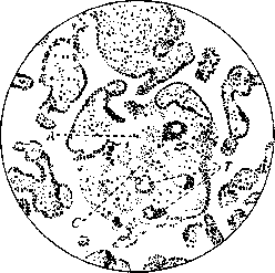
Figure 2. Tuberculosis of the villi; in the middle is granulation tissue (C) with tubercles (T); A is the site of penetration into the villi (after Schmorl-Ockeghem).
and its effect on the process, especially when performed repeatedly, is far from indifferent, the prevention of pregnancy through the use of contraceptive measures is necessary. Laryngeal tuberculosis during pregnancy usually proceeds extremely acutely and is an absolute indication for its interruption regardless of the gestational age. But even the termination of pregnancy does not improve the process; the majority of those who fall ill die in the very first months or in the first year after the pregnancy. Syphilis (for details see Syphilis). Pregnancy has no particular effect on syphilis. On the other hand, the effect of syphilis on pregnancy and on the fetus is enormous, as a result of which it is necessary to carry out rational specific treatment from the very beginning as a measure to combat congenital syphilis and its consequences. Prophylaxis consists in the early diagnosis of syphilis (which often proceeds in a latent form), the mandatory performance of the Wassermann reaction and other control serological reactions, and subsequent treatment as early as possible. Of a certain significance for establishing the diagnosis are also the anamnesis, clinical symptoms, examination of the father, and macroscopic and microscopic data on the placenta and fetuses from past pregnancies. Carrying out this prophylaxis is one of the tasks of the consultation clinic for pregnant women and the venereological dispensary. In diagnosing
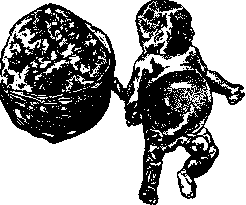
Figure 3. Syphilitic fetus (9 months) who died during labor: widespread syphilitic exanthema; maceration in places; significant ascites; large placenta (after W. Stoeckel).
syphilis, it is necessary to take into account the time of infection (whether it occurred before pregnancy or already after its onset and at what time). In the first case, syphilis always leads to disease of the fetus; in the second, the fetus may remain healthy if the mother fell ill at the end of pregnancy. Consequently, with maternal syphilis there can be a healthy fetus, but there can be no fetal syphilis in a healthy mother. Syphilis is the cause of frequent

Figure 4. Numerous spirochetes in the adrenal gland of a syphilitic newborn (according to W. Stoeckel). Habitual interruption of pregnancy leads to abortions, premature births (10% of premature births, 30% of intrauterine death of the fetus, and 40% of macerated fetuses--are, according to Tsangmeister, due to syphilitic infection--see figures 3-5). Children born alive are frail, weak, with insufficient development, low weight (various fetal malformations are also common in syphilis); in severe cases--with obvious external manifestations of syphilis and with typical lesions of internal organs (pemphigus, severe jaundice, etc.). In most cases, diagnosing congenital syphilis in newborns is difficult (due to often negative results of serological reactions and lack of clinical data). Recently, radiography has been helpful in this regard. In such doubtful cases, examination of the placenta, both macroscopic and microscopic, also becomes very important. Along with a significant increase in weight, with significantly expressed petrification, with paleness, softness and great thickness of the placenta, changes in blood vessels and the formation of nodes similar to gummas are observed in it. In addition, for the diagnosis of syphilitic placenta, the finding of the pale spirochete is also important (and in dead and macerated fetuses--anatomical examination of all organs). Syphilis is not a medical indication for abortion, and its presence rather serves as a social indication for abortion. Therapy for syphilis is the usual, as in the absence of pregnancy. Malaria. Pregnancy in prolonged, severe forms of malaria occurs rarely. Depending on the nature and severity of the disease, if pregnancy occurs, malaria can give serious complications, taking a more acute course and at the same time adversely affecting the pregnancy. On the other hand, pregnancy itself can be an agent causing an exacerbation of a latent malaria. In severe (and even moderately severe) cases, abortion (more often late) often occurs, and intrauterine death of the fetus is also observed; children born alive in premature births or even at term often show signs of congenital weakness and underdevelopment. There are no exact data regarding changes in the placenta in malaria; the malaria plasmodium can pass through the placenta and affect the fetus. The causes of interruption of pregnancy and intrauterine death of the fetus are the same as in other acute and chronic infections (see above). Heart diseases (details--see Heart). The heart, like other organs, during pregnancy is in a state of physiologically increased activity, manifested by changes [partly mechanical, partly chemical in nature (passing in the postpartum period); they consist, mainly, in hypertrophy of the heart muscle, in the appearance of dull tones (so-called myopathia gravidarum) and physiological heart murmurs, in approximately 20-25% of all cases of pregnancy. If there have been any deviations in the past, especially organic diseases, this physiological load should manifest to a greater extent: a diseased heart reacts more strongly to the changes occurring in the pregnant woman's body. One must always take into account the age of the patient, the number of pregnancies and births she has had and the nature of their course. This is important for the prognosis of the course of pregnancy and the act of delivery, and mainly for deciding on the need to interrupt it. Various heart diseases (valvular defects, lesions of the heart muscle and combined forms) in combination with pregnancy occur quite often (in 1-2%). Recent data (according to Stoeckel--2-5% mortality) largely refute the old opinion of great danger and a high mortality rate (50-60%) with this complication. Tsangmeister believes that the overall mortality of cardiac patients is 2.5%, with uncomplicated defects--0.5%, with complicated ones (on average) reaching 12%. In diagnosis and prognosis, especially in the early stages of pregnancy, the greatest individualization is necessary. It is necessary to approach not the disease, not this or that type of heart defect, but to approach the patient herself, because often the importance lies not in the severity of this or that process, but in the arising and accompanying other pathological phenomena. This must be especially taken into account with early sometimes decompensation. The living conditions of the pregnant woman are also of no less importance. The better she is placed in favorable conditions, the more she is preventively protected from harmful influences and from the possible appearance of this or that specific disease (toxemias--kidney, liver of pregnant women, etc.) on the basis of pregnancy itself, the more favorable (sometimes even regardless of the severity of the main process) the pregnancy will proceed. Prevention is necessary for cardiac patients to avoid subsequent complications after the end of pregnancy; it should also consist in careful, rationally justified management of labor and in possible protection from purely postpartum diseases. A poor prognosis depends both on the developing phenomena of decompensation and on the addition of toxemias to the heart disease. The latter often give an acute picture of cardiac insufficiency and are accompanied by a much higher mortality rate. Along with heart defects, one sometimes has to deal with so-called small heart, with hypoplasia of the entire vascular system (status hypoplasticus), which can be considered on a par with infantilism of the whole body. Some believe that these deviations do not affect either the course of pregnancy or the act of delivery itself, because, on the contrary, pregnancy, causing hyperfunction, acts beneficially on the indicated shortcomings of the vascular system. In certain cases, lesions of the heart muscle and heart defects serve as a definite indication, sometimes even absolute, for interrupting pregnancy. Prevention of the normal course of pregnancy in cardiac patients consists in careful observation of them, limitation of labor, especially physical, in dietary regimen and periodic visits to the pregnant consultation. Diseases of the endocrine glands (see Internal secretion). During pregnancy, the activity of individual endocrine glands changes to a large extent. Along with this, only with the onset of pregnancy do individual components of the endocrine glands begin to function; hormones that are characteristic and specific only to pregnancy appear. Finally, the placenta is also included in this common link of this closed network, which by many authors, according to the nature of its activity and effect on other glands, is also classified as an endocrine gland. As a result, new chemical relationships, a new correlation in the endocrine system arise. Of course, in all deviations from the physiological hyperfunction of pregnancy, the endocrine glands take the closest part; in some of them, in certain toxemias, characteristic changes have recently been discovered. Along with physiological changes and pathological deviations on the basis of pregnancy itself, the onset of pregnancy in dysfunction of this or that endocrine gland is of no less importance. This dysfunction can either directly be the cause of reduced ability to conceive (be the cause of infertility) or (if fertilization occurs) can affect the correct course of pregnancy (abortion, premature birth, underdevelopment of the fetus) and give exacerbations of the main process. Diseases of the adrenal glands. In relation to Addison's disease, it is necessary to distinguish whether it is of primary or secondary origin; in primary--pregnancy never occurs, in secondary--rarely (due to significantly reduced ovarian function or even their atrophy). Secondary Addison's disease arises on the basis of some infections (syphilis, tuberculosis) of the gland itself or its neoplasms. Addison's disease as a complication of pregnancy is observed extremely rarely; as a rule, under the influence of pregnancy the disease worsens and can lead to premature interruption of pregnancy, cause atonic bleeding. Artificial interruption of pregnancy is not indicated, as it does not improve the course of the disease. Death can occur directly in the postpartum period. Diseases of the thyroid gland. Depending on whether there is hypo- or hyperfunction of the gland, both the possibility of pregnancy occurring and its course, as well as the reaction of the diseased gland to pregnancy itself, are determined. In hypothyroidism (cachexia strumipriva, myxoedema, cretinism) pregnancy is rarely observed, as hypothyroidism usually causes ovarian insufficiency. During pregnancy, there is always a worsening of the disease, but hypothyroidism, as a rule, is not an indication for interrupting pregnancy. The indicated special predisposition to eclampsia is not justified. A certain therapeutic effect from the use of thyreoidin has been noted. In hyperthyroidism (thyrotoxicosis, basedowismus, morbus Basedowi) pregnancy occurs more often, but even with it, a certain reduced ability to conception is noted due to weakened ovarian activity.
Pregnancy often has no effect on the course of hyperthyroidism (in 60-70%; according to other data - only in 40%); in some cases (5-10%) the condition worsens and in 4-5% even ends fatally due to complications, mainly from the heart (insufficiency) and kidneys (toxic nephritis). In milder cases, a number of pathological symptoms are noted (palpitations, tachycardia, etc.); hyperthyroidism is observed mainly in the first months and may be an indication for termination of pregnancy. After childbirth, improvement almost always occurs. It should be remembered that these patients have a great tendency to bleeding in the postpartum period. In the presence of a goiter, the most rational intervention is strumectomy, which is not dangerous for the mother; if strumectomy is ineffective, abortion or premature labor is indicated. Diabetes (diabetes mellitus) may exist before pregnancy but can first occur only during pregnancy. In the latter cases, it must be differentiated from the frequently (8-12%) observed (so-called simple, or physiological) glycosuria of pregnant women (or lactosuria). Diabetes during pregnancy is rarely encountered, since due to significantly weakened ovarian function, the ability to conceive is greatly reduced in diabetes (according to Tsangeyster, pregnancy is observed in only 5% of diabetes cases in women aged 20 to 40 years). The effect of diabetes on pregnancy and pregnancy on diabetes is extremely unfavorable. Diabetes progresses, worsens, complications occur from internal organs; diabetic coma often occurs in the postpartum period or even during pregnancy. With such complications, the mortality rate is also high (a large number of women die in the months following the end of pregnancy). The effect of diabetes on the fetus is also detrimental, late abortions, intrauterine death, and polyhydramnios are common; cases of congenital diabetes have been described. Patients should be under observation on a very strict diet. There are as yet no decisive observations regarding the use of insulin. In mild cases of diabetes, pregnancy can be continued; in severe cases, abortion is absolutely indicated. Even rarer during pregnancy are pituitary obesity (dy-strophia adiposo-genitalis), status thymico-lymphaticus and status hypoplasticus. In the latter cases, it is necessary to consider the possible dangers of postpartum bleeding, as well as to avoid the use of anesthesia (chloroform). Diseases and abnormalities of the genital sphere and the ovum (fetus, its membranes and placenta). Abnormalities of the genital sphere may consist of various developmental defects (generally, local and general infantilism), incorrect positions of the genital organs and neoplasms. All these abnormalities, which are often the cause of various complications during pregnancy, also cause the sometimes observed infertility.- In some types of developmental defects, diagnosis can be very difficult. Pregnancy can occur with complete doubling of the genital organs or with doubling only of the uterine cavity. In most cases, following usual hygienic rules, pregnancy proceeds normally, goes to term, and no deviations are noted during labor. But often (almost always depending on the degree of defect) premature termination of pregnancy, incorrect positions of the fetus (especially with abnormal uterine forms, e.g., uterus arcuatus-saddle-shaped uterus), bleeding during pregnancy, and incorrect attachment of the placenta (especially its location on the septum between two uterine cavities) may be observed. During the act of labor, the most careful observation of labor activity and uterine contractions (danger of uterine rupture) is necessary. Incorrect contractions and secondary labor weakness are noted more frequently than normal. Sometimes surgical intervention is required to remove one or another septum in the vagina that interferes with delivery. Complications in the postpartum period are also common, complications in terms of incorrect position of the placenta (see above) and subsequent atonic bleeding are possible. The most unfavorable is pregnancy in the so-called rudimentary horn of the uterus. The only method is surgical intervention. (Diagnostic errors are very common.) With forward and backward bends, deviations in the normal course of pregnancy and during labor may occur. Forward bending of the uterus has no special significance: miscarriage with it, if it occurs, is very rare. With artificially created forward bending of the uterus after operations (vagino-ventrofixatio), termination of pregnancy occurs much more frequently. Complications during labor are possible; for their prevention, some even recommend cesarean section (Gubarev, Pobedinsky).- It is also necessary to note the tendency of the pregnant uterus to tilt forward. This occurs with a narrow pelvis (the so-called pendulous abdomen-venter propendens forms) and with significant stretching of the abdominal walls (large fetus, multiple pregnancy, polyhydramnios). For prevention, restriction and sometimes complete prohibition of physical labor and mandatory wearing of a special bandage are necessary.- Much more complications are caused by tilting and bending of the uterus backward (retroversio et retroflexio uteri gravidi). Such a position of the uterus, although not often, can still be a direct cause of infertility. Backward bending of the uterus usually (in the absence of adhesions and fusions) corrects itself (recommended: daily lying on the stomach, do not sleep on the back). Accompanying symptoms-frequent urination or, conversely, retention of urine, constipation, pain in the sacrum-are sometimes tormenting for patients. Artificial correction requires extreme caution (possibility of miscarriage, shock). Insertion of various types of rings to hold the uterus hardly seems rational. A serious danger is the presence of adhesions that prevent possible self-correction of the position and threaten incarceration. The above symptoms progress; complete retention of urine develops with significant stretching of the bladder, persistent constipation, severe pain in the sacrum and lower back. Among the serious consequences, cases of uremia, pyemia and even peritonitis were previously described (there were also case reports of bladder rupture). Incarceration of a retroflexed pregnant uterus in a number of cases is an absolute indication for abortion. Sometimes the usual method is difficult, and puncture of the uterus with a trocar through the posterior fornix to remove amniotic fluid and reduce the uterus, or incision of the posterior vaginal wall and uterus is recommended. At present, cases of successful use of the operation hysterotomia vaginalis have also been described. Prolapses, prolapses, as well as hernias of the pregnant uterus are rare. Among hernias, hernias of the linea alba are more common in multiparous women. Prenatal hernia of the pregnant uterus is extremely rare; in such cases, the uterus can move into the hernial sac even before pregnancy (see Figure 6). Pregnancy terminates prematurely; spontaneous labor may occur (Rozanov); sometimes the hernia is also an indication for abortion and cesarean section. Various neoplasms of the genital sphere are of considerable importance; on the one hand, they can be the cause of infertility in a certain percentage of cases, in a number of cases they can cause miscarriage or premature labor, and finally even be the cause of the development of ectopic pregnancy. Fibromyoma of the uterus, mainly in the form of small nodules, is quite common during pregnancy and in most cases does not affect its course or the position and development of the fetus. Sometimes these fibromyomas have a significant tendency to grow and soften; sometimes, on the contrary, under the influence of pregnancy, their significant reduction is observed. Malignant degeneration, as well as complete necrosis due to impaired nutrition, is rare. Extremely rare is the twisting of the entire uterus with subsequent phenomena of acute peritonitis. In terms of prognosis, for a favorable course, the location, size and multiplicity of fibromyomas are important. The highest percentage of termination of pregnancy (as well as incorrect positions of the fetus) is caused by submucous fibromyomas. Labor usually proceeds normally, sometimes incorrect positions of the fetus, weakness of uterine contractions and bleeding in the postpartum and postnatal periods are observed. Atonic bleeding is rare. Sometimes in the postpartum period, the so-called "birth" of a fibroma occurs; sometimes fibromyomas are the cause of severe septic infection. In a number of cases, depending on in which part of the uterus the tumor is located (also depending on its size and number of nodes), surgical intervention is indicated. Surgical intervention may consist of conservative cesarean section with enucleation of nodes, or the Porro operation, or total hysterectomy. In some rare cases, conservative myomectomy during pregnancy is also indicated.
The presence of fibromyomas is one of the medical contraindications for performing an artificial abortion. - Cancer of the cervix (cancer of the body of the uterus cannot occur during pregnancy) occurs rarely (1 case in 2,000-3,000 births); during pregnancy, it is characterized by special malignancy, as it occurs in younger women (according to some latest data, this is not always observed). Cancer often leads to abortion. If cancer is diagnosed during pregnancy, therapy depends on whether the cancer is operable or not. In inoperable cases, X-ray irradiation is recommended followed by extirpation (at the end of pregnancy), or vice versa. In operable cases--immediate radical abdominal operation. - Tumors of the ovaries (cysts, more often dermoids-60%) occur in combination with pregnancy relatively often (0.1-0.3%). Due to the danger of twisting, subsequent irritation of the peritoneum, the possibility of complications during childbirth and in the postpartum period, surgical removal of ovarian tumors is indicated. It is best to operate in the first months; the percentage of pregnancy interruption after this operation ranges from 5 to 15%.

Figure 6. Inguinal hernia of the pregnant uterus (Rozanov's case).
Bleeding during pregnancy can occur with premature termination of pregnancy (abortion, missed abortion), with ectopic attachment of the fertilized egg, with its diseases and degeneration, with incorrect attachment and premature separation of the placenta. Diseases of the fetus, membranes and placenta (see also corresponding words). In the placenta, changes are observed in the sense of anatomical structure (e.g., placenta marginata, circumvallata, bipartita, etc.), diseases on the basis of acute and chronic infections, deep attachment and its adhesion to the uterine wall. New growths are also often observed, especially cysts; they have no special influence on the development of the fetus, nor do they cause premature termination of pregnancy. - From the side of the fetal membranes, hydramnios is quite often encountered, much less frequently oligohydramnios, rare cases of graviditatis extraamnialis (Kolosov) have been described. - Abnormalities from the side of the umbilical cord can have as their consequence complications during pregnancy (sharp and repeated entanglement leads to intrauterine death of the fetus) or, mainly, during childbirth (short, long, eccentric attachment, insertio velamentosa, false and true knots). Among the diseases of the fetus, intrauterine death on the basis of chronic and acute diseases of the mother is most often noted, with subsequent phenomena (maceration). A large section of this group of pathology of pregnancy consists of deformities and diseases, which in recent times are attributed to intrauterine constitutional processes. - Lit. see above- Normal pregnancy.
S. Selishchii. Pregnancy Psychoses are connected either with auto-intoxication due to metabolic disorders during pregnancy (changes in the blood, disorders of the function of the sex glands, pituitary gland, thyroid gland, liver, kidneys, etc.), or with psychological factors. Depending on the constitution, heredity, there is a greater or lesser predisposition to nervous-psychological disorders. In the first period of pregnancy, a series of disorders of the nervous system are very often observed, namely: vasomotor disorders, chills, cramps of the calves, tendency to vomit, dizziness, loss of appetite, disturbance of taste sensations, increased knee reflexes, distraction, fluctuations in mood, irritability, hypochondriasis. Of more serious disorders, one can note dizziness, severe neuralgias (sciatic and trigeminal nerves), klepto- and pyromaniacal tendencies. In some pregnant women, on the contrary, an improvement in the nervous-psychological state is noted; they become calmer, more balanced. - True psychoses are observed in the second half of pregnancy, usually in persons over 25 years of age. The most severe forms of psychoses are observed in women who have given birth several times, with the exception of 'chorea of pregnancy' and hysteria, which more often occur in primiparas. There are no specific psychoses of pregnancy. Manic-depressive psychosis is observed, in particular, the depressive phase, and often each pregnancy is accompanied by attacks of depression. If pregnancy is accompanied by disorders of a schizophrenic type, then often relapses of such attacks with recurring pregnancy give an increasingly worsening psychological picture. Among psychogenic diseases, reactive depression is most often encountered (due to fear of childbirth, fear of material and other worries about future offspring, with ideas of self-blame, etc.). These reactive depressions differ from endogenous ones in that the depression is limited to the period of pregnancy and there are no ideas of sinfulness, while affectivity here is more amenable to external influence. Due to the danger of suicide, such patients should be under careful supervision (according to Pilcz's statistics, among 426 women-suicides, 19.9% were pregnant). - Sometimes hypochondriacal, obsessive-neurotic reactions are observed, as well as paranoid (jealous delusions). Epileptic seizures are observed less often; it is interesting to note that sometimes pregnancy has a beneficial effect on epilepsy existing before pregnancy. - Among the psychoses that are more often observed in primiparas, hysteria and chorea should be noted. Chorea usually occurs at the age of 18-23 years, in the 4th month of pregnancy. The psychological disorders here are sharper than in childhood chorea; marked irritability and excitement are noted, there is confusion, stupor, a state close to delirium and 'acute confusion'. In 25% of cases, the outcome is fatal. Those cases where chorea was also noted in childhood give a better prognosis. - Eclamptic psychoses (in the second half of pregnancy in primiparas) are encountered much less frequently, usually 3-6 days after the onset of convulsions. As prodromal phenomena, dizziness, insomnia, irritability, paresthesias, confusion and excitement (characterized by massive hallucinations of all kinds), disturbance of attention, distractibility, incoherence, motor excitement, amnesia (in general, a state close to epileptic twilight state) are observed. If patients do not die from eclampsia, the psychosis itself has a good prognosis. - According to statistics, 3% of all psychological disorders in women fall on the period of pregnancy. Therapy depends on the type of psychotic disorders. The question of terminating pregnancy in connection with psychoses is generally answered negatively, since termination of pregnancy usually does not affect the course of the psychosis. Only in rare cases, if each pregnancy is accompanied by severe organic symptoms (like pseudotumor cerebri, etc.), or by severe emaciation (in psychogenic depressions), or by an increase in the number of epileptic seizures, or in cases of severe chorea, uncontrollable vomiting, when the mother's life is in danger--abortion is indicated. Abortion for eugenic reasons is not indicated, since direct inheritance of severe mental anomalies is not proven. Progressive paralysis is not an indication for abortion, since the children of such patients are usually not suffering from syphilis. In individual cases, pregnancy is prohibited for preventive purposes.
M. Sereyskii. Ectopic Pregnancy. In humans, pregnancy always begins extrauterinely; subsequently, the fertilized egg enters the uterus, where it implants. In ectopic pregnancy, the fertilized egg implants and begins to develop not in the uterus, but on the path from the ovary to the uterine opening of the tube; depending on where the egg gets stuck, three main forms of ectopic, or extrauterine pregnancy (graviditas extrauterina s. ectopica) are distinguished: 1) ovarian pregnancy (gr. ovarialis), 2) primary abdominal pregnancy (gr. abdominalis), and 3) tubal pregnancy.
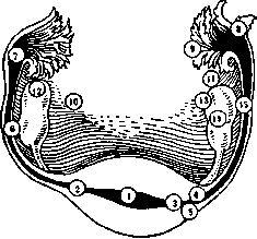
Figure 1. Topical varieties of ectopic pregnancy: 1-graviditas uterina; 2-gr. interstitialis propria; 3-gr. tubo-uterina; 4-gr. tubo-interstitialis (Popov); 5-gr. intramuralis (Rosenthal); 6-gr. isthmica; 7-gr. ampullaris; 8-pregnancy in an accessory tube; 9-gr. tubo-abdominalis; 10-gr. abdominalis; 11-gr. fimbriae ovaricae; 12-gr. ovarialis; 13-gr. epioophoralis (Kushchalov); 14-gr. ovarialis interstitialis (Serebrennikova); 15-pregnancy in the ovarian sac. (gr. tubaria). Tubal pregnancy, depending on the anatomical section of the tube in which the egg implanted, is divided into interstitial or isthmic (gr. interstitialis), isthmic (gr. isthmica), and ampullar (gr. ampullaris). Some distinguish minor variants, such as gr. tubo-ovarialis abdominalis (part of the developing egg faces the lumen of the tube, while the other part faces the abdominal cavity), gr. tubo-ovarialis (the egg is partly in the ovary and partly in the dilated tube), gr. fimbriae ovaricae (pregnancy on the ovarian fimbria), gr. intramuralis (interstitial pregnancy in which the egg develops in the thickness of the myometrium), etc. (see Figure 1). Of all the listed varieties, in practice, tubal pregnancy occurs in the vast majority of cases [according to Werth (R. Werth), at least 3% of all gynecological diseases]: more often ampullar, less frequently isthmic, and very rarely (1-3%) interstitial. Other forms are observed extremely rarely (see below). Tubal pregnancy Etiology. When determining the etiology of ectopic pregnancy, it is necessary to consider the physiological conditions present in the sexual organs during fertilization of the egg (see Fertilization). Under normal conditions, after the rupture of a mature follicle, the egg almost immediately enters the ampullar part of the tube. The further movement of the egg along the tube to its implantation site in the uterus occurs mainly due to peristaltic contractions of the tube (Mikulicz-Radecki), with the impulse for initiating such peristaltic movements coming from ovarian hormones, respectively, the corpus luteum. The ciliated epithelium, to which the so-called ciliary theory was formerly attributed primary importance in all stages of egg movement, according to modern views plays a secondary role. Passing through the narrow isthmic part of the tube, equipped with a special sphincter (Reinberger, Arnstam), the egg performs a series of movements and changes, which in a distant analogy resemble childbirth: it changes its configuration in a certain way and only after that passes through pars isthmica. The entire process of egg movement takes 7-8 days in humans. During this period, the egg undergoes segmentation, with the formation of the trophoblast (the outer layer of the ectoblast) being a particularly important moment, from which implantation depends, as it must dissolve the zona pellucida of the egg and the underlying maternal tissue (precisely where the fertilized egg will be at that moment). Normally, this occurs when the egg enters the uterus. But this moment may occur earlier (ectopic pregnancy will develop) or later than the normal term (placenta praevia will form). A priori causes that can lead to ectopic pregnancy may lie: 1) in the egg itself, in its abnormal development (reduced ability to move) or excessively increased biological energy, which accelerates the formation of trophoblast, and consequently, implantation (ovulogenic theory--Poorten's); or 2) in conditions disrupting the above 'transport means' necessary for egg movement. The ovulogenic theory has not yet received scientific justification. Thus, the causal factors that can be linked to disruption of the transport mechanism remain. Here, inflammatory processes of the tubes (catarrhal salpingitis), especially gonorrheal, should be placed in the first place, although the nature of the pathogen apparently does not play a special role. Supporters of the inflammatory theory explain the mechanism of ectopic pregnancy development in salpingitis in different ways: some attribute the essential role to insufficiency of the ciliated apparatus of the tubes, damaged during inflammation (which is unlikely); others put forward mechanical factors hindering egg movement (swelling of the mucosa, adhesion of folds, adhesions, formation of false cavities, adhesions and constrictions on the basis of perisalpingitis); others reduce the role of inflammatory processes in the tubes to damage to the tubal musculature, which should also affect the main factor of egg movement-peristalsis. In addition to catarrh of the Fallopian tubes, other factors also play a role in the etiology of tubal pregnancy. Freund considers one of them to be the infantile state of the tubes (infantile theory). In this case, the unusual tortuosity of congenitally insufficient tubes, excessive abundance of folds of the tubal mucosa, as well as inadequacy of the musculature causing weak peristalsis (the most important) apparently have significance. Obstacles to the movement of the fertilized egg can arise purely by chance (diverticula in the tubes, accessory tubes, polyps and other tumors, migratio ovi externa, etc.). Some include lactational atrophy of the tubes (disruption of the peristaltic apparatus) among the factors contributing to the development of ectopic pregnancy. Recently, intrauterine injections of iodine tincture according to Grammaticati (a contraceptive measure) have been mentioned, which may favor the development of tubal pregnancy. The effect of these factors essentially comes down to disruption of tubal peristalsis, and consequently, ultimately, to delay of the egg in the tube. For the delayed egg to implant here, in addition to the trophoblast, a favorable soil for implantation is required. There are still insufficiently developed hypotheses that here the so-called endometrioid heterotopias (the presence in the tube and other organs of the abdominal cavity of a special epithelial tissue, microscopically resembling the uterine mucosa) play an essential role. As is known, endometrium-like epithelium functionally manifests itself as uterine epithelium, giving the same cyclic changes (heterotopic menstruating organ). In the presence of a fertilized egg, endometrioid tissue undoubtedly undergoes a decidual reaction, with the proliferating decidual tissue creating a favorable soil for the arrest of the fertilized egg. Path. anatomy of tubal pregnancy. In ectopic pregnancy, the same changes are observed in the woman's entire body and in her sexual organs as in uterine pregnancy; in the uterus, a decidua forms (it contains only no chorionic villi and syncytial giants, see Figure 2), hypertrophy and hyperplasia of its muscular elements occur, and a noticeable increase in the size of the organ. Until the end of the second month, the uterus enlarges as in normal pregnancy; from the beginning of the third month, its growth ceases to correspond to the duration of pregnancy. The decidual reaction, along with hypertrophy and hyperplasia of the myometrium, is a response to irritation coming from the egg implanted in the tube. As a result of the same irritation, a decidual reaction (the so-called ectopic decidua) also occurs in other organs of the abdominal cavity (on the surface of the ovary, in the omentum, lymph nodes, spleen, on the peritoneum of the Douglas space, in the area of the vermiform appendix, etc.). The most essential anatomical changes in tubal pregnancy are in the tube in which the egg implanted. The egg that has stopped here implants either between two tubal folds (insertio intercolumnaris) or on the crest of one of the main folds (insertio columnaris), which more often occurs in ampullar pregnancy. At implantation, the covering
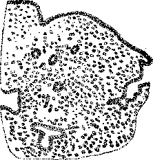
Figure 2. Decidua in the uterus in ectopic pregnancy.
the powerfully developed layer of trophoblast melts, with the help of enzymes, the underlying tissue, at which point it immediately enters the subepithelial layer directly adjacent to the muscular layer of the tube (the submucosal layer is absent in it) - a circumstance extremely important for the further course and outcomes of tubal pregnancy. Subsequent changes in the tube to some degree resemble the changes that occur in the uterus during normal pregnancy (formation of a decidua with hypertrophy and hyperplasia of the muscular layer), but there is also a substantial difference. Due to the absence of a submucosal layer and glands, decidua basalis and decidua reflexa cannot form in the tube in the same way as in the uterus. Regarding decidua reflexa, most authors hold the opinion that during tubal pregnancy it generally does not develop (others allow for the possibility of its formation in ampullary pregnancy). As for decidua basalis, it is also absent here, since there is no layer in the tube from which it could develop. The egg immediately after implantation lies on the muscularis, which is unable to react to this implantation with the development of a decidual reaction. The trophoblast, coming into contact with the muscular elements of the tube and not encountering here a protective layer of cells of the decidua, destroys the underlying tissues, resp. the wall of the tube, along with the dilated blood vessels passing through it (sometimes right down to the peritoneum itself). In the area of the implanted egg, large cells can be found, the so-called pseudodecidual cells (according to most authors, cells of Langhans' layer and syncytial giants). In those places where elements of trophoblasts grow into the wall of the tube, and later also the chorionic villi, the maternal tissue usually undergoes coagulation necrosis, as a result of which a layer similar to Nitabuch's (canalized fibrin) is formed, separating the maternal tissue from the fetal tissue. If a decidua vera forms during tubal pregnancy, it also only partially, in separate islands (see Figure 3), and not in all forms and stages of tubal pregnancy. Hypertrophy of the muscular layer of the tube during pregnancy is observed, but not as powerful as during normal pregnancy, and not throughout the entire tube; sometimes, on the contrary, there is atrophy of the tubular musculature due to the stretching of the walls by the growing egg.

Figure 3. Decidual reaction in a fold of the pregnant tube outside the site of implantation of the fertilized egg.
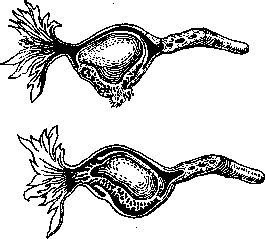
Figure 4. Internal (below) and external (above) ruptures of the gestational sac in tubal pregnancy (schematic according to Bumm).
usually in the 4th-6th week. According to Werth, two main outcomes are distinguished in the interruption of tubal pregnancy: internal rupture of the gestational sac (in former terminology - tubal abortion) and external rupture (in former terminology - rupture of the tube; see Figure 4). Apparently, internal rupture occurs more often; sometimes both are observed. Tubal pregnancy often terminates without external trauma, although in many cases its termination coincides with the moment of some trauma (lifting heavy objects, falls, jolts, coitus, medical examination, uterine curettage, intrauterine injections according to Grammaticati, etc.). The main cause of rupture of the gestational sac lies in the ingrowth of the cellular columns of Langhans (villi) with subsequent erosion of the fetal membranes and the wall of the tube. - In internal rupture of the gestational sac, only the fetal capsule ruptures (the fragile, thin decidua reflexa tears) with blood flowing into the gestational sac itself. A so-called tubal hematoma mole (mola haematomosa tubaria; see Figure 5) is formed. When the abdominal opening is closed off or when it is blocked by a blood clot, the blood accumulating in the tube stretches the tubal wall, forming a haematosalpinx, in which the blood, unlike haematosalpinx of other origins, is usually clotted. Sometimes haematosalpinx also forms in the other, non-pregnant tube, obviously due to excessive hyperemia of the tissue leading to rupture of vessels and bleeding. If in internal rupture of the gestational sac the blood flows out through the abdominal opening, accumulation of blood mass within the fimbrial end of the tube can occur, with the development of a peculiar blood tumor, the nucleus of which consists of the fertilized egg and the fimbrial end of the tube (haematocele peritubaria; see Figure 6); if, however, the blood, in larger quantity, flows into the Douglas' space, then a so-called retro-uterine blood tumor (haematocele retrouterina; see Figure 7) is formed. - In external rupture of the gestational sac, the bleeding is always more abundant than in internal rupture, and the detached egg is carried away along with the blood mass into the abdominal cavity, where it usually perishes, and the spilled blood accumulates in the same Douglas' pouch (haematocele retrouterina). Small retro-uterine blood tumors are resorbed, while larger ones become encapsulated: along their periphery (due to irritation of the pelvic peritoneum) there is proliferation of connective tissue, and the adjacent intestines, uterus, and omentum form an arch, separating the entire blood mass from the abdominal cavity. Rupture of the tubal wall sometimes occurs toward the mesosalpinx; blood, accumulating in the cellular tissue, forms a so-called broad ligament hematoma (haematoma ligament. lati; see Figure 8). Other, but rarer, outcomes of tubal pregnancy are also observed. They are connected, mainly, with the fate of the egg itself, resp. the fetus. In the vast majority of cases, when tubal pregnancy is interrupted, the fertilized egg perishes; very rarely the pregnancy, despite the rupture of the gestational sac, progresses further, and the fetus, maintaining in the abdominal cavity a connection with the placenta, continues to develop even to full maturity. These are cases of so-called secondary abdominal pregnancy. In it, the fetus may be in the abdominal cavity either without any fetal sac and without
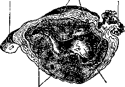
Figure 6. Haematocele peritubaria. A probe has been inserted into the tube (according to Eden).

3 ^^
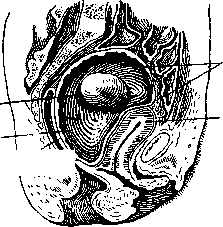
Figure 7. Haematocele retrouterina on a sagittal section: 1 - blood-soaked egg; 2 - cavity of haematocele filled with blood clots; 3 and 4 - walls of haematocele; 5 - fundus uteri (according to Bumm). amniotic fluid, or develops in an amniotic sac, or finally, a so-called secondary fetal sac is formed from false membranes and adhesions that arise as a result of reactive irritation of the peritoneum. Rarely the fetus remains alive. In the literature, about 200 cases of progressive ectopic pregnancy with a mature or almost mature live fetus have been described (Russian gynecologists have described about 30 cases). Much more often, even in secondary abdominal pregnancy, the fetus perishes. It can then undergo aseptic preservation (mummification), can petrify (petrification) with deposition of lime salts in the superficial layers of the fetus (lithokelyphopaedion) or turn into a so-called petrified fetus, lithopaedion (the entire body becomes petrified). Women can live for a long time with such petrified fetuses (in Leinzell's case, a 94-year-old old woman carried such a fetus for 46 years), become normally pregnant and give birth. If the egg membranes become petrified, a so-called lithokelyphos is obtained. For mummification and petrification, aseptic conditions are required; in their absence and in the presence of infectious agents, maceration, suppuration, putrefactive and ichorous decomposition of the fetus itself, of the entire gestational sac, and of the formations associated with the interruption of tubal pregnancy (haematosalpinx, broad ligament hematoma, retro-uterine blood tumor) can occur. Products of decomposition are expelled directly outward or are secreted through neighboring organs (bladder, rectum, vagina, etc.). Purulent-decomposed parts and detached bones of the FETUS MAY BE EXCRETED through the abdominal wall or through the "genital organs"
Figure 8. Rupture of the tube into the broad ligament with formation of a hematoma: 1 - hematoma. 2 - gestational sac
face. After many years. Clinical picture, symptomatology and diagnosis of tubal pregnancy. Typically, tubal pregnancy proceeds with the following phenomena: the woman considers herself pregnant (delayed menses, nausea, tendency to vomiting, etc.), often with infantilism or inflammatory changes in the genital sphere. Usually, she has infertility, absolute or relative, of the Einkindsterilitat type, and even more often-a long interval between the current pregnancy and the previous one (on average, 4-7 years). At the 4-6th week after cessation of menstruation (often during complete well-being), the pregnant woman suddenly has a stroke with all the signs of abdominal shock: extremely sharp pain, sometimes of a cramping nature, initially unilateral, localized in one or another groin, often radiating to the corresponding shoulder or scapula (the so-called phrenicus-symptom-pain from irritation of the thoraco-abdominal nerve). The disease immediately takes on a threatening character. The signs of internal bleeding come to the fore: fainting state, characteristic paleness of the face with a bluish tint on the lips; pulse above 100, small, rapid, of weak filling, sometimes barely palpable or not felt at all (not so much from blood loss as from shock); temperature normal, less often-subfebrile and even less often-subnormal; limbs and tip of the nose cold; very soon symptoms of peritonitis appear-bloating of the abdomen, especially below the navel, sharp tenderness of the abdominal wall, sometimes nausea and vomiting. Reflex urinary retention (anuria according to Pischashek) is often noted. Sometimes such an attack can occur after some moments to which patients usually point (she lifted something, stumbled, walked a lot, medical examination, sexual intercourse, etc.). Soon after the onset of the attack, bloody discharge from the vagina usually appears. External bleeding, as a rule, is moderate, resembling menstruation on the first or last day, with the blood being brown in color, and later completely dark, like tar. Sometimes membranes are discharged with the blood, and very rarely, accompanied by intensified pain and bleeding, the decidua membrane (the discharge of the shedding membrane is noted no more often than in 25% of cases). Attacks in ectopic pregnancy can occur repeatedly. Cullen and Hellendall in fresh shock cases noted a special discoloration of the skin in the navel area: blue color with all transitions from greenish-yellow to orange. The noted phenomenon is based on the absorption by numerous lymphatic vessels through the stretched anterior abdominal wall of the blood that has entered the abdominal cavity. It is rarely possible to prove the presence of blood in the pelvic cavity. With significant blood loss (1-2 liters), the blood accumulating in the lateral parts of the abdomen (in the horizontal position of the patient) can be determined by percussion (dulling of the intestinal tone); but in other cases (and this happens more often) even with the help of puncture of the posterior fornix, which is not indifferent for the further course of ectopic pregnancy (introduction of infection), a definite result is not always obtained. Recently, a simple diagnostic method for determining hematine in the patient's serum has been frequently used-the so-called Antoshina-Egorov pyramidone test. Its technique is as follows: 2-3 cubic cm of blood are taken sterilely from the cubital vein of the patient, placed in a thermostat at 37° for 1 hour (preferably for a day), the serum is aspirated, centrifuged twice, and one drop of it is mixed with a special reagent [3 cubic cm of 5% alcoholic (96°) solution of pyramidone, 8 drops of 50% acetic acid and 8 drops of fresh hydrogen peroxide]. In the presence of hematine, a beautiful purple (amethyst) coloring appears after 1-2 minutes, lasting 5-8 minutes. The reaction is not specific, but it can serve as a good auxiliary method in the diagnosis of ectopic pregnancy. The data of objective examination, and in part the entire clinical picture of tubal pregnancy, will vary depending on its particular stage, which is important for differential diagnosis. In progressive, uninterrupted early tubal pregnancy, there is a picture of uterine pregnancy; on bimanual examination, a tumor is determined, located next to the uterus, somewhat to the side and behind it, mobile, of a sausage-like shape, of unelastic consistency, slightly painful, with a pronounced pulsation of the vessels. The size of the tumor depends on the duration of pregnancy: at the end of the fourth week it is the size of a pigeon's egg, at the sixth week it reaches the size of a chicken's egg, and by the end of the tenth week it is almost equal to the size of a goose's egg. In 70-90%, a positive reaction with floridzin (matrine) is obtained. The blood picture is normal, the sedimentation reaction (SR) is as in uterine pregnancy (1 hour 30 min.-2 hours). It is very difficult to make a correct diagnosis at this stage (4-6 weeks). Unlike uterine pregnancy complicated by some process in the area of the tubes, resp. appendages (oophoritis, retention tumors, ovarian cysts, etc.), one can recommend the method used by Banki: if two fingers are inserted into the posterior fornix of the vagina and the cervix, resp. uterus, is lifted by them, bringing it closer to the symphysis, the patient will experience severe pain. Obviously, the egg, before the onset of intracapsular rupture, by embedding itself into the wall of the tube, causes small hemorrhages here, and the blood entering the Douglas space leads to irritation of the peritoneum. According to some data (Gubarev), in tubal pregnancy, unlike uterine pregnancy, the so-called new sign of Hegar is not obtained. To establish a final diagnosis, it is best to place the patient in a hospital setting, where through repeated examinations it is possible to establish or reject the growth of the uterus corresponding to the development of pregnancy. For later stages of progressive ectopic pregnancy, starting from the 4th month, X-rays can be used. Leiser managed to determine parts of the fetal skeleton on the 14-20th week of tubal pregnancy using X-rays. Some have used the application of pneumoperitoneum. The picture of interrupted tubal pregnancy, in its clinical course, resembles a number of diseases with a similar abdominal symptom complex (perforating peritonitis, ileus, appendicitis, twisting of the stalk of ovarian tumors, rupture of ovarian cysts and other diseases characterized by the so-called "acute abdomen"). Only a carefully collected and properly interpreted anamnesis and comprehensive objective examination make it possible to sort out the confusing picture of the disease. In peritonitis, in general, there is a more pronounced picture of inflammation (high temperature, more severe vomiting, sharp defense musculaire, significant leukocytosis) in the absence of pronounced anemia (normal picture of red blood cells). More detailed reference points for differential diagnosis are given in the table on p. 251 (at the top). In particularly confusing cases, one can use the Promptov method. In appendicitis, examination of the patient through the rectum gives severe pain in the Douglas pouch area, while lifting the uterus with a finger in the rectum, in the area of its external os, does not cause pain or it is minimal. In ectopic pregnancy (and in acute inflammation of the appendages), the reverse relationships are obtained: there is no pain in the Douglas pouch, and on the contrary, it is sharply expressed when attempts are made to lift the uterus upward. Examination technique: the finger is inserted into the rectum and advanced upward, toward the bottom of the Douglas pouch, strictly along the median line, but Tubal abortion Maternal abortion 1. More often observed between the 4th and 6th weeks of pregnancy, rarely later. 2. The Solley attack is of a more violent nature (sudden onset, shock, collapse, fainting state, signs of peritoneal irritation). 3. External bleeding is small, sometimes completely absent, the blood is dark, liquid, brown in color; sometimes membranes are discharged. 4. The symptoms of hemorrhage and shock are much stronger than the visible blood loss. Pregnancy. The uterus is slightly enlarged, not quite corresponding to the duration of pregnancy; the os is closed.
A tumor on one side of the uterus, characteristic of ectopic pregnancy (see above). 7. More noticeable increase in pulse rate (above 100 beats per minute). 1. Most commonly between 8 and 12 weeks. 2. Slow, gradual increase in regular pains, similar in nature to labor pains. The pains of moderate intensity are localized in the central part above the symphysis. 3. External bleeding is profuse, sometimes torrential, the blood is bright red with clots; sometimes placental tissue is discharged, with a necrotic odor. 4. Symptoms of hemorrhage are proportional to visible blood loss. 5. The uterus is enlarged according to the month of pregnancy, the os is often slightly open. 6. Next to the uterus are either normal adnexa or inflammatory formations, almost always bilateral, less painful, better defined. 7. Less noticeable increase in pulse rate, deviating neither to the right nor to the left. To lift the uterus upward, the inserted finger is slightly withdrawn downward and brought to the external os of the cervix. - Sometimes a ruptured ectopic pregnancy (tubal abortion) can be confused with a miscarriage, which can have particularly serious consequences (different therapy). Data for differential diagnosis are presented in the table on p. 251 (at the bottom). In some cases, dirty, small bloody discharge ('spotting') is observed also in inflammatory processes of the ovaries and can simulate ectopic pregnancy. In addition to the above, it is recommended (Wagner) in such cases to try an injection of pituitrin. Cessation of bleeding after injection speaks more in favor of an inflammatory process (the uterus will contract); and conversely - with continuing bleeding (bloody discharge comes from the tube) ectopic pregnancy can be suspected. The discharge of the detached membrane from the uterus makes the diagnosis of ectopic pregnancy indisputable. Some (Lider, Ott) resort to diagnostic curettage of the uterus. The diagnostic value of this procedure (in ectopic pregnancy elements of the ovum should be absent) is not so great as to justify its use, considering the associated risk (cases of fatal outcome after diagnostic curettage have been described). - Regarding the recognition of retro-uterine blood tumor, the following must be kept in mind: in the circle of diagnostic considerations in this case, various tumors (ovarian, tubal) and exudates occupying the space behind the uterus should be included. Here, in differential diagnosis, one has to resort to a trial puncture through the posterior fornix, which is far from safe but is the only one that in complicated cases can lead to the right path. More often a retro-uterine blood tumor is confused with a retroflexed pregnant uterus; this error has more than once served, during the correction of a supposed retroflexion, as the cause of fatal bleeding from a ruptured retro-uterine blood sac. The differential diagnosis of these two pathological conditions is given in the table on p. 253. Treatment of tubal pregnancy. A progressive, undisturbed, viable ectopic pregnancy of the first half should be operated on as soon as its diagnosis is made. This view is shared by most gynecologists, joining Verth, who equates ectopic pregnancy with malignant neoplasm: the earlier the operation, the better. Most prefer the abdominal route for the operation, but it can also be done through the vagina. The choice of incision is not of major importance. The position put forward by Bert, that every recognized ectopic pregnancy must be operated on immediately, regardless of the child or the stage of pregnancy, is not satisfactory to all at present. Many gynecologists, especially French ones, find it possible to demand that when indications for operation are established, the fate of the living child in ectopic pregnancy during its second half should be taken into consideration on equal terms with the fate of the mother. Most, however, still considers Verth's requirement for immediate operation of recognized ectopic pregnancy correct also for the second half of pregnancy. Treatment of ruptured ectopic pregnancy should also be surgical. The operation should be performed, if possible, immediately, since any waiting (allowing the patient to recover from shock) can not only be harmful but also dangerous. It is necessary to operate even in extremely serious condition of the patient. By means of operation it is often possible to save the patient in cases where apparently there is no hope. When operating on a ruptured ectopic pregnancy, the abdominal route is preferable, and during the operation the gestational sac and the entire pregnant tube are removed. The proposal of some surgeons - to necessarily remove the second, non-pregnant tube (according to Verth in 5% of cases a repeated ectopic pregnancy occurs in the second tube) - finds few followers at present. It is often observed after ectopic pregnancy that a normal uterine pregnancy ensues (according to some statistics - in 50%), which makes one adhere to a conservative method of operating: to remove the second tube only in case of its pathological changes or if there is haematosalpinx (see above). The proposal of some surgeons to limit the operation to removing only the ovum, without removing the tube itself (the egg is either expressed through the ampullary end or, after a longitudinal incision along the length of the tube, is removed by scraping with a sharp curette) has not found wide acceptance. The remaining tube can soon become a source of a new stroke (viable villi may remain in the walls), and later - a predisposing factor for repeated ectopic pregnancy. The blood that has poured into the abdominal cavity should be removed as much as possible. Many believe that retro-uterine blood tumor does not require immediate operation, since such a haematocele often heals spontaneously. At present, in this stage, most prefer surgical intervention, considering the possibility of repeated hemorrhage and the tendency of such blood tumors to suppuration and disintegration. In such cases, suppuration is always more dangerous than the operation (Bumm). In case of suppuration or gangrenous disintegration of such a tumor, surgical intervention in the form of posterior colpotomy with drainage of the cavity with gauze is indicated. If it is impossible to operate for ectopic pregnancy, the patient has to be treated symptomatically (morphine, ice, subcutaneous infusions of physiological solution, cardiac agents, etc.). Rare forms of ectopic pregnancy. Multiple ectopic pregnancy. If we speak of cases of true multiple ectopic pregnancy, i.e. when both ova are products of the same ovulatory period, then it occurs in ectopic pregnancy no more often than in uterine. Verth reduces all cases of multiple ectopic pregnancy to three forms, of which the most common is that in which one fetus is in the uterus and the other develops ectopically (about 300 such cases have been collected in the literature, and in only 9 were both children extracted alive). A rarer form of multiple ectopic pregnancy is that in which both fetuses are in one tube. Only 25 such cases have been collected [once - triplets, the case of Nijhoff]. The third form is the so-called bilateral tubal pregnancy, in which each tube contains one fetus, and pregnancy developed and arose simultaneously in them. Only 32 such cases are known in the literature. - Interstitial pregnancy, graviditas interstitialis, belongs to the rarest forms of tubal pregnancy. In the entire world literature, no more than 70 cases have been described. Anatomically, a distinction is made: true interstitial pregnancy - tubo-isthmic (the isthmic part is involved) and utero-isthmic (it adjoins the uterine cavity). The conditions for the development of the decidua in the tube, hypertrophy of the tubal musculature, and development of decidua in the uterus are the same as in other forms of tubal pregnancy. Outcomes: more often an external rupture of the gestational sac with threatening hemorrhage into the abdominal cavity occurs (intracapsular rupture is less common); cases of carrying the fetus to term in interstitial pregnancy (secondary abdominal pregnancy) or (significantly more often) its death (lithopedion, maceration) have been described.
Distinctive signs of true interstitial pregnancy: displacement of the fundus of the uterus, due to the growth of the pregnant horn, into the non-pregnant side; typical displacement of the round ligaments and tubes (they lie on the healthy side lower than on the diseased side, and on the outer, lateral side of the gestation sac); broad base of the gestation sac and presence of a groove (constriction) between the tumor and the uterus. Recognition is very difficult. Often only microscopic research clarifies the diagnosis. Treatment is surgical (excision of the pregnant horn with subsequent suturing and peritonization, sometimes using the round ligament). Ovarian pregnancy, gr. ovarialis (see figure 9), has several topographical variations: intrafollicular ovarian pregnancy (development of the egg inside the follicle, resp. corpus luteum), epiovarian ovarian pregnancy (the egg implants on the outer surface of the ovary), and interstitial ovarian pregnancy.
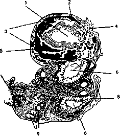
Figure 9. Ovarian pregnancy: 1-egg cavity; 2-chorionic villi; 3-egg sac formed by the wall of the corpus luteum; 4-site of rupture of the egg sac; 5-blood clots; 6-diverticula of the corpus luteum; 7-hilus ovarii; 8-opening of the corpus luteum with fibrin deposits; 9-follicles (according to Tissenbrock).
ovarian (the egg is immersed in the ovarian stroma). Reliable cases of ovarian pregnancy have been described very rarely. Some believe that the so-called ovarian hematomas, which are often encountered, frequently result from ovarian pregnancy. Decidua does not develop in ovarian pregnancy. Outcomes: usually ovarian pregnancy is interrupted at 6-8 weeks by the type of external rupture of the gestation sac, with formation of hematoma inside the gestation sac (death of the egg, development of hematoma), but sometimes it goes to full term. The latter occurs more often than in other forms of ectopic pregnancy (abundance of blood vessels, extraordinary ability of ovarian tissue for rapid and excessive proliferation of cellular elements, relatively greater capacity of the cavity in intrafollicular implantation of the egg, etc.). Topographic diagnosis of ovarian pregnancy is impossible. Primary abdominal pregnancy is extremely rare (at one time it was completely denied). In recent years, several reliable cases have been described. The fertilized egg must primarily implant on the peritoneal endothelium [in the Douglas pouch, on the broad ligament (see figure 10)]. To prove primary implantation of the egg on the
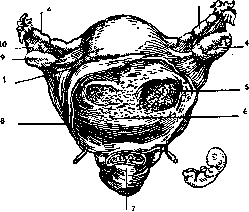
Figure 10. Primary abdominal pregnancy: 1-adhesion of the capsule to the posterior wall of the uterus; 2-left tube; 3-right tube; 4-right ovary; 5-fundus of the placental bed; 6-bed of the afterbirth; 7-rectum; 8-capsule; 9-left ovary; 10-mesosalpinx.
peritoneum is possible only in the early stages of pregnancy (presence of viable villi on the peritoneum, absence of microscopic signs of pregnancy in the tubes and ovary). Pregnancy in a rudimentary accessory horn of the uterus (see figure 11) is a rare anomaly of pregnancy (only 22 cases have been described in Russian literature). The rudimentary accessory horn is almost always connected to the uterus (normally developed horn) by a compact broad stalk, which departs from the uterus at the level of the internal os. In 85% of cases, the stalk is not canalized ('closed' horn). The muscular tissue in the rudimentary horn is poorly developed; connective tissue predominates with poorly developed blood vessels. Pregnancy in such a horn (hypatretic pregnancy, according to Sänger) can occur by
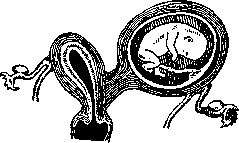
Figure 11. Pregnancy in a rudimentary horn of a bicornuate uterus.
external migration of the egg (migratio ovi externa) or spermatozoa (unlikely). Anatomically, it proceeds according to the type of ectopic pregnancy: a decidual membrane develops (only more powerful), the muscular tissue hypertrophies, in the empty horn (in the uterus) its own shedding membrane develops, etc. Due to more powerful hypertrophy and better decidual protection, pregnancy in the rudimentary horn relatively often (25%) goes to full term, but even more often (45%) it is interrupted, only much later than tubal pregnancy (usually at 4-5 months). In addition to going to term and rupture of the gestation sac, postmaturity (25%) is observed with subsequent death of the fetus (maceration, lithopedion). Interruption of pregnancy in the rudimentary horn occurs by the type of external rupture of the gestation sac, which is more dangerous in its consequences than rupture in tubal pregnancy. In recognizing pregnancy in the rudimentary horn, it is often confused with other types of ectopic pregnancy. Distinctive signs are considered: the round ligament extending outward (laterally) from the gestation sac (in tubal pregnancy it extends medially), presence of a thick fleshy stalk, clear contractions of the fetal sac under the hand in the presence of an 'empty' uterus, as well as absence of painful sensations and complete mobility of the gestation sac (Abuladze). Unlike interstitial pregnancy (in which the round ligament also extends laterally), in pregnancy in the rudimentary horn there is a well-expressed stalk connecting the gestation sac with the separately lying uterus. Among the differential diagnostic possibilities, one should also remember about ovarian cyst during normal pregnancy; in such cases, it is necessary to wait to confirm the progressive growth of the 'tumor'. Pregnancy in the rudimentary horn can be suspected in the presence of a double vagina or double cervix. The position of the non-pregnant developed horn can be determined by sounding with simultaneous use of X-ray. Treatment is surgical. Recently, for progressive pregnancy in the accessory horn, in the interests of the child, an expectant method is recommended (under clinical conditions). In case of rupture, immediate surgery is necessary. During laparotomy, especially in young individuals, the greatest possible conservatism should be exercised: excise only the pregnant horn together with its tube. In such cases, the distal end of the round ligament is sutured to the fundus of the remaining uterus. The mortality rate for this operation is about 5.5% (Beckman).
M. Malinovsky. Pregnancy from a forensic medical perspective. The main questions from a forensic medical point of view regarding P. are: recognizing P. at a given moment, determining its term, and more often determining a past P. (that ended in childbirth) in cases involving infanticide, child abandonment, child substitution, paternity, alimony, abortion, etc. In these cases, the expert must, besides confirming P., determine its duration, the time of onset and completion of childbirth, abnormalities of P., the causes of its termination, as well as establish the connection between P. and the cause of death, the possibility of P. not being recognized by the woman herself, and a number of other questions. The well-known signs of P. sometimes have significant deviations and present considerable difficulties for the expert from a forensic medical perspective; information obtained by questioning the woman about intimate aspects related to P. rarely causes doubt about its truthfulness; the expert, however, should cautiously rely on the testimony of the pregnant woman as an interested party, and should be guided only by objective data. One of the essential signs of P.—cessation of menstruation—despite its well-known significance for obstetricians and the public, does not serve as absolute proof for the expert, since P. can occur in non-menstruating women, and the cessation of menstruation itself may depend on other causes; on the other hand, the proper appearance of menstruation is sometimes observed after conception and even throughout the entire P. (Casper-Liman A., Mayer, Lewy, Hofmann, and others). Enlargement of the uterus, and consequently of the abdomen, arouses suspicion of existing P.; however, it is clear that abdominal enlargement can also depend on other causes. Moreover, it can be concealed, and in other cases, conversely, simulated. Obstetric, and moreover repeated, examination of the uterus provides the most reliable proof of P., especially in its second half, when it becomes possible to feel the movements of the fetus and its parts and to hear its heart tones (see Obstetric examination). Although the signs of P. mentioned above seem the most reliable, the most experienced obstetricians do not deny the possibility of gross errors on the part of both the physician and the person being examined. Changes in the breasts, in the form of the appearance of pigmentation of the nipples and, even more so, of the areolas, occur during the first two months of P. and reach a significant degree in its second half. Swelling of the follicular glands in the area of the areola from the second month of P. is also (according to Faye) a very constant phenomenon. Other signs of P., such as: coloring of the vaginal mucosa to the color of wine yeast, swelling of the genital parts and lower extremities and other phenomena, can only confirm the presence of P. The duration of P. can only be determined with accuracy in rare cases due to the impossibility of establishing the day of conception. Overdue childbirth has long caused conflicting opinions among experts: some deny P. beyond 40 weeks, while others allow for a duration of P. of 320 days or more. Our legislation, as before, has established the term of P. as 306 days; this term is considered short by Olshgausen. If the exact day of birth has not been established by the investigation, the expert must determine the day of childbirth, which is especially important in cases of simulated overdue childbirth; in fresh cases, examination of the mother and child can provide sufficient grounds for establishing the time of past childbirth and can reveal false statements by the mother. When discussing such cases, data rarely exists that would support the possibility of intercourse on the very day of the husband's death or shortly before the termination of marital relations in divorce. Of the abnormalities of P., from a forensic medical perspective, secondary conception, implantation pregnancy, and ectopic pregnancy are of interest. The most common form of ectopic pregnancy is tubal, usually ending suddenly with rupture of the tube between the 2nd and 3rd months of P., with a fatal outcome from internal hemorrhage, which has more than once aroused suspicion of poisoning (a case in Prague; in Moscow after eaten sausage; in Petersburg after lunch—the son accused his mother of poisoning his wife). Sometimes recovery with an outcome in lithopedion, etc., is possible. It is believed that P. can result from fertilization of several eggs after one as well as after repeated intercourses. Despite numerous examples of multiple births of different developments, the question of secondary conception remains controversial to this day. - When examining a corpse, the question of a past P. often arises in connection with the death of a woman from sepsis after an induced miscarriage. Finding the ovum or parts of it, the site of attachment of the placenta, and the size and properties of the uterus provide sufficient data to confirm P. After the rupture of the Graafian follicles in the ovaries, false yellow bodies are formed. These differ from the true yellow body, which forms at the site of the follicle from which the egg that underwent fertilization emerged; the true yellow body differs from the false one in size (like a cherry or a hazelnut), its yellow color, and the fact that it occurs in only one ovary. This sign is very valuable but not unconditional. After childbirth, the uterus weighs 1 kg, after 2 days—0.75 kg, after a week—0.5 kg, after 2 weeks—375 g, and only after 5-6 weeks it reaches its original weight of 80-120 g in women who have given birth (Vibert). The uterus after childbirth sharply decreases in size, and its fundus descends slightly below the navel, lowering daily by about a centimeter and disappearing behind the pubic symphysis on the 10-12th day. The uterine os is widely open, with tears; the vaginal part is flaccid, funnel-shaped, and passable for a finger to the internal os for a week; then, from the second week, the vaginal part elongates and by the end of the second week forms a soft cylinder. Postpartum lochia during the first 3-4 days consist almost entirely of pure blood (lochia cruenta), then in the second half of the week they take the appearance of meat slops (lochia serosa), and finally thicken, becoming more and more cloudy-whitish (lochia alba), sometimes yellowish or greenish in color; their duration is 2-3 weeks, and of all postpartum lochia about 4-6 weeks, depending on the nature of both the childbirth itself and the postpartum period (the possibility of past female diseases must also be taken into account). Other signs of a past P. help in recognizing past childbirth: flaccidity and stretching of the abdominal walls, which disappear at different times, depending on the woman's age and the number of past births (scars on the abdomen of a pregnant woman remain for life). Pigmentation of the areolas and the white line of the abdomen disappears slowly, the mammary glands in non-nursing women remain coarse-lobulated and secrete a small amount of milk a month or more after childbirth. Signs of recent childbirth become unreliable after a month and finally disappear after 6 weeks. The question of repeated childbirth is extremely difficult to resolve. P., not recognized by the pregnant woman herself, has more than once been the subject of legal proceedings when intercourse was performed with a woman in an unconscious state, sometimes in a state of extreme intoxication. This possibility must also be admitted in young individuals who have not reached sexual maturity, those limited intellectually, not to mention the mentally deficient and mentally ill. Abnormalities of menstruation, the climacteric, chronic diseases resembling the signs of P., especially if these signs are maintained by the attending physician, can mislead non-pregnant women regarding the presence of pregnancy.
Related articles
Mentioned in
Cite this page
“Pregnancy.” Soviet Medical Encyclopedia. English translation of Bolshaya Meditsinskaya Entsiklopediya, 1st ed. (Moscow, 1928–1936), ed. N. A. Semashko. https://sovietmedicalencyclopedia.pages.dev/article/pregnancy/