Yolk
Historical document, translated for reference. It reflects medical knowledge of the 1920s–30s and is not medical advice.
Summary
This article defines yolk as the nutritive substance within an egg cell, detailing its classification based on distribution (alecithal, telolecithal, centrolecithal) and its structure in avian eggs. It further explains the biological function of yolk as a nutrient reserve for the developing embryo and defines related embryological terms.
Encyclopedia article (1928–1936)
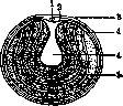
YOLK (vitellus, lecithus), a name initially given to the granular, opaque, sometimes colored substance of an egg (ovum); later, when it was proven that the egg is a cell, the name yolk was transferred to the entire body of the egg cell. Reichert was the first to distinguish between formative yolk (vitellus formativus), corresponding to the part of the egg rich in protoplasm, and nutritive yolk (vitellus nutritivus), which represents deutoplasm. The distribution of these types of yolk in different egg cells is not uniform and allows them to be divided into three main groups (Balfour): alecithal, with a small amount of evenly distributed yolk (mammals); telolecithal, with a polar distribution of formative yolk at the top and nutritive at the bottom (fish, reptiles, birds); and centrolecithal, with nutritive yolk in the center and formative yolk at the periphery (arthropods). At the present time, the name yolk is applied almost exclusively to nutritive yolk; its quantity determines, first and foremost, the size of the egg cell. The nutritive yolk of a boiled chicken egg, on a cross-section passing through both poles, reveals two modifications: yellow and white yolk (Fig. 1); the former is denser and forms the main mass, while the latter is liquid, covers the surface of the yellow yolk in a thin layer, accumulates in greater quantity under the germinal disc (Pander's nucleus), and penetrates from there toward the center of the egg in the form of a flask-shaped thickening (latebra—hidden area); in addition, it forms thin concentric layers in the yellow yolk. Under a microscope, yolk consists of formed elements—yolk globules or plates of oval shape. The white yolk of a chicken egg (Fig. 2) consists of soft, large globules (up to 100 µ) containing tiny grains, while the yellow yolk consists of smaller globules with larger, strongly refracting grains.
Yolk represents a reserve of nutritive material consumed by the embryo during development; the composition of chicken yolk includes: vitellin, lecithin, cholesterol, neutral fats, mineral salts, and the yellow pigment lutein. As development proceeds, the formed elements of the yolk dissolve and are utilized by the cells of the embryo; in amphibians, they remain visible in the cells of the larvae for a long time.
Vitelline membrane, or skin (membrana vitellina)—the primary egg membrane secreted by the egg cell itself. Yolk cells—nutritive cells containing yolk in the complex eggs of flatworms; they are formed in the vitellarium, a special section of the reproductive apparatus. Yolk nuclei—large nuclei (merocytes) in the upper sections of the yolk adjacent to the germinal disc in eggs with partial cleavage; yolk nuclei arise as a result of cleavage and, accumulating in large numbers along the edges of the disc, form a yolk syncytium (periblast). Balbiani's yolk nucleus—see Cell. Rusconi's yolk plug—see Blastopore. Yolk vesicle, yolk duct, yolk vessels—see Embryo.
V. Karpov. Figure 2. Chicken egg cell: 1—germinal vesicle; 2—protoplasm (formative yolk); 3—vitelline membrane; 4—white yolk; 5—yellow yolk.
Related articles
Mentioned in
Cite this page
“Yolk.” Soviet Medical Encyclopedia. English translation of Bolshaya Meditsinskaya Entsiklopediya, 1st ed. (Moscow, 1928–1936), ed. N. A. Semashko. https://sovietmedicalencyclopedia.pages.dev/article/yolk/