Embryo
Historical document, translated for reference. It reflects medical knowledge of the 1920s–30s and is not medical advice.
Summary
This article defines the embryo in zoological, botanical, and microbiological contexts, with a detailed focus on early human embryology. It covers the stages of development from fertilization and implantation to the formation of the blastoderm and embryonic membranes.
Encyclopedia article (1928–1936)
EMBRYO, a) E. in zoology (embryo)—an animal in the period from the beginning of egg cleavage to the moment of exit from the egg membranes, respectively, from the mother's body. Nutrition of the E. occurs in oviparous animals at the expense of nutritional reserves (yolk) of the developing egg, and in viviparous animals, in which the circulatory system of the E. communicates with the circulatory system of the mother—at the expense of the maternal organism. The E. of mammals in the uterus is usually called a fetus. b) E. in botany—the rudiment of flowering plants, consisting of all main organs (root, stem, and leaves) and enclosed in the seed. The main mass of the E. is formed by fleshy cotyledons, i.e., embryonic leaves, which contain reserves of nutritional material (starch, oil) for the nutrition of the germinating embryo. c) In popular microbiological literature, the term E. (German Keim, French germe) is often used to denote any living bacterial microorganism in general, regardless of its stage of development (e.g., anthrax E.).
Human embryo. The human E., or embryo, denotes (in human embryology) the initial stages of development from the moment of fertilization of the egg cell to the end of the 2nd month of intrauterine life, when the developing organism, reaching a length of about 2 cm, acquires characteristic human features. Further stages of development from 3 months to birth are called a fetus. Embryonic stages include cleavage, implantation into the mucous membrane of the uterus, formation of the blastoderm and primary organs, the laying down of definitive organs and the beginning of histogenesis, formation of the E. body, development of limbs, head, and face. Simultaneously with this, the formation of embryonic membranes occurs: the trophoblast, amnion, yolk sac, allantois, and chorion; only the development of the placenta occurs later, from the 3rd month.
The very first stages of development of the human E. have not been observed, and they have to be constructed on the basis of the development of other mammals and theoretical considerations; then follow stages known from isolated observations; it is customary to designate them by the names of the authors who described them. From the end of the 1st month, human E.s have been studied quite fully, although much in them is still unclear (e.g., age). The rule of His, according to which "the age of the E. is the time elapsed from the first day of the first missed menstrual period," turned out to be devoid of any foundation, and at the present time, the designation of the E. by age has been abandoned. But on the other hand, the designation of human E.s by their size (occipito-coccygeal length, from the occipital to the coccygeal tubercle) accepted at the present time does not give a proper concept of the degree of development, since these dimensions can vary within fairly large limits. Therefore, the American embryologist Mall proposed designating human E.s from 2 mm to 22 mm with letters (from N to U); this rational proposal, however, did not enter into new textbooks.
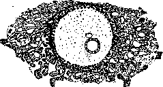
Fertilization of the egg cell occurs in humans, in all probability, in the upper ampullary part of the tube, where sperm penetrate already a few hours after coitus. The fertilized egg begins to divide immediately; cleavage, as one can assume, is complete, uniform, and as a result, a morula stage arises [Figure 1—morula stage—about 10 days at the moment of implantation. The outer layer—the trophoblast (darker)—is distinguished from the central cell cluster, the so-called embryoblast (diagram according to Grosser)]. During cleavage, the egg moves along the tube into the uterus partly due to the movement of the cilia of the ciliated epithelium lining the tube, and partly by peristaltic contraction of its muscular wall. The movement lasts 7-8 days, after which the embryo, having reached the uterus, begins to implant into its mucous membrane (see Decidua, Pregnancy), which takes, as is assumed, another three days. The process of implantation has not been directly observed, but judging by the known early stages of implanted human embryos, as well as the pictures of implantation of the guinea pig embryo (Graf Spee), one can assume its active penetration, accompanied by histolysis of the mucous membrane with the help of enzymes and, perhaps, its amoeboid movements.
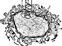
The E. at this time is probably in the morula stage, in which a division into a surface layer of cells, the future trophoblast, and an inner cell mass—the embryonic node (Figure 1)—is outlined; according to another assumption, the E. has already managed to turn into a vesicle. The site of implantation is plugged with a fibrinous plug, and the tissue surrounding the E. disintegrates and often dissolves, forming a nutritional mass—embryotroph; behind it follows a reactive zone with dilated vessels and emigration of leukocytes. The first well-studied human E. is the Bryce-Teacher E. (1908; Fig. 2), obtained by spontaneous abortion. It consists of a vesicular trophoblast (0.7-0.5 mm), from which reticular cellular outgrowths extend, immersed in disintegrating tissue, and in some places—into blood lacunae. The cavity of the trophoblast is filled with a delicate network of mesenchyme and contains two vesicles: the larger—the amnion vesicle (or ectodermal) and the smaller—the yolk (or endodermal) vesicle; the blastoderm has not yet managed to form. The age of this E. was estimated differently—from 13 to 15 days. A key to understanding the Bryce-Teacher E. is provided by the Peters E. (1899; Fig. 3), obtained by autopsy of a suicide (a similar E. was described by Mollendorff; 1921). The trophoblast is of larger size (internal dimension 1.6-0.9 mm), with mesenchymal strands in the cavity. The E. consists of two vesicles pressed against each other; the upper—amniotic, the lower—yolk; between them is the blastoderm, formed by two germ layers:
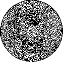
ectoderm from cylindrical cells and endoderm from flattened ones. On the sides are two cavities bounded by mesenchyme (extraembryonic body cavity, or exocoelom); mesenchyme also penetrates between the layers. The age of this embryo is from 14 to 18 days. The described E.s clearly show that human development proceeds differently than that of most mammals (rabbit, dog): the amnion is formed not by folds on the surface of the trophoblast, but by splitting (formation of a cavity) in the depth; in the same way, the blastoderm is formed inside from adjacent sections of the amniotic and yolk vesicles. Something similar is observed in the development of the hedgehog and lemurs, and these data allow us to reconstruct the first stages of development of the human E. It is possible to assume that the morula turns into a vesicle, the wall of which forms the trophoblast, and the embryonic node remains inside, not adhering to the surface, as, for example, in the rabbit; at the same time, early formation of mesenchyme occurs in the cavity of the trophoblast (morula-mesoderm of authors; Fig. 4). This stage probably corresponds to the SCH Mollendorff embryo (1924), the earliest of those described (12-15 days; Fig. 5); unfortunately, it was obtained in a deformed state, and views on it diverge. Further, the embryonic node probably disintegrates into two clusters of cells, from which one gives rise to the amnion vesicle by the formation of a cavity, the other—to the yolk vesicle, whereby the formation of the latter can also proceed by the folding of the cell plate into a vesicle (in lemurs). Stages later than Peters, e.g., Spee Gl. or Eternod I (Eternod; Fig. 6), show an oval trophoblast (chorion; 10-9 mm), which can be separated entirely from the mucous membrane and receives the name of the human egg; in it, the E. has the appearance of an oval blastoderm 1.3-1.5 mm in length, the ectoderm of which passes into a low amnion, and the endoderm—into a more voluminous yolk vesicle (syn. yolk sac) with protein content. The E. is covered with mesenchyme, which thickens at the posterior end of the blastoderm, forming a short cord attaching the E. to the chorion, the so-called abdominal stalk (Haftstiel, Bauchstiel); the allantois rudiment enters it, growing from the posterior end of the yolk vesicle in the form of a glove finger.
The development of the human blastoderm proceeds, as in other mammals, according to the type of chick development. Reconstruction of the embryo 1.17 mm (Frassi) (Fig. 7) shows at the posterior end of the blastoderm the primitive streak with Hensen's node; from it extends forward the head process. The primitive streak-a proliferation of ectoderm-gives rise to lateral outgrowths of mesoderm, the middle germ layer; in the region of the node all three layers are fused into one mass, which is penetrated by a canal extending obliquely from behind forward-canalis neurentericus. The head process, fused with the endoderm, gives rise in its axial part to the notochord, and to its sides also to mesoderm. At approximately the same time, the nervous system is laid down in the form of two brain ridges, or folds of ectoderm, running parallel to each other on either side of the head process. Subsequently they begin to fuse into a tube, starting from the middle. With further growth of the blastoderm, the primitive streak comes to lie at its posterior end; a thickening appears in it-the tail bud, which serves as a source for the development of the posterior part of the body with limbs and tail (Fig. 8). 3. described- Fig. 4. The trophoblast has increased significantly, the embryoblast has differentiated into the embryonic node and the mesoderm of the morula (schema according to Grosser). of structure, not yet having somites, are considered early stages and are designated by the names of authors; in all 35 have been described (summary by Grosser; 1927). At the end of this period (about 20 days) the rudiments of the circulatory system appear, first in the form of islands in the wall of the yolk sac (blood islands), which merge into networks, and then in the lateral parts of the 3. in the form of two aortas and a paired rudiment of the heart, which gradually connects into one tube. In the embryo of Eternod 1.3 mm there is already primary circulation (see color plate, Fig. 2), which however has recently been disputed (Ogovveg). Subsequent stages are characterized by 1) segmentation of the mesoderm, 2) closure of the neural tube, and 3) formation of the head and tail gut. A well-studied embryo of Eternod 11, 2.11 mm (stage of N. MaI'ya; Figs. 9 and 10) can serve as an example. On the dorsal side somites (primary vertebrae) are clearly visible, located on both sides of the midline (8 pairs). Cross sections of the corresponding stage (embryo 2 mm MaI'ya) show on either side of the neural tube the somites (Fig. 11), and outward from them the formation of the body cavity (coelom) by splitting the mesodermal layer into splanchno- and somatopleura. The neural tube of the Eternod embryo in the middle part is closed, the anterior parts of it (brain rudiments) have greatly proliferated but are not yet closed into vesicles; the caudal part is also not closed, in the region of which lies canalis neurentericus. On the ventral side
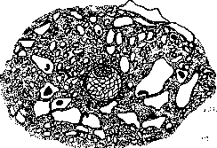
Figure 5. Stage corresponding to the embryo : of Mellendorff (schema according to Gropser'y). The trophoblast has turned into a network of struts; the mesoderm of the morula has become mesenchyme due to the formation of slits. (From Broman's.)
in the wall of the yolk sac (blood islands), which merge into networks, and then in the lateral parts of the 3. in the form of two aortas and a paired rudiment of the heart, which gradually connects into one tube. In the embryo of Eternod 1.3 mm there is already primary circulation (see color plate, Fig. 2), which however has recently been disputed (Ogovveg). Subsequent stages are characterized by 1) segmentation of the mesoderm, 2) closure of the neural tube, and 3) formation of the head and tail gut. A well-studied embryo of Eternod 11, 2.11 mm (stage of N. MaI'ya; Figs. 9 and 10) can serve as an example. On the dorsal side somites (primary vertebrae) are clearly visible, located on both sides of the midline (8 pairs). Cross sections of the corresponding stage (embryo 2 mm MaI'ya) show on either side of the neural tube the somites (Fig. 11), and outward from them the formation of the body cavity
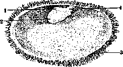
Fig. 6 Human egg at the beginning of the 4th week (opened according to Eternod'y). 1-amnion; 2-yolk sac; 3-chorionic villi; 4-belly stalk. (From Broman's.) (endocoelom) by splitting the mesodermal layer into splanchno- and somatopleura. The neural tube of the Eternod embryo in the middle part is closed, the anterior parts of it (brain rudiments) have greatly proliferated but are not yet closed into vesicles; the caudal part is also not closed, in the region of which lies canalis neurentericus. On the ventral side Figure 1 (A and B). Two diagrams showing the connection of the egg membranes and the formation of the umbilical cord (ectoderm is marked in black; mesoderm in red, endoderm in black dotted line): 1-intestine; 2-embryo; 3-amnion; 4-amniotic cavity; 5-allantois; 6-belly stalk; 7-neck of the yolk sac; 8-yolk sac; 9-external body cavity (exocoelom); 10-chorion; 11-embryo (its neural tube); 12-mesoderm of the belly stalk; 13-yolk duct; 14-oral opening. Figure 2. Vascular system of the embryo (1.3 mm) according to Eternod'y. A- area embryonalis and belly stalk from above; B-the same vessels from the left side. 1-edge of the amnion incision; 2-heart; 3-aorta dextra; 4-sinus umbilicalis ensiformis; 5-canalis neurentericus; 6-vena umbilicalis impar; 7-amnion-chorion incision; 8-allantois; 9-arteria umbilicalis sinistra; 10-vena chorio-placentaris; 11-*-belly stalk; 12-arteria et vena umbilicales; 13-yolk sac; 14-chorion (with cut-off villi). Figure 3. Diagram of pupillary fibers. 1-nervus opticus; 2-center nervi oculomotorii; 3-parasympathetic pupillary fiber; 4-circular muscle of the pupil; 5-radial muscle of the pupil; 6-nervus sympathicus; 7-ganglion sympathicum cervicale superius; 8-ganglion sympathicum cervicale medium; 9-nervus oculomotorius; 10-lens; 11-Zinn's ligament; 12-musculus ciliaris; 13-cortical influences. (Figure 1 and 2-from Bromnn's, figure 3-from Meyer's.) To the illustration art. Embryo, Pupillary fibers, reflexes, centers,
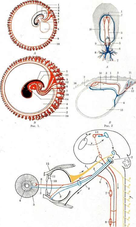
Figure 3. To art. Embryo. Pupillary fibers, reflexes, centers. 1st fig.: t."|' S09
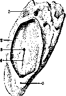
Figure 7. Reconstruction of the embryo 1.17 mm (according to Frassi): 1-yolk sac; 2-edge of the amnion incision; 3-canalis neurentericus; 4-primitive streak; 5-allantois (in the severed belly stalk). (From Broman's.)
on the anterior and posterior ends of the blastoderm, which begin to isolate the body of the 3. from the yolk sac and at the same time form the head gut and tail gut, while the middle part remains in communication with the yolk sac (which from this time is sometimes called the umbilical sac -vesicula umbilicalis) by a wide opening. The changes outlined here continue to develop further; in the embryo of 2.5 mm (approximately 31/^ weeks), the reconstruction of which is given by Thompson (Thompson; Fig. 12), a considerable number of somites (more than 23) are found, closure of the brain vesicles and their strong bending forward and almost complete isolation of the intestinal canal, which remains in connection with the yolk sac by means of the yolk stalk (intestinal navel). Also striking is the strong protrusion of the heart (cardiac bulge) and the appearance in the region of the pharyngeal gut of pharyngeal, or / 2 : j ; * \ :> branchial arches. Here are noticeable the mandibular arch, the hyoid arch, and the first branchial arch; between them pharyngeal clefts, according to some authors never breaking through. Under the forebrain vesicle is the oral pit, bounded on the sides by jaw processes and separated from the pharyngeal gut by a membrane (membrana bucco-pharyngea), which ruptures at about this time. Above the III branchial arch is an indentation of the ectoderm, the auditory pit. The caudal end of the body is strongly curved. In the embryo of 3 mm [reconstruction by Broman's; see separate plate (art. 511-512), Fig. 1] eye vesicles are clearly visible; the auditory vesicle has already pinched off; the heart is large and divided into atrium and ventricle; under it is a fairly large liver.
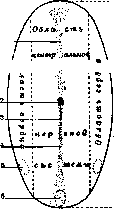
Figure 8. Topography of the embryonic disk (schema according to Holmdahl'io): 1-head process of the primitive streak; 2-canalis neurentericus; 3-head node (Hensen's); 4-primitive streak; 5-rudiment of the arm; 6-tail bud (rudiment of the caudal part of the body with hind limbs and tail); 7-region of neck and body; 8-region of head. (From Broman's.)
The intestinal canal is significantly differentiated; rudiments of tongue and lungs are visible, the stomach and rudiments of the pancreas are outlined; the yolk stalk has turned into a narrow canal. The allantois arises from the hindgut, behind it there is a small indentation of the ectoderm-the cloacal pit. Outside appear the rudiments of the upper limb. From the stage of 2.5-3 mm begins the rapid growth of the 3., so that in a few days by the end of the 1st month (i.e. the 4th week) the 3. reaches a length of 8 mm. During this interval it
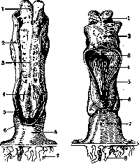
Figure 9.
Figure 10. Figure 9. Human embryo 2.11 mm (according to the model of E1ernoda'): 1-open neural tube in the region of the brain; 2-edge of amnion; 3-neural tube suture; 4-open neural tube of the caudal part; a-canalis neurentericus; b-primitive streak; 7-chorionic villi; 8-belly stalk. Figure 10. Human embryo 2.11 mm from the ventral side (according to the model of E1ernoo?a): 1-open end of the neural tube; 2-cardiac bulge; 3-head gut; 4-chordal slit; 5-edge of the yolk sac wall; 6-caudal gut; 7-belly stalk; 8-chorionic villi. (From Corning's.) very strongly bends, so that the tail almost touches the head; the brain vesicles increase significantly; at the anterior end of the head the olfactory pits are outlined; the number of branchial arches increases, so that 4 are visible from the outside, and rudiments of the lower limbs appear. The dimensions of the 3. in this pe
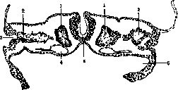
Figure 11. Section of embryo 2 mm with 7 somites'(according to Keibel-MaI'yu): 1-third somite; 2-endocoelom; 3-exocoelom; 4-right aorta; 5-notochord; 6-yolk sac cavity. (From Broman's.) riod, as in subsequent ones, by no means always correspond to the degree of its development, which greatly complicates their description, and the data
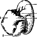
Figure 12. Human embryo 2.5 mm (reconstruction according to Thompson): 1-yolk sac; 2-belly stalk; 3-edge of amnion (umbilical fold); 4-cardiac bulge; 5-auditory pit. (From Broman's.)
the authors often differ. Usually it is accepted that at the beginning of the 2nd month (see separate table, fig. 2) the human embryo is about 8 mm in length and has typical features of mammalian embryos (it differs from bird embryos in the weak development of the eyes, and from reptiles in the shorter tail). The curvature of the body decreases, the number of somites is 40-43; on the head, due to the bending of the brain vesicles, three bulges protrude—frontal, parietal, and occipital; the eyes protrude in the form of a small convexity and are barely noticeable, since the pigmentous rim around them is not yet visible; the development of the lens begins; the olfactory pits have deepened considerably. The arches visible are: the maxillary process, the mandibular and hyoid; the branchial arches begin to close with a fold (operculum), and the cervical sinus is outlined. Directly under the head is the prominent cardiac bulge, behind it the hepatic; the skin (skin-yolk) navel has already formed. The limbs bend, and the upper limb receives a plate-like expansion. The changes occurring during the second month consist in that the embryo grows from 8 mm to 2 cm (approximately) and acquires the characteristic human form (see separate table, figs. 3 and 6). This is primarily due to the enhanced growth of the head, which by the end of the month is almost equal to the trunk part. In the first half of the month, the bulges corresponding to the different parts of the brain are still visible from the outside, in the second half the head becomes rounded and the cervical fossa is outlined. The eyes become pigmented (in the 10 mm embryo) and therefore become clearly visible; they are bordered by the folds of the eyelids and begin to move to the facial part; from the dorsal end of the 1st branchial cleft, the auditory opening is formed; ear hillocks appear at its edges. On the sides of the olfactory pits, which are turning into slits, the nasal, frontal, and maxillary processes grow, forming the nose and mouth opening. The head is raised and the neck is outlined; the branchial clefts disappear. In the trunk part, the straightening of the back and the strong protrusion of the abdomen due to liver development occur; the heart grows relatively less. Under the abdominal protrusion is the umbilical cord, behind it at the cloaca opening is the genital tubercle. The tail, which reaches its greatest development by the end of the 1st month, decreases during the 2nd month, becoming part of the trunk. The limbs develop strongly: all their parts are outlined, and the fingers separate from the webbing; the development of the lower limb lags somewhat behind the upper. The limbs face each other with their palmar sides; by the end of the 2nd month the arms cross at the upper part of the abdomen. The embryo, having thus assumed the human form, is called a fetus (fetus) from the beginning of the 3rd month. Definition of the human embryo according to Mall. (The numbers indicate the average length in mm) Stages HIJKLMNOPQRSTU 2 3 4 5 6 8 10 12 14 16 18 20 22 21 H—from the first appearance of somites to the appearance of the limb rudiments. I—rudiments of the upper limb, 3 clearly visible branchial arches. J—4 branchial arches. K—the number of branchial arches decreases to 3. L—3 branchial arches; the bend of the elbow is indicated; the olfactory pit reaches maximum development. M—2 branchial arches; the lower limb is divided into thigh and leg; the lacrimal groove is clearly expressed; the digital plate of the hand is well noticeable. N—2 branchial arches; the thumb of the hand is outlined, the plate on the foot; the ear and cervical sinus are well visible. O—a little more developed than N; the big toe appears. P—the branchial arches have disappeared; the ear is well formed; the fingers of the foot are clearly outlined. Q—the arms reach the middle of the abdomen (doubtful stage). R—a little more developed than Q. S—the arms touch each other; the legs are extended, their big toes are clearly visible. T—the arms cross, the legs are formed. U—fetal form; the head is well developed.
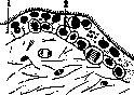
Embryonic membranes of man. The embryonic membranes include 1) membranes in the proper sense: trophoblast, amnion, chorion, and the modified mucous membrane of the uterus—the 'deciduous' (decidua); 2) the main organ of nutrition of the fetus formed by their modification—the placenta or child's place, and 3) embryonic organs: the yolk sac, allantois, abdominal stalk, and the umbilical cord formed with their participation. The formation of the trophoblast, amnion, yolk sac (yolk sac, otherwise the umbilical sac), and allantois is closely connected with the early stages of development of the human embryo and has already been described. At first after implantation, the outgrowths of the trophoblast represent a spongy mass with nuclei, devoid of cell boundaries (syncytium, or plasmodium), by means of which the absorption of disintegrated maternal tissue (embryotroph) and blood (primary villi; figs. 2, 4 and 5) occurs. Then the mesenchyme lining the wall of the trophoblast from within begins to penetrate into the villi, and with it the vessels going to the abdominal stalk with the allantois; such vascularized villi are called secondary, and the trophoblast (or primary chorion) becomes the definitive chorion. The basis of the developed villus is formed by delicate connective tissue with blood capillaries; from the outside it is covered with two layers of epithelium: the inner one with clear cell boundaries (Langhans' layer) and the outer syncytial; on its surface there is a brush border resembling the border of intestinal epithelium (fig. 13). By the end of the 1st month the chorion can be separated from the uterine mucosa in the form of a sac covered on all sides with villi (egg; see separate table, fig. 4); by the end of the 2nd month the villi on the side of the sac facing the uterine cavity begin to disappear (chorion laeve), on the other side they continue to grow and branch (chorion frondosum); they later become part of the placenta. At the site of implantation of the sac (usually on the posterior wall of the uterus, at the top) the decidua splits (Vol. III, art. 211, fig. 2); the part covering the growing sac (decidua capsularis, s. reflexa) becomes thin, the part lying between the sac and the muscular layer of the uterus, on the contrary, hypertrophies (decidua basalis, s. serotina) and later becomes part of the placenta. The remaining part receives the name decidua vera; between it and decidua capsularis there is a slit for the first two months; on the 3rd month these two layers come into contact and fuse.
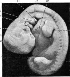
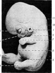
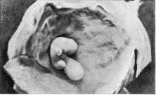
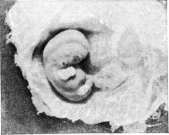
Figure 13. Section of a chorionic villus at the 2nd month (according to Bennet): j—syncytium (covering layer); k—Langhans' layer. (From Corning.)
Figure 1. Reconstruction of a 3 mm embryo (left half of the body removed): 1—hindbrain with neuromeres; 2—auditory vesicle; 3—midbrain; 4—Rathke's pocket; 5—eye vesicle; 6—forebrain; 7—oral pit; 8—heart (atrium); 9—abdominal stalk; 10—point of origin of the allantois; 11—hindgut; 12—notochord; 13—coelom; 14—yolk stalk; 15—yolk sac; 16—liver stalk; 17—dorsal rudiment of the pancreas; 18—position of the left lobe of the liver (dotted line); 19—stomach; 20—rudiment of the left lung; 21—unpaired tubercle. Fig. 2. Human embryo 7.7 mm: 1—branchial fold; 2—hyoid arch; 3—mandibular arch; 4—maxillary process; 5—olfactory pit; 6—parietal bend; 7—tail; 8—leg; 9—arm; 10—cardiac bulge; 11—occipital bend. Figure 3. Human embryo 10.5 mm in length: 1—eye; 2—nose; 3—umbilical hernia sac; 4—plate-like part of the leg; 5—inguinal region; 6—hepatic bulge; 7—hand plate; 8—mouth opening; 9—ear; 10—neck; 11—occipital bend. Figure 4. Human egg 8 mm (natural size). Figure 5. Human embryo 9 mm with yolk sac (enlarged 3/2). Figure 6. Human embryo 16 mm (2nd month, enlarged 2/2). (Figure 1, 2 and 3—from Brönn's, figs. 4, 5 and 6—according to photographs by Lyakhovetsky.)
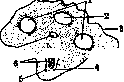
While the embryo is in the blastoderm stage, the amnion covers it in the form of a sac (see separate table, fig. 4). The yolk sac is located under the embryonic shield and is connected to the embryo by the yolk stalk (vitelline duct). The allantois grows from the hindgut and extends into the extraembryonic celom, where it expands. The abdominal stalk connects the embryo with the chorion. Figure 14. Section of the umbilical cord according to His: 1—a. umbilicalis; 2-v. umbilicalis; 3—amnion; 4
precise vessels; 7-allantois. (From Broman.) it from above into the stalk of the yolk sac; yolk;5-ecocoelom; in-shell PRECISE POUCH HAS the appearance of a cup, the opening of which is closed by the blastoderm. As the folds (head, tail and lateral) form and the intestine develops, the yolk sac takes the form of an oval sac, connected by a narrow yolk duct to the walls of the intestine (intestinal navel; see separate table, fig. 5). The amnion at this time begins to cover the E. in the form of a bladder, rather closely adjacent to the body [see separate table (pp. 507-508), fig. 1.4]. Due to the accumulation of fluid, the amniotic bladder inflates and approaches the chorion; its head and tail parts, coming closer, press the yolk duct (which elongates) to the abdominal stalk and envelop them in a common sheath [see separate table (pp. 507-508), fig. 1B]. Thus, the umbilical cord is formed, which includes the allantois, vessels (2 art. and 2 venae umbilicales, of which v. umbilicalis dextra soon disappears) and the yolk duct with yolk vessels (fig. 14). The basis of the umbilical cord is mucous tissue that developed from the mesenchyme of the abdominal stalk (Wharton's jelly); the surface is covered by the epithelium of the amnion. During the 2nd month, the amnion reaches the inner surface of the chorion and fuses with it; between them remains the yolk sac in the form of a small sac. For the further fate of the membranes and the development of the placenta, see Fetus, Placenta.
Related articles
Mentioned in
Cite this page
“Embryo.” Soviet Medical Encyclopedia. English translation of Bolshaya Meditsinskaya Entsiklopediya, 1st ed. (Moscow, 1928–1936), ed. N. A. Semashko. https://sovietmedicalencyclopedia.pages.dev/article/embryo/