Branchial Arches
Historical document, translated for reference. It reflects medical knowledge of the 1920s–30s and is not medical advice.
Summary
This article from the 1928–1936 Soviet Medical Encyclopedia describes the anatomy and evolutionary origin of the branchial arches and clefts in vertebrates. It explains how these visceral arches transform from respiratory structures in fish into components of the hyoid apparatus and laryngeal cartilages in terrestrial vertebrates, noting that their failure to close during embryogenesis leads to branchial fistulae.
Encyclopedia article (1928–1936)
BRANCHIAL ARCHES AND CLEFTS. The branchial arches constitute the skeleton of the respiratory apparatus of lower aquatic vertebrates, i.e., cyclostomes and fishes. The number of branchial arches in the vast majority of modern fishes is limited to five. Only in some lower sharks does it rise to seven. However, there is no doubt that even this number is already reduced. In vertebrate embryos, a series of initially completely identical visceral arches is laid down, located between adjacent visceral clefts. The anterior of these arches gives rise to the jaws of lower vertebrates and is therefore called the mandibular arch. The second arch suspends the jaws in lower vertebrates, while in terrestrial forms it gives rise to the main part of the hyoid skeleton and is therefore called the hyoid arch. The subsequent arches bear gills in fishes. However, in view of the similarity in the development and structure of all visceral arches together with their muscles, vessels, and nerves, it is conceivable that the first two visceral arches also once bore a branchial function.
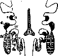
The number of branchial arches was originally no less than a dozen, and only gradually did their total number begin to decrease. The branchial arches of fishes are segmented on each side usually into 4 sections, movably connected by synarthroses. On the ventral side, they are connected by a longitudinal series of unpaired elements, and on the dorsal side, the ends of successive arches of each side are connected by ligaments, often with the participation of intermediate skeletal elements. Thus, all the arches together form a single integral movable system supporting the system of gills and the branchial clefts between them. In terrestrial vertebrates, with the loss of branchial respiration, the corresponding arches are also lost—their rudiments become part of the hyoid apparatus, forming part of its body and posterior horns, and participate in the formation of the laryngeal cartilages. In mammals, the posterior (in humans, "greater") horns of the hyoid bone represent the remnants of the first branchial arch, and the thyroid cartilage of the larynx is the result of the fusion of the second and third arches. The material of the last branchial arches goes into the formation of the other laryngeal and tracheal cartilages, with the exception of the epiglottis (see figure). The non-closure of embryonic branchial clefts is the cause of the formation of branchial fistulae in higher vertebrates, including humans.
Related articles
Mentioned in
Cite this page
“Branchial Arches.” Soviet Medical Encyclopedia. English translation of Bolshaya Meditsinskaya Entsiklopediya, 1st ed. (Moscow, 1928–1936), ed. N. A. Semashko. https://sovietmedicalencyclopedia.pages.dev/article/branchial-arches/