Eustachian Tube
Historical document, translated for reference. It reflects medical knowledge of the 1920s–30s and is not medical advice.
Summary
This article from the 1928-1936 Great Medical Encyclopedia covers the anatomy, embryology, and physiological significance of the Eustachian tube, detailing its osseous and cartilaginous parts, associated muscles, blood supply, and innervation.
Encyclopedia article (1928–1936)
EUSTACHIAN TUBE (tuba auditiva Eustachii), named after the Italian anatomist Eustachio who discovered it in 1563, is a channel connecting two cavities: the middle ear and the nasopharynx. In its origin, the Eustachian tube is a part of the first visceral cleft located between the mandibular and hyoid visceral arches. In lower fishes, this first cleft opens outward like the gill slits following it, via a special opening called the spiracle, and still contains a rudimentary gill. In terrestrial vertebrates, it is always closed from the outside early on, and its cavity forms various kinds of outgrowths that subsequently give rise to the tympanic cavity. The latter, however, always retains its original connection with the pharynx in the form of a pair of Eustachian tubes. The latter only in certain cases (in aglossal amphibians, in crocodiles, and birds) fuse at their ends to form a single unpaired opening. Anatomy. The Eustachian tube consists of two parts: osseous and cartilaginous-membranous. Its length is on average 3.5 cm; of these, 2.5 cm belong to the cartilaginous-membranous section, and 1 cm to the osseous section. In the channel of the tube, there are two openings: the tympanic opening (into the middle ear cavity) and the pharyngeal opening (into the nasopharynx); the width of the tympanic opening is 3.3 mm, the height is 4.5 mm. The pharyngeal opening of the tube has the shape of a vertically standing slit about 1 cm high; during swallowing or speaking, this slit-like shape changes into a triangular one due to the contraction of the tensor veli palatini muscle, its elevation, and the displacement by it of the folds limiting the slit-like opening. Two folds of the pharyngeal opening of the tube (plica salpingo-palatina and plica salpingo-pharyngea) are called the lips of the pharyngeal opening of the tube—labia tubae Eustachii; of these, the posterior one, located medially, protrudes into the nasopharynx cavity, lifting the mucous membrane, as a result of which a depression is formed behind it (recessus pharyngeus Rosenmulleri; Fig. 1). The direction of the Eustachian tube from the tympanic cavity to the pharyngeal opening is forward and downward; with respect to the horizontal plane, the Eustachian tube forms an angle of 40°; the pharyngeal opening of the tube is located lower than the tympanic one by an average of 2.5 cm. Along its length, the lumen of the tube is not uniform: at the place of transition of the osseous part into the cartilaginous one, it forms a narrowing, reaching here a height of 3 mm and a width of 0.25 mm (Fig. 2); this narrowed part of the tube is called its isthmus (isthmus tubae Eustachii). The osseous part of the Eustachian tube is a continuation of the upper wall of the tympanic cavity (tegmen tympani); the lower wall of the tube is sharply demarcated from the lower wall of the tympanic cavity (Fig. 1). The diameter of the osseous Eustachian tube is about 2 mm. Above the Eustachian tube, separated from it by a bony plate, runs the middle ear muscle—tensor tympani muscle (Fig. 2); below and medial to the tube runs the canal of the carotid artery (canalis caroticus). The cartilaginous-membranous part of the Eustachian tube consists of two parts—cartilaginous and membranous; the upper-medial part of the tube is cartilaginous, and the lower-lateral part is membranous. The oval main mass of the cartilaginous tube, narrowed in the upper part in the form of a hook, continues and curves around the upper part of the lumen of the tube. The cartilage of the tube is basically hyaline, and in the central areas it is reticular. The mucous membrane lining the tube is covered with ciliated epithelium, the movement of the cilia of which is directed toward the nasopharynx; acinous glands are located in the subepithelial layer, most abundantly near the pharyngeal opening; in the osseous section,
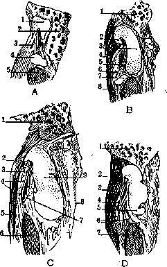
Figure 1. A-Lateral side of the cartilaginous
parts: 1-cart. tubae audit.; 2-pars petrosa oss. tympani; 3-m. tensor veli palat.; 4-cartilaginous part; 5-m. levator veli palat. B-Border between the lateral and middle thirds: 1-pars petrosa oss. tymp.; 2-lam. lat. cartil.; 3-lam. med. cartil.; 4-m. tensor veli palat.; 5-tun. mucosa; 6-lam. membran.; 7-cartilaginous part; 8-m. lev. veli palat. C-Section near the pharyngeal opening of the auditory tube: 1-pars petr. oss. tempor.; 2-lam. lat. cartil.; 3-lam. membran.; 4-gland. mucosae; 5-m. tens. veli palat.; 6-m. levat. veli palat.; 7-tunica mucosa; 8-rec. pharyng. (Rosenmulleri); 9-lam. med. cartil. D-Border between the middle and medial thirds: 1-pars petrosa oss. tymp.; 2-lam. lat. cart.; 3-m. tens. veli palat.; 4-tunica mucosa; 5-lam. med. cart.; 6-gland. mucosae; 7-m. levat. veli palat. (After Spalteholz.) they are extremely sparse. The mucous membrane of the cartilaginous section of the tube forms folds, predominantly in the medial wall of the tube. Lymphoid tissue is especially abundant at the pharyngeal ostium, where its clusters are sometimes so powerful that there is every reason to isolate them into a special tonsil—amygdala tubaria. Muscles closely related to the Eustachian tube: 1) levator veli palatini muscle (m. petro-salpingo-staphylinus), 2) tensor veli palatini muscle (m. spheno-salpingo-staphylinus). The levator veli palatini muscle originates from the area of the petrous bone near the canalis caroticus, runs parallel to the tube under its base and under the pharyngeal ostium of the tube, goes to the soft palate, where it ends (Figures 3 and 4). The tensor veli palatini muscle starts from the lower surface of the sphenoid bone from the greater wing of the pterygoid process
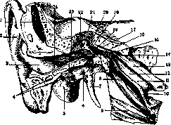
Figure 2. Right external and middle ear: 1-squama tempor.; 2-auricula; 3-concha auric.; 4-cart. meatus acust. int.; 5-proc. mast.; 6-pars tymp. oss. temp.; 7-proc. styl.; 8-m. lev. veli palat.; 9-m. constr. pharyng.; 10-ost. pharyng.; 11 and 12-pars cartil.; 13-cartil.; 14-isthmus; 15-pars ossea; 16-ost. tympan.; 17-m. tensor tymp.; 18-cavum tymp.; 19-membr. tymp.; 20-tendo m. tens. tymp.; 21-capit. mallei; 22-rec. epitymp.; 23-meat. acust. ext. oss.; 24-meat. acust. ext. cartil. (After Spalteholz.)
and partly from the hook of the cartilage of the tube and its membranous wall; then the muscle curves around the hook of the pterygoid process (hamulus processus pterygoidei) and further ends in the soft palate. Blood supply of the Eustachian tube. Arterial blood comes from the ascending pharyngeal artery, middle meningeal artery, and internal maxillary artery. The venous plexus of the tube, with which the
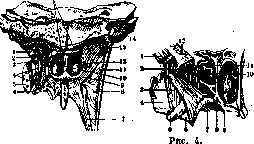
Figure 3.
Figure 3. Muscles of the pharyngeal wall: 1 and 9-m. lev. veli palat.; 2-cart. tubae audit.; 3-pars. lat. tubae (membr.); 4-m. tens. veli palat.; 5-m. pteryg. int.; 6-ham. pteryg.; 7-m. pharyngo-palat.; 8-m. constr. phar.; 10-sept. nasi; 11-fascia pharyngo-basilaris; 12-ost. phar. tubae; 13-tuba auditiva; 14-sin. sigmoid.; 15-tons. pharyngea. (After Braus.) Figure 4. Model of the Eustachian tube with tensor and levator veli palatini muscles: 1-m. lev. veli palat.; 2-torus tubar.; 3-m. tens. veli palat.; 4-fossa pterygoidea; 5-ham. pter.; 6-aponeur. palat.; 7-m. pharyngo-palat.; 8-spina nasal. post.; 9-mucous membrane of the hard palate; 10-pars membran. tubae audit.; 11 and 12-cartil. tubae auditivae. (After Braus.) veins of the tympanic membrane and nasopharynx anastomose, pours into the cavernous sinus. Nerves: the levator veli palatini muscle is innervated by a branch of the facial nerve—greater superficial petrosal nerve; the tensor veli palatini muscle by a branch of the third branch of the trigeminal nerve. The physiological significance of the Eustachian tube should be evaluated from two points of view: 1) the significance

its role in sound conduction (acoustic role) and 2) its importance as a channel through which infection can spread to the tympanic cavity from the nasopharynx and vice versa. To understand both roles of the Eustachian tube, it is necessary to know in what state the tube is during quiet breathing, nasal and oral breathing, swallowing, and speaking (phonation). Although the immobile bony part of the tube is in an open state according to anatomical data, however its lumen in the isthmus is so insignificant that with even slight swelling of the mucous membrane, for example with even minor venous congestion, this lumen can close. The lumen of the cartilaginous-membranous part is open in normal condition, however the muscles of the tube change this state. When the m. levatoris veli palatini contracts, the floor of the Eustachian tube is elevated in the form of a ridge, whereby its pharyngeal opening takes a triangular shape; by elevating the floor of the tube, closure of the lumen is achieved; thus this muscle keeps the tube in a closed state (this is supported by a number of observations). When the m. tensoris veli palatini contracts, the lateral wall of the tube is pulled laterally, i.e. the lumen of the tube is opened. Thus both these muscles are antagonists; the degree of their contraction determines one or the other state of the lumen. In normal condition the Eustachian tube is in a closed state when the m. levatoris et tensoris veli palatini are at rest; during quiet nasal breathing it is open to varying degrees depending on the state of contraction of these muscles; during ordinary oral breathing it is closed; during swallowing the tube opens. The different states of the lumen of the tube affect sound perception, since when the Eustachian tube is open, sound waves penetrate into the middle ear not only through the external auditory canal and the bones of the skull, but also through the Eustachian tube channel. That sound conduction through the Eustachian tube plays a role in the act of hearing is indicated by observations of the hard of hearing: when listening they open their mouth and tense the soft palate to expand the Eustachian tube. During swallowing both muscles contract - the m. tensor and the m. levator, but the m. tensor with greater force, therefore during swallowing the tube opens (figure 5). If during swallowing one carefully analyzes the sensation in the ears, the movement of the eardrum inward is perceived, accompanied by a slight sound; this movement and sound are stronger if swallowing occurs with the external nasal openings tightly closed (Toynbee's experiment); a much stronger sensation is obtained in Valsalva's experiment - strong exhalation through the nose when it is closed; during this air is forced through the tube into the middle ear and the eardrum is displaced
Figure 5. Pharyngeal opening of the tube during swallowing: 1 - torus tubarius; 2 - m. levat. veli palatini; 3 and 4 - concha inferior et superior (According to Braus). Pushing the palate outward; after performing these two experiments, it is easy to catch the difference in sensations and realize that during the Valsalva experiment the eardrum moves outward, while during the Toynbi experiment it moves inward. In paralysis of the soft palate, when the nasopharynx is not separated from the pharynx, liquid food can enter the nasopharynx and be thrown through the open tube into the middle ear, causing inflammatory processes here. During the Valsalva experiment, along with air, pus-causing agents can be pushed into the middle ear along with mucus, resulting in inflammation of the ear. The significance of the E. t. as a channel through which infection from the nasopharyngeal cavity can spread is exceptionally great: infection of the middle ear with an intact eardrum is possible only through the tube; however, in its normal state, infectious agents cannot penetrate to the middle ear: the ciliated epithelium hinders them, expelling them into the nasopharyngeal cavity. The usual infectious agents - strepto- and staphylococci - can enter the middle ear when they are thrown by an air stream during sneezing, strong nose blowing, vomiting; the latter occurs especially in infants during regurgitation. When secret accumulates in the middle ear cavity, the E. t. acquires the significance of a drainage, although some authors deny this significance of the tube. Diseases of the E. t. are inflammatory and non-inflammatory. Non-inflammatory diseases of the tube are extremely rare. The most common are muscular noises. With each swallowing, the lateral wall of the tube is pulled away from the medial by the contraction of m. tensoris veli palatini, at which time a sound is produced that is usually not noticed, but with other ear ailments, especially in nervous individuals, it can be perceived. A sound of a different nature is obtained with clonic contractions of the muscles of the E. t.; it is exactly the same as can be obtained when the nail of one finger slips off the nail of another. The force of the sound is so great that it is heard not only by the patient himself but sometimes by others. In some cases, patients perceive this sound as distressing: if these clonic contractions are prolonged, they not only strongly irritate the patient but also greatly tire him. In some cases in such patients, careful observation of the soft palate can reveal small contractions of it. For treatment, massage (with a finger) of the soft palate and the area of the tube, galvanization and faradization of the soft palate are recommended. Foreign bodies in the tube, if we do not consider pieces of food that get there during choking, coughing, sneezing, are very rare. Cases of breaking off pieces of bougies during bouginage of the E. t. have been described. The usual outcome is the exit of foreign bodies into the nasopharyngeal cavity, but cases of a fragment of a bougie passing into the middle ear cavity have been described. When foreign bodies enter the tube, one should limit oneself to waiting and prescribing instillations into the nose of a 2% cocaine solution with adrenaline (1%0) of 3-4 drops (on the corresponding side), expecting that the resulting contraction of the mucous membrane of the tube's opening will facilitate the expulsion of the foreign body from the tube. If a foreign body enters the tympanic cavity and is found there, it can be easily removed by irrigating the middle ear with a warm (body temperature) 2% boric acid solution; if it has not yet completely left the tube - it is better to wait until it comes out completely; then it can be removed by irrigation, while when grasped with forceps in the depth, it can be pushed back. The open state of the tube is accompanied by so-called autophony, i.e. strong sounding of one's own voice; if this state is prolonged, it strongly irritates and disturbs the patient. This pathological condition is the result of rapid emaciation of the patient, when the fatty tissue located in the walls of the tube and serving as a cushion supporting the walls of the tube in contact disappears; such an open state of the tube is also possible with toxic (rheumatic) pareses of m. levatoris veli palatini. The treatment of such patients should be directed according to etiology: in emaciation - to improve general nutrition; in rheumatic lesions - salicylates; and in both cases - massage of the pharyngeal opening of the tube with a finger or a cotton ball soaked in 1/4% iodine-glycerin; often electrification of the tube with a faradic current in several sessions eliminates the gaping of the tube. Injury to the tube most often occurs during catheterization of the tube and during bouginage; these injuries usually consist in violation of the integrity of the mucous membrane of the tube, not requiring medical intervention, but unquestionably requiring cessation of these manipulations (until the mucous membrane heals). Gunshot injuries to the tube are extremely dangerous, as neighboring organs (a. carotis) are damaged at the same time. Inflammatory diseases of the tube. The mucous membrane of the E. tube, being a continuation of the nasopharyngeal mucous membrane, by this very fact always participates to one degree or another in inflammatory processes of the nasal mucosa and nasopharynx; however, subjectively and objectively this pathological condition of the tube is not manifested at first; only when the process spreads to the middle ear cavity does the patient begin to notice hearing loss, of one kind or another ear noises, a tingling sensation in the ear, a feeling of stuffiness in the ear; therefore, patients usually turn to the doctor already with chronic changes, and not only in the tubes but also in the middle ear cavities; in these cases, both objective symptoms - changes in the eardrums (their retraction, change in color, atrophic and calcareous spots) - and subjective symptoms - noises in the ears, hearing loss of the same nature as in lesions of the sound-conducting, and often sound-perceiving apparatus - can be established. In these cases, it is not only difficult but mostly impossible to separate the symptoms of inflammation of the E. t. from the symptoms of inflammation of the middle ear: here there is a combination of both these pathological forms. From what has been said, it is clear that although inflammation of the mucous membrane of the E. t. exists in three stages: acute, subacute and chronic, in practice, cases with chronic inflammation of the E. t. are more often observed; acute inflammations either proceed without symptoms or the latter are absorbed by the accompanying symptoms from the nose and middle ear. In accordance with the latest views of foreign authors, narrowing of the E. t. (stenosis, strictura tubae Eustachii) should be singled out as a separate nosological unit. Narrowings can be both congenital and acquired. The causes of congenital narrowings are usually clefts of the hard and soft palate, accompanied by inactivity or insufficient activity of the muscles of the soft palate (m. tensor and m. lev. veli palat.). The causes of acquired narrowings are: various inflammatory processes of the nasal mucosa, nasal cavities and nasopharynx; ulcerative processes in the area of the pharyngeal opening of the tubes - syphilitic, tuberculous, lupous, scleromatous, diphtheric, smallpox (these processes end with scars that pull the E. t. until their complete closure); benign and malignant neoplasms of the nasopharynx. The most common and usual is the so-called adenoid, i.e. hypertrophy of the pharyngeal (III) tonsil. Then come nasopharyngeal fibromas, sarcomas, cancers; finally exostoses and hyperostoses of the tube, an abnormally wide canal for m. tensor tympani, exostoses of the septum of unusually large size reaching the opening of the tube, and congenital atresia of the choanae. Symptoms of closure of the E. t. consist of general and local symptoms. The general symptoms include hearing impairment, subjective noises and balance disorders. Hearing impairment consists of phenomena from the tube itself and from the middle ear, which, as already indicated above, always participates in the process. From closure of the E. t., hearing is impaired only to a small degree, and the nature of the impairment is the same as in lesions of the sound-conducting apparatus: elevation of the lower border of hearing, elongation of bone conduction, shortening of air conduction; the upper border is within normal limits; subjective sensations (auditory) consist in a feeling of stuffiness in the ear, sometimes a splashing in the ear when changing the position of the head, noises, mostly of low tone, and sometimes autophony. Objectively, from the side of the eardrum, its retraction is found with characteristic changes in the appearance of individual parts of the eardrum: protrusion of the short process, perspective shortening of the handle of the malleus with a more horizontal direction of it and expressiveness of the posterior and anterior folds; the reflex is either completely absent or it is not conical but thinner or interrupted in the middle; the color of the eardrum is changed: it is duller, sometimes with chalky, sometimes with atrophic spots.
From the side of the vestibular apparatus, phenomena of increased irritability of the semicircular canals are noted: voluntary nystagmus, often more pronounced in one particular direction, and dizziness. More severe conditions are observed as a result of complete closure of the tube; the secretion of the mucous glands of the E. t. and the excretion products of the mucous membrane itself together with the desquamating epithelium of the tube, having no outlet into the nasopharyngeal cavity through the closed tube, move in the direction accessible to them—into the cavity of the middle ear, forming accumulations here, which, although not reaching significant sizes, are nevertheless sufficient for nourishing the pathogens of suppuration that penetrate the middle ear by hematogenous route during infectious diseases (influenza, tonsillitis, measles, scarlatina), as a result of which otitis develops with all its consequences. Local symptoms are: subjectively - weakening of hearing and autophony, objectively - non-passage of air through the tube. The diagnosis of closure of the E. t. is based on subjective sensations and objective data of research—otoscopic and functional (cochlear and vestibular functions). Objective evidence of closure of the tube is the detection of scars, adhesions at the openings of the tube during posterior rhinoscopy or—even better—salpingoscopy. Closure of the lumen along the length of the tube can also be diagnosed by the absence of characteristic noises during catheterization of the tube; with certainty, however, closure is indicated by bouginage of the tube, which succeeds easily in those cases when it is straight and has no folds. During bouginage, a bougie of appropriate thickness should be selected; it should be borne in mind that a thicker bougie passes more easily, as it is not detained by narrow pockets in the mucous membrane where thinner bougies get stuck; therefore, one should start with thicker ones and proceed to thinner ones; if the bougie does not pass, often pushing it out and turning it around its axis helps; in this case, the direction of its head changes, and the bougie often passes through the place where it previously stopped. To facilitate bouginage, the opening of the tube should first be cocaineized by smearing it with a 10% solution of cocaine (with 1%0 adrenaline), and then the bougie should be dipped into vaseline oil. Often the bougie does not enter the tube, because the catheter is incorrectly inserted: not into the opening of the tube, but somewhere nearby. It is better to take a horn catheter, as it is more elastic, flexible, and causes the least unpleasant sensations to the patient during insertion. The best bougies are made of whalebone. Having inserted the bougie into the catheter to the distal end of the catheter, a mark is made by tying a silk thread; then the silk thread is tied, retreating 2.5 cm from the proximal end of the bougie (the length of the cartilaginous part of the tube), and another one—1 cm from the last one; then the catheter is inserted into the nose of the patient and positioned at the opening of the tube, and the bougie is inserted into the catheter; the advancement of the bougie to the first mark indicates its passage through the entire length of the catheter, to the second mark—reaching the bony part of the tube; passage to the third mark indicates reaching the tympanic opening of the tube. One cannot go further, as one may enter the tympanic cavity and traumatize it. The entire procedure must be performed very carefully, without force, as otherwise the mucous membrane can be traumatized and infected. Bouginage is the only indisputable proof of closure of the tube, since noises during catheterization can often occur in those cases when the catheter is incorrectly inserted into the opening of the tube and consequently air cannot in any way pass into the tube. The prognosis in case of closure of the tube depends mainly on the nature of the closure: if it is caused by scars in the tube or persistent narrowings over a large extent—restoring the lumen of the tube is extremely difficult, and sometimes impossible, or after a temporarily achieved success, closure quickly forms again. The treatment of narrowings of the E. t. should first of all be causal, i.e., directed at treating the process that caused the narrowing. As for the treatment of the narrowing itself, bouginage finds the greatest application in it. In significant narrowings, the bougie is left in the tube for 5-10 minutes. Massage of the tube with a bougie is applied with great benefit in two forms: either the bougie is moved outward and inward for 2-3 minutes, or bouginage is combined with vibratory massage of the auricle according to Preobrazhensky. As for climatic treatment, sojourn on the seashore can be recommended only in those cases where there are no subjective noises, and that far from the sea shore; whereas sojourn in the mountains (Kislovodsk, Nalchik, Tiberda) has a very beneficial effect on the process, and treatment there gives better results.
Related articles
Mentioned in
Cite this page
“Eustachian Tube.” Soviet Medical Encyclopedia. English translation of Bolshaya Meditsinskaya Entsiklopediya, 1st ed. (Moscow, 1928–1936), ed. N. A. Semashko. https://sovietmedicalencyclopedia.pages.dev/article/eustachian-tube/