Retropharyngeal Space
Historical document, translated for reference. It reflects medical knowledge of the 1920s–30s and is not medical advice.
Summary
The retropharyngeal space is a narrow cellular cleft located behind the pharynx and in front of the prevertebral fascia. The article details its anatomical boundaries, contents including lymph nodes, and pathological conditions such as acute and cold retropharyngeal abscesses.
Encyclopedia article (1928–1936)
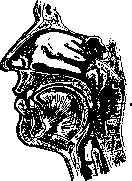
RETROPHARYNGEAL SPACE (spatium retropharyngeum), a narrow cleft filled with loose connective tissue and located in front of the cervical part of the spine. This space is bounded posteriorly by the deep cervical fascia, or prevertebral aponeurosis (fascia colli profunda, s. fascia praevertebralis) (Figure 1), which covers the deep neck muscles (m. longus colli, m. longus capitis, and m. rectus capitis ant. et lateralis), and anteriorly by the fibrous membrane of the posterior wall of the pharynx. The nature of the tissue
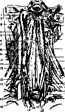
of the retropharyngeal space changes sharply due to the appearance of dense fibrous bundles connected on one side with the posterolateral
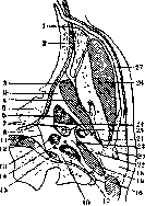
angle of the pharynx and the prevertebral aponeurosis, and on the other with the sheath of the main vessels. The fibrous bundles extending from the outer aponeurosis of the pharynx, near the posterolateral angle of the pharynx, to the prevertebral aponeurosis (ligamentum Charpy), form the outer boundary of the retropharyngeal space and separate it from the posterior section of the pharyngomaxillary space, the so-called presubvascular or retrostyloid space (spatium retrostyloideum, espace retrostylien Testut-Jacob), in which the main vessels and nerves are located (Fig. 2). In the upper part, the outer boundary of the described space is formed by the muscles that tense and elevate the soft palate, and the Eustachian tube lying between them. The tissue of the retropharyngeal space begins at the base of the skull and within the specified boundaries continues into the retroesophageal tissue and then into the posterior section of the mediastinum. The retropharyngeal space contains arterial branches originating from the ascending pharyngeal artery and a venous plexus. Two groups of lymph glands are located in the retropharyngeal space: the lateral group
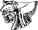
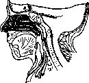
gillette's lateral retropharyngeal glands (lgl. retropharyngeales laterales Gillette'a) in newborns (Fig. 3) consist of one or two glands located symmetrically on both sides near the posterolateral angle of the pharynx at the level of the atlas. Less commonly, there are 3 or more glands, which are then arranged vertically one above the other. In children, this group of glands is constantly present and lies medially from the internal carotid artery, mostly near the place of its entry into the carotid canal. In adults, these glands are usually present on both sides, but sometimes they are absent on one side. The medial group (lymphoglandulae retropharyngeales mediales) lies medial to the previous ones and is found only in newborns or small children, while later these glands atrophy and completely disappear in adults. The size and number of these glands are extremely variable; sometimes they are very small. The medial group of lymph glands is interposed along the course of the lymphatic vessels collecting lymph from the posterior and upper parts of the epipharynx, and some of these lymphatic vessels, bypassing the medial group of lymph glands, empty either directly into the deep cervical lymph glands or preliminarily cross the lateral group of lymph glands. This group of lymph glands receives afferent lymphatic vessels from the lateral wall of the epipharynx, lying behind the pharyngeal opening of the Eustachian tube, from the posterior parts of the nasal cavity and the mucosa of the Eustachian tube and middle ear (Fig. 4). The efferent lymphatic vessels penetrate through the posterior wall of the pharynx into the retropharyngeal space near the midline and sharply change their direction outward, with part of the vessels, having gone behind the internal carotid artery and jugular vein, directly pouring into the deep cervical lymph nodes or preliminarily crossing the medial and lateral retropharyngeal glands. Pathology of the retropharyngeal space. The tissue of the retropharyngeal space serves as a site for the formation of abscesses, which can have various origins. Most often, retropharyngeal abscesses develop with acute inflammation of retropharyngeal lymph glands (lymphadenitis retropharyngealis) resulting in suppuration. Usually, one of the lateral retropharyngeal glands is affected, and only rarely the medial group of glands. Therefore, acute retropharyngeal abscesses in children are mostly located on the posterior wall of the pharynx, slightly to the side of the midline, and only gradually, as the abscess spreads, do they cross
the midline. Rarely do they occupy a median position, and in such cases, they originate from the medial group of retropharyngeal lymph glands (Bokay). Suppuration of these glands is caused by infection penetrating through lymphatic pathways from the roof, posterior and lateral walls of the epipharynx lying behind the Eustachian tube, from the posterior parts of the nasal cavity, and from the mucosa of the Eustachian tube and middle ear. The cause of retropharyngeal lymphadenitis is inflammatory diseases of the upper respiratory tract, to which young children are especially predisposed. The etiology of inflammatory processes can be various: rhinitis, rhinopharyngitis, retronasal angina, congenital syphilitic coryza, scarlatina, measles, influenza. Inflammatory swelling of the lymph glands does not always lead to suppuration. Retropharyngeal lymphadenitis in 9.5% of cases can spontaneously disappear (McLean and von Hofe). Chronically proceeding retropharyngeal abscesses occur in the form of so-called cold abscesses. Cold abscesses, developing with tuberculous disintegration of lymph glands in both children and adults, are extremely rare. Mostly they belong
to the category of tuberculous abscesses originating from the cervical vertebrae (vertebral caries).
Clinical picture. The symptoms of acute retropharyngeal abscesses are very characteristic. The disease begins with fever, often with chills, and difficulty in swallowing. The child refuses the breast or food, breathes heavily, with a snort, and sometimes stridor occurs. The head is often fixed and tilted back or to the healthy side. Voice changes: it becomes nasal, hoarse, or barking. With large abscesses, suffocation attacks can occur. When examining the pharynx, a more or less significant protrusion of the posterior wall is found, usually unilateral, covered with hyperemic, edematous mucosa. Palpation of this protrusion reveals a soft, fluctuating swelling.
Complications and course. Untreated retropharyngeal abscesses can spontaneously open into the pharynx, which usually brings relief, but can be dangerous due to the entry of pus into the respiratory tract. Opening into the large vessels of the neck (internal carotid artery, internal jugular vein) can lead to fatal bleeding. Extension of the abscess down into the posterior mediastinum leads to fatal mediastinitis. Chronic cold abscesses have a sluggish course, are often accompanied by signs of bone tuberculosis, and open outwards through fistulas.
cold abscesses. They are formed as a result of a caries process of the upper cervical vertebrae and the bones of the skull base, more often in spondylitis tuberculosa (Fig. 5) and rarely in gummatous syphilis. Often the source of a retropharyngeal abscess is purulent processes in the cavities of the middle ear. Infection can penetrate through lymphatic vessels from the middle ear and Eustachian tube to the lateral group of retropharyngeal lymph glands. More often in this case, we are dealing with cold abscesses arising as a result of the movement of pus along certain (preformed) pathways: 1) pus from the mastoid process through the incisura mastoidea can descend into the pharyngeal-maxillary space and from there into the retropharyngeal space; 2) with abundant development of pneumatic cells at the bottom of the tympanic cavity, suppuration can penetrate into the retropharyngeal space along the cellular tissue along the Eustachian tube; 3) in some cases, the cellular tissue surrounding the parotid gland serves as a conductor of suppuration. Pus penetrates either from the cartilaginous auditory canal through slits in its anterior wall (incisura Santorini) or from the incisura digastrica and from there along the posterior belly of the digastric muscle sequentially captures the posterior section of the pharyngeal-maxillary space and finally the retropharyngeal space. Retropharyngeal abscesses of otitic origin occur in both acute and chronic mastoiditis. Empyema of the sphenoid sinus is very rarely the cause of a retropharyngeal abscess. Finally, retropharyngeal abscesses can develop (Figure 5. Retropharyngeal abscess in tuberculosis of the cervical vertebrae) as a category of so-called mechanical damage to the posterior wall of the pharynx (by foreign bodies, during esophagoscopy). As for bacteriology, in acute retropharyngeal abscesses in children, it is almost always a streptococcal infection. In tuberculosis of the lymph glands, tubercular bacilli were found. Clinical symptomatology. Acute retropharyngeal abscesses sometimes develop rapidly—within a few days, but more often from 10 to 14 days. At the beginning of the disease, symptoms are poorly characterized; the first sign is hoarse breathing during sleep. When an abscess forms, symptoms of difficult breathing and swallowing come to the foreground, the degree of which depends on the position and size of the abscess. Sharp difficulty in breathing is expressed by inspiratory dyspnea. Unlike stenotic breathing (e.g., in true and false croup), inhalation has the character of a moist rattle, gurgling in the throat, especially during sleep. Breathing difficulty increases with the vertical position of the head. The voice takes on a nasal shade, becomes coarse, dull, hoarse, but is never aphonic. Cough is either completely absent or insignificant. Sharp disorder of swallowing is caused not only by a mechanical obstacle and inflammatory edema around the abscess, but also by acute pain, as a result of which children completely refuse food and drink. Especially characteristic of retropharyngeal abscesses is the forced position of the child's head; to reduce pain sensations, the head is thrown back somewhat and tilted to the affected side. Almost always, swelling is noted on the neck behind the angle of the lower jaw in front of the sternocleidomastoid muscle. In the initial period, fever is absent or there is a slight increase in temperature. The formation of an abscess is always accompanied by a high remittent temperature (up to 39–40°). Retropharyngeal abscesses are more common in children of the lymphatic and exudative type under the age of two years. Chronic retropharyngeal abscesses in tuberculosis of the retropharyngeal lymph glands are mostly accompanied by insignificant pain. Retropharyngeal cold abscesses accompanying spondylitis of the upper cervical vertebrae develop extremely slowly and can remain undetected for a long time. Respiratory and swallowing disorders occur only with a significant volume of the abscesses. In this case, the impossibility of lateral movements of the head and pain upon pressure on the spinous processes of the cervical vertebrae come to the foreground. Sometimes cold abscesses have the character of rapidly growing abscesses with secondary infection. Diagnosis. The above-mentioned symptoms arouse suspicion of the existence of a retropharyngeal abscess; however, an accurate diagnosis can be made only on the basis of finger examination, which must be resorted to in doubtful cases. A simple examination of the pharynx in infants and small children is extremely difficult and, due to the enormous amount of mucus accumulating in the fauces and obscuring the field of vision, often causes an erroneous diagnosis. Upon finger examination, a retropharyngeal abscess gives the sensation of a smooth, elastic, fluctuating tumor located on the posterior wall of the pharynx, somewhat to the side of the midline. If it is possible to perform an examination per os, it is possible to detect a bulging of the posterior wall of the pharynx, which mostly occupies a lateral position, less often located along the midline; with further development of the retropharyngeal abscess, the swelling can spread to the entire visible part of the pharynx, protruding the soft palate forward and filling the epipharynx. The mucous membrane is mostly strongly reddened. Benign tumors (lipomas, strumas, Busch's retropharyngeal tumors) are rare in childhood and do not give fluctuation upon palpation. It is difficult to confuse a retropharyngeal abscess with a peritonsillar abscess (angina phlegmonosa), in which the inflammatory swelling occupies the lateral wall of the pharynx, i.e., mainly the region of the soft palate corresponding to the supratonsillar fossa. Prognosis in acute retropharyngeal abscesses depends on correct diagnosis and mainly on the speed of the surgical intervention undertaken. Timely performed opening usually gives a favorable prognosis. An abscess left to its own course can entail life-threatening complications. It can spontaneously open into the pharynx and cause death from suffocation (asphyxia) due to the flow of a large amount of pus into the larynx, especially during sleep, or entail septic aspiration pneumonia. Large abscesses represent a serious danger of suffocation due to simple compression of the larynx by the tumor or collateral edema of the entrance to the larynx. Rare complications include a) fatal hemorrhages due to the erosion of large vessels of the neck and b) cold abscesses in the posterior mediastinum, to which purulent pleurisy and pericarditis mostly join. Prognosis is poor in chronic (cold) abscesses depending on the caries process of the spine, since the underlying disease mostly gives an unfavorable outcome. The prognosis for cold abscesses of otitic origin is closely dependent on the underlying disease. Operative intervention on the mastoid process (in the absence of intracranial or labyrinthine complications) generally gives a good prognosis. Treatment of acute retropharyngeal abscesses consists of their earliest possible opening through the oral cavity. The use of anesthesia and mouth gags should be avoided. The opening is made with a sharp-pointed scalpel, which is guided by the index finger of the right hand to the posterior wall of the pharynx; at the same time, the tongue is pushed downwards by the index finger of the left hand. To avoid wounding neighboring parts, the blade of the scalpel is wrapped halfway with adhesive plaster. The incision is made along the midline of the abscess in the direction from bottom to top, about 2 cm long. In view of the danger of pus flowing into the larynx, it is necessary immediately after the incision to strongly tilt the patient's head forward and down. As a result of timely opening of the retropharyngeal abscess, rapid healing usually occurs. The danger of wounding large vessels is completely excluded, since the internal carotid artery and internal jugular vein are located in the posterior section of the pharyngeal-maxillary space and are largely pushed outward by the abscess. Cases of sudden death after opening a retropharyngeal abscess due to collapse are among rare phenomena. Retropharyngeal cold abscesses developing as a result of tuberculosis of the cervical vertebrae are not recommended to be opened by incision, since opening is followed by ichorous disintegration of the cavity, often with a fatal outcome. It is more advisable to open them by puncturing with a syringe and injecting iodoform emulsion into the cavity, except in cases with secondary infection. As for surgical techniques aimed at opening access to the retropharyngeal space from the lateral surface of the neck, they find few supporters, at least in uncomplicated cases of acute retropharyngeal abscesses in children, since the possibility of wounding the neck vessels is not excluded in this case. According to Bókay's statistics, in 129 cases of idiopathic acute retropharyngeal abscesses, opening was never performed externally, and yet a fatal outcome was observed in only 5 cases. External surgery is indicated when it is impossible to open a retropharyngeal abscess due to jaw tightening (trismus) or too deep a position of the abscess, as well as in cases of complication by phlegmon of the pharyngeal-maxillary space (phlegmona parapharyngeale). As for the access route to the retropharyngeal space, an incision of the soft tissues along the anterior edge of the sternocleidomastoid muscle (Burckhardt) deserves preference.
Related articles
Cite this page
“Retropharyngeal Space.” Soviet Medical Encyclopedia. English translation of Bolshaya Meditsinskaya Entsiklopediya, 1st ed. (Moscow, 1928–1936), ed. N. A. Semashko. https://sovietmedicalencyclopedia.pages.dev/article/retropharyngeal-space/