Human Muscles
Historical document, translated for reference. It reflects medical knowledge of the 1920s–30s and is not medical advice.
Summary
This article provides a detailed anatomical description of human muscles, including their Latin and Russian names, synonyms, origins, insertions, innervation, and variations. It covers various muscle groups from the larynx to the lower extremities.
Encyclopedia article (1928–1936)
HUMAN MUSCLES
80. Latin and Russian names. Synonyms. Form and position. Origin and insertion. Innervation and relation to segments. Thyreo-epiglotticus (thyroid-epiglottic muscle). Syn.: thyreo-epiglotticus inferior, s. major, thyreo-membranosus inferior, dilatator vestibuli laryngis, stratum thyreo-membranosum. Part of m. ary-epiglottici, namely those fibers which extend from the thyroid cartilage to the epiglottis. Origin: thyroid cartilage-facies interna. Insertion: epiglottis. N. vagus-n. laryngeus inferior, its anterior branch. Thyreo-epiglotticus major and minor (large and small thyroid-epiglottic muscles). Name for separate parts of the thyreo-epiglotticus. N. vagus-n. laryngeus, its anterior branch. Thyreo-hyoideus impar (azygos) (Soemmering) (upper unpaired thyroid-hyoid muscle). [Variation.] See m. thyreo-hyoideus. Thyreoideus superior (par), s. minor (Morgagni) (upper paired thyroid-hyoid muscle). [Variation.] See m. thyreo-hyoideus. 46) Thyreo-glossus lateralis (Fürbringer) (lateral thyroid-glossus muscle). [Variation.] See m. thyreo-hyoideus. Thyreo-mediastinalis (Macalister) (thyroid-mediastinal muscle). [Variation.] See m. sterno-thyreoideus. Thyreo-syndesmicus (Soemmering) (thyroid-syndesmic muscle). [Variation.] See m. thyreo-hyoideus. Thyreo-triticeus (Macalister). [Variation.] See m. thyreo-hyoideus. Tibialis anterior (anterior tibialis muscle). Syn.: tibialis anticus, hippicus, tibiaeus, catenae. Long prismatic muscle located on the lateral surface of the leg directly on the tibia. The muscle begins tendinous and on the leg forms a flat long terminal tendon which passes to the dorsum of the foot. The muscle lies directly under the skin and fascia cruris, on the lateral side first adjacent to m. extensor digitorum longus, then to m. extensor hallucis longus, covering m. tibialis posterior through the interosseous membrane. Origin: thigh-condylus lateralis and facies lateralis femoris, fascia cruris and interosseous membrane to its lower third. Insertion: First cuneiform and first metatarsal bones. N. peronaeus profundus. Liv (v) and Si. Tibialis posterior (posterior tibialis muscle). Syn.: tibialis, s. tibiaeus, s. tibicus posticus, s. internus, nauticus. Bipennate prismatic long muscle, located on the posterior surface of the leg under m. triceps surae on the interosseous membrane between m. flexor digitorum longus and flexor hallucis longus, partially covered by it. Flat long tendon curves around malleolus medialis (sulcus malleolaris) and through the medial edge passes to the sole; here on the bones the tendon forms a whole 'bouquet' of tendons to the tarsal and metatarsal bones. Origin: interosseous membrane and adjacent parts of tibia and fibula to the distal quarter and deep fascia cruris. Insertion: navicular bone-tuberositas ossis navicularis, I (II and III) cuneiform bones- their plantar surface, talus-sustentaculum tali and II, III and IV metatarsal bones, basis ossis metatarsalis II, III and IV. N. tibialis. Ly and SKii). Trachealis (Guysse) (M. tracheae). Tunica muscularis parietis membranaceae of the respiratory tract. Tracheo-mastoideus minor (Luschka) (trapezius-sternomastoid muscle). Syn.: trachelo-mastoideus accessorius-accessory trapezius-sternomastoid muscle). [Variation.] See m. longissimus. Tragicus (tragus muscle). Syn.: tragi. Small plate of vertical, sagittal, frontal muscle fibers on the anterior surface of the tragus. Origin: tragus-below tragus. Insertion: auricle-tragus, its outer surface. N. facialis. Blood supply. Action. Variations. Pulls the epiglottis forward and thereby increases the entrance to vestibulum laryngis. Tarin describes two muscles: a large one from the lateral part of the thyroid cartilage and a small one from its medial part. A. tibialis anterior and a. recurrens tibialis anterior. Dorsiflexes (extends) the foot. 1) The tendinous part of the muscle may partially or wholly divide into two bundles: 2) from the muscle often a bundle departs to lig. cruciatum,-this is m. tensor fasciae dorsalis pedis; 3) M. may attach to articulatio metatarso-phalangea; 4) may have a musculo-tendinous connection with m. extensor longus digiti II and m. extensor hallucis brevis. A. tibialis posterior and a. peronaea. Plantarflexes the foot, supinates and adducts. i) Absence of the muscle is described; 2) the muscular part of the muscle may duplicate-m. tibialis secundus; 3) may attach: a) to II, III and IV os metatarsale; b) and os cuboideum; c) to m. flexor hallucis brevis; d) to the fifth toe; e) to the tendon of m. peronaei longi. Function unknown. 80. Latin and Russian names. Synonyms. Form and position. Origin and insertion. Innervation and relation to segments. Transversalis cervicis anticus (Luschka) (anterior transverse muscle of the neck). Syn.: singularis in collo, transversarii cervicis anteriores, intertransversarius lateralis longus, transversarius cervicis medius. [Variation.] See ram. intertransversarii ventrales. Transversus abdominis (transverse muscle of the abdomen). Syn.: transversalis abdominis, abdominis intimus. The muscle is a flat, broad sheet of parallel transverse muscle fibers, having the form of a triangle with a truncated apex; the apex is directed dorsally, while the base is neutral. The muscle is located on the lateral surface of the abdominal wall, in the space between the costal cartilages and the ilium, covered by both oblique muscles of the abdomen and lies internally against fascia retroperitonaealis and against the peritoneum. The muscle fibers behind and in front pass into tendinous expansions. The posterior aponeurosis in the form of a quadrangular plate attaches to the transverse processes of the lumbar vertebrae. The anterior aponeurosis begins at the semilunar line (linea semilunaris Spigelii); in the upper two-thirds this aponeurosis goes to form the posterior wall of the sheath of the rectus abdominis muscle, and in the lower third-its anterior wall; therefore in it there is an oval slit, into which passes m. rectus abdominis-this is linea semicircularis Douglasii. At its origin from the ribs, the muscle stands in close connection with m. transversus thoracis. Origin: VII-XII costal cartilages- their inner surface, fascia lumbodorsalis-deep leaflet (lumbar vertebrae-their processus transversus), ilium-crista iliaca, its labium internum and ligamentum inguinale. Insertion: linea alba. Spinal nerves nn.
intercostales (VII-XII) and nn. hypogastricus and inguinalis. Dyj - XII and Li-in Transversus auriculae (transverse M. of the auricle). A long series of short parallel muscle fibers passing into tendon fibers; located on the posterior surface of the auricle between eminentia conchae and the beginning of cauda helicis. Origin: auricle, posterior surface of it-eminentia conchae. Insertion: auricle-its edge, beginning of cauda helicis. N. facialis Transversus colli (Luschka) (transverse M. of the neck). [Variation.] See m. sterno-thyreoideus Transversus glabellae (Rouge) (transverse M. of the glabella). [Variation.] See m. orbicularis oculii Transversus linguae (transverse M. of the tongue). Syn.: stratum musculare linguae transversum. Numerous muscle bundles located in the thickness of the tongue in a transverse direction between the upper and lower longitudinal M. of the tongue (m. longitudinalis linguae sup. and inf.). Origin: septum linguae. Insertion: lateral edges and dorsum of the tongue. N. hypoglossus Transversus manus (Halette) (transverse M. of the hand). [Variation.] See m. adductor pollicis Transversus menti (transverse M. of the chin). [Very frequent variation, see also m. triangularis.] Transverse muscle fibers, which go from the lower jaw along the distance between tuberculum mentale and fossa digastrica of both sides, ending posteriorly along linea submentalis. N. facialis Transversus nuchae (Schultze) (transverse M. of the nape). Syn.: corrugator posticus, occipitalis teres ac minor (Santorini), suboccip- Origin: linea nuchae superior and protuberantia occipitalis externa. Insertion: skin of the lateral part of the occiput. N. facialis Blood supply Action Variations A. circumflexa ilium interna and a. epigastrica inferior from a. iliaca externa and aa. musculo-phrenica and epigastrica superior from a. mammaria interna Participates in the formation of the abdominal press 1) Complete absence of the M. was observed very rarely; partially the portio inguinalis may be absent; 2) the number of rib bundles may vary, increasing to seven and eight and decreasing to five; 3) fusion of the portionis inguinalis of the M. with m. obliquus internus abdominis is possible; 4) cases of partial doubling of the M. have been observed; 5) the M. may extend caudally beyond funiculus spermaticus even to spina pubica, then it is pierced by the spermatic cord; 6) tendon bridges have been described by only one author; 7) the M. may be connected with m. intercostalis intermedius and m. intercostalis internus, with lateral fibers of m. recti abdominis; 8) additional bundles in the area of foveae inguinalis are often observed: these are m. interfovealis (Hofmann)-bundles of muscle fibers along the lateral edge of m. recti abdominis, located ventrally from vasa epigastrica inferiora interna; these fibers may go from the inferior surface of m. transversi abdominis or from its aponeurosis or between the two layers of the aponeurosis and may reach lig. inguinale reflexum and lig. lacunare; the same M. is described under other names (see m. interfoveolaris); m. tensor fasciae transversalis-bundles of muscle fibers, which may go, starting from lig. lacunare up to the height of lineae semicircularis (Douglasii), and pass into fascia transversalis, sometimes it can be seen that this M. lies dorsally from vasa epigastrica inferiora interna; m. tensor laminae posterioris vaginae m. recti et fasciae transversalis abdominis (Gruber)-three bundles of muscle fibers, which began tendinous on the superior surface of fasciae transversalis or, diverging cranially under the last bundle of m. transversi abdominis, ended in thin tendons in the fascia, with the medial ones reaching linea semicircularis Brings the helix closer to the auricle dorsalis profunda linguae Protrudes the tongue upward The place to which the M. attaches varies particularly significantly; this may again be linea nuchae superior, or the dorsal edge of m. sterno- №№ Latin and Russian name. Synonyms. Form and position Origin and insertion Innervation and relation to segments n and i o И o o. И tails. [Frequent variation (27-72% according to different authors).] Muscle bundles of very different shape and size in the nape area under the skin Transversus perinaei profundus (deep transverse M. of the perineum). Syn.: transversus perinaei anterior, s. superior, constrictor urethrae membranaceae.iscaio-urethralis, prostaticus internus, transverso-analis, triangularis perinaei, s. trigoni urethralis, Wilsoni, Guthrie. A muscular plate lying in the perineum between both layers of the perineal fascia-fasciae diaphragmatis uro-genitalis superioris and inferioris in the space of trigoni uro-genitalis. Muscle fibers, which are stretched between the pubic bones-rami inferiores and interlace with the fibers of the opposite side and with fascia diaphragmatis uro-genitalis. N. pudendus Transversus perinaei superficialis (superficial transverse M. of the perineum). Syn.: transversus perinaei medius, s. posterior, ischio-perinaealis, transverso-analis. A small muscular ribbon, located transversely in the perineum immediately under the skin and fascia perinaei superficialis, along the posterior edge of trigoni uro-genitalis, between the ischial bone and a fibrous plate near raphe bulbi; both antimera pass into each other at the midline. Origin: ischial bone-ramus inferior, its medial surface and fascia superficialis perinaei. Transversus radicis nasi (Eisler) (transverse M. of the root of the nose). [Variation.] See m. orbicularis oculi Transversus thoracis (transverse M. of the thorax). Syn.: transversus anterior, s. pectoris, infracostalis anterior, sterno-costalis, triangularis sterni, intracostales. It is a series of flat muscle bundles located on the medial surface of the sternum and cartilaginous ribs (II-VI), obliquely in the upper bundles and transversely in the lower ones. Origin: sternum-corpus sterni and processus xiphoideus; their posterior surface, cartilage VII (VI) of the rib. Insertion: II-VI rib cartilages. Nn. intercostales II- VI Trapezius: pes ardescendens, pars transversa and pars ascendens (trapezius or hood M., M. of the capuchins). Syn.: cucullaris, mensalis. It is a large flat broad triangular M., located on the dorsal surface of the neck and thorax, directly under the skin and the superficial fascia of the back and neck. The cut-off apex of the triangle is located laterally on the shoulder, and the broad base on the back along the midline on the vertebral column. Three portions are distinguished according to the direction of the fibers, between which however there are usually no definite boundaries. The origin of all portions is tendinous, but particularly so is the so-called tendinous mirror (speculum Helmonti); the insertion is also tendinous. The M. in turn covers mm. semispinalis capitis, splenius, levator scapulae, supraspinatus, rhomboides, serratus posterior superior and sometimes mm. latissimus dorsi and infraspinatus. Origin: pars descendens occipital bone-linea nuchae superior, protuberantia occipitalis externa, lig. nuchae and I-IV vertebrae-processus spinosus; pars transversa: VII cervical and I (II) thoracic vertebra-processus spinosus; pars ascendens: II-XI (XII) thoracic vertebrae-processus spinosus. Insertion: pars descendens: clavicle-lateral part of it; pars transversa-clavicle-extremitas acromialis, scapula-processus acromialis and spina scapulae, its upper edge; pars ascendens-scapula-trigonum spinae scapulae. N. accessorius Willisii and branches of cervical nerves (II), III and IV. C(ц), ш и IV Blood supply Action Variations cleido-mastoidei or fascia superficialis colli, or m. occipito-parotideus, or m. auricularis posterior-this is m. auriculo-iniacus; the ventral end of the M. may pass into the most cranial bundles of m. platysmatis or atypical muscle formations, which may go to os hyoideum into the pharynx or into m. sterno-cleido-mastoideus; once a M. was found consisting of three portions, all of them originated from protuberantia occipitalis externa and went: one together with m. auricularis posterior, another to fascia parotidea, and the third to the dorsal edge of m. sterno-cleido-mastoidei and ended in fascia superficialis colli. The existence of this M. is not recognized by all anatomists (see.
(Le Double) 1) Sometimes the muscle may be absent; 2) the beginning and end of the muscle may split into two muscles--this is the transversus perinaei alter; 3) it may begin on the lower lamina of the fascia perinaei, fascia obturatoria, fascia diaphragmatis pelvis; 4) some bundles may pass: a) into the m. sphincter ani externus; b) into the bulbo-cavernosus tissue; 5) it may receive accessory bundles from the aponeurosis of the gluteal muscles--this is the m. glutaeo-perinaealis 1) It may be completely absent or replaced by tendon fibers; 2) it often divides into separate bundles or into two completely separate portions or separated from each other only cranially; 3) cranially it may extend to the VII rib; 4) the number of bundles may vary from six to two or even one; 5) from the II rib a bundle may pass through the third rib to the fourth; 6) once a two-layered muscle was observed on both sides; 7) once bundles were observed to the linea alba and aponeurosis of m. transversi abdominis A. transversa colli and a. transversa scapulae from a. subclavia, rr. dorsales from aa. intercostales and a. occipitalis from a. carotis externa All portions, acting together, bring the scapula closer to the spine; pars descendens raises the scapula and clavicle; pars ascendens lowers the scapula 1) Often or even constantly both antimera are unevenly developed, with the right half usually longer, wider and more massive; 2) the origin on the occipital bone on one side may be greatly spread out or on the other side completely absent, when the muscle attaches to a tendon arch stretched from protuberantia occipitalis externa to the tendon of m. sternocleidomastoidei; 3) the origin of the cervical part may have a wide tendon mirror--cranial continuation of the speculum Helmonti; 4) the origin of partis ascendens may be reduced to the VIII thoracic vertebra (even V and IV); 5) the caudal tendon triangle may merge with the initial tendon of m. latissimi dorsi; 6) the attachment to the clavicle may diminish to complete disappearance; 7) more often there is elongation of this part to fusion with m. sternocleidomastoideus; 8) once a tendon bundle was observed from the ventral edge of the muscle to sternum; 9) there is a transition of tendon fibers into m. deltoideus; 10) division into two independent muscle layers was observed; 11) possible division of the muscle into three independent muscles; 12) pars descendens occasionally has an inscriptio tendinea; 13) with athletic development there may be division into separate layers; 14) this same layering can explain the existence of mm. subcucullarium, which go from speculum rhombo- №№ Latin and Russian name. Synonyms. Form and position Origin and attachment Innervation and relation to segments "a? « n o И O a I Triangularis (triangular muscle of the lower lip). Syn.: triangularis inferior, s. menti, s. labii inferioris, depressor labiorum communis, s.anguli oris, angularis oris inferior, pyramidalis menti. Muscle fibers in the form of a triangle, located under the skin and fascia superficialis on the face between the lower edge of the jaw (base of the triangle) and the angle of the mouth (apex of the triangle). Under the muscle lie fibers of m. quadrati labii inferioris Origin: lower jaw-its lower edge, laterally from tuberculum mentale. Insertion: skin of the angle of the mouth and upper and lower lip N. facialis-rr. buccales Triceps brachii: caput longum, caput laterale and caput mediale (triceps muscle of the arm). Syn.: triceps antibracliii, s. cubiti, extensor antibracliii, s. cubiti magnus, s. posticus. Caput longum=m. anconaeus longus; caput primum=omo-anconaeus; caput mediale=caput tertium, s. parvum, s. internum, anconaeus internus, s. brevis, vastus internus; caput laterale=caput externum, s. magnum, s. secundum, vastus externus, anconaeus brevis, s. externus. A large muscular mass occupying the entire posterior surface of the arm, from the scapula to the olecranon process of the ulna,-lies directly under the skin and superficial fascia of the arm. The muscle is formed from three heads. The long head on the dorsal side is covered by a tendon stretching, to which laterally at the middle of the arm also approach tendon fibers of the lateral head, and closer to the distal end fibers of the medial head also flow in, thus forming a strong terminal tendon Origin: caput longum: scapula-tuberositas infraglenoidalis scapulae; caput mediale: humerus-facies posterior distally from sulcus n. radialis, septa intermuscularia to epicondylus lateralis humeri; caput laterale: humerus-facies posterior upper part-laterally from sulcus n. radialis and to tuberculum majus, in the lower half from septum intermusculare laterale. Insertion: ulna-olecranon ulnae, fascia antibrachii and posterior surface of capsula articulationis cubiti N. radialis. Cvi. VII and VIII Uretericus (Barkow) (ureter muscle). Syn. triangularis infundibuli vesicae urinariae Longitudinal bundles in trigonum vesicae Ureterum(Barkow) (ureter muscle). Syn. lig. elasticum interuretericum Longitudinal muscle bundles of the lower end of the ureters Urethralis transversus (Krause) (transverse muscle of the urethra) Upper part of m. sphincteris urethrae membranaceae Uvulae (muscle of the uvula). Syn.: azygos, s.levator, s. motor uvulae, palato-staphylinus. A very small muscle bundle (paired) on the dorsal surface of the uvula Origin: palatine bone-spina nasalis posterior and tendon plate of m. tensoris veli palatini. Insertion: apex of the uvula N. vagus-r. pharyngeus Vastus intermedius (intermediate vastus muscle of the thigh). Syn.: vastus medius, s. cruralis, s. crureus, s. femoralis, Muscle mass on the anterior surface of the thigh between vastus medialis and lateralis under m. rectus femoris; anterior surface of the muscle is covered by a tendon aponeurosis, which passes into the terminal tendon Origin: thigh-facies anterior and lateralis from the middle of lineas intertrochantericae to the beginning of the lower quarter of the thigh. Insertion: patella-basis patellae together with the tendon of t. vasti medialis and t. articularis genu (more correctly genus) to the capsule of the knee joint N. femoralis. Ltt-iv Blood supply Action Variations ideum or from the spinous process of the III thoracic vertebra to the tendon of m. trapezii; 15) occasionally bundles were observed on the dorsal surface of the muscle as remnants of the dorsal musculature of the trunk-m. dorso-fascialis-these are bundles from the spinous processes of thoracic vertebrae to speculum rhomboideum; 16) a bundle of muscle fibers-m. subtrapezius was also observed under m.trapezius, which with good development begins from the lower surface of the tendon of m. trapezius at the level of IV-VI thoracic vertebrae-processus spinosus and attaches to the base of spinae scapulae A. labialis inferior and a. submentalis from a. maxillaris externa and a. mentalis from a. maxillaris interna Pulls the angle of the mouth backward and downward 1) The size of the muscle can vary greatly; 2) sometimes it divides into several portions, more often into three; 3) lateral bundles may pass into the facial part of m. platysmatis; 4) individual bundles may pass into mm. buccinator, zygomaticus, caninus, quadratus labii superioris; 5) there is a connection of the medial bundle under the chin with the antimere in the form of a loop from one angle of the mouth to the other-this is m. transversus menti; but this name is also sometimes given to a well-developed muscle that begins in the area of tuberculi medialis and goes under the chin toward the opposite side. This m. transversus menti (Santorini) divides both parts of the "double chin" A. circumflexa humeri posterior from a. axil-laris Extends the arm at the elbow joint. Caput longum additionally moves the entire arm backward (extensio) 1) Very rare cases when the origin of capitis longi may extend along margo axillaris to angulus inferior or descend on humerus (shoulder capsule); 2) The muscle may turn into a quadriceps and an additional bundle may begin: a) from processus coracoideus; b) from the shoulder capsule; c) from the tendon of m. latissimi dorsi; d) from collum chirurgicum humeri; 3) most often connection (even as a rule) of capitis longi with the tendon of m. latissimi dorsi; when this connection contains muscle fibers the muscle is called m. dorso-epitrochlearis; 4) closer relationships are observed with the following muscles: a) m. infraspinatus, b) teres major, c) anconaeus, d) flexor carpi ulnaris; 5) The muscle may turn into a quadriceps also due to division of capitis longi into two muscles, if this division is complete-more often it is incomplete Pulls the uvula upward and backward A. circumflexa femo-ris lateralis and rr. perforantes from a. profunda femoris Extends the leg at the knee joint 1) The place of its origin can vary greatly within the limits from lineaintertrochanterica in cases of strong development and almost to articulatio genu-in case of weak development; 2) closer relationships are possible: a) with m. vastus medialis; b) vastus lateralis; c) rectus femoris and d) articulationis genu №№ Latin and Russian name. Synonyms. Form and position Origin and attachment Innervation and relation to segments Vastus lateralis (lateral vastus muscle of the thigh). Syn.: vastus externus. extensor crus vaptus.
Mass of muscle fibers running obliquely from top to bottom and from outside forward along the lateral surface of the thigh. The anterior surface in the lower part is aponeurotic and then passes into the terminal tendon, which divides into several bundles. Origin: thigh-greater trochanter, its lateral surface, intertrochanteric line, tuberosity of the gluteus, and lateral lip of the linea aspera of the femur. Insertion: patella-lateral and upper angle. Nerve: femoral nerve. Vastus medialis (medial vastus muscle of the thigh). Synonym: vastus internus. Mass of muscle fibers directed obliquely from top to bottom and from inside forward along the medial surface of the thigh. The tendon, which began inside the muscle, then emerges outward, lies adjacent to the tendon of the rectus femoris muscle, partially crosses in front of it; partially attaches to the patella. Origin: thigh-linea aspera of the femur, its medial lip, below the intertrochanteric line, terminal tendons of the adductor longus and magnus muscles. Insertion: patella-medial edge, to the common tendon mirror of the quadriceps femoris. Nerve: femoral nerve. Veli palatini et faucium See mm. glossopalatinus, levator veli palatini, pharyngopalatinus, tensor veli palatini, uvulae Ventricularis (muscle of the laryngeal ventricle). Synonym: arytaenoepiglotticus inferior, compressor sacculi laryngis, thyreoarytaenoideus minor, s. superior, s. medialis. Muscle bundle of the aryepiglotticus muscle. Origin: arytenoid cartilage-its lateral edge. Insertion: epiglottis-lateral edge of its lower part. Nerve: vagus-n. laryngeus inferior. Verticales linguae (vertical muscles of the tongue). Bundles of muscle fibers penetrating the entire thickness of the tongue from the upper to the lower surfaces of the tongue-in its free part. Origin: dorsum of the tongue. Insertion: lower surface of the tongue. Nerve: hypoglossus. Wilsoni See m. sphincter urethrae membranaceae. Vocalis (vocal muscle). Synonym: thyreoarytaenoideus inferior major, s. internus, aryvocalis. Part of the muscle fibers of the thyreoarytaenoideus muscle. Origin: thyroid cartilage-inner surface of the lamina. Insertion: arytenoid cartilage-vocal process. Nerve: vagus-n. laryngeus inferior, its anterior branch. Zygomaticus (zygomatic muscle). Synonym: zygomaticus major, malaris. A flat, narrow ribbon of parallel muscle fibers on the lateral surface of the cheek, immediately under the skin and superficial fascia, in the space between the zygomatic bone and the angle of the mouth. Origin: zygomatic bone-facial surface of the zygomatic bone. Insertion: skin of the angle of the mouth. Nerve: facial nerve-zygomatic branches. Zygomaticus accessorius (accessory zygomatic muscle). [Variation.] See m. zygomaticus. Zygomaticus minor=caput zygomaticum m. quadrati labii superioris See m. quadratus labii superioris. Ypsiloformis. [Variation.] See m. extensor pollicis longus. Blood supply Action Variations A. circumflexa femoris lateralis and rr. perforantes from a. profunda femoris 1) Usually the terminal tendon divides (Le Double, Gegenbaur, Poirier) into 2, sometimes into 4; their own muscle bundles approach them; sometimes they may be more independent and doubling of the muscle results; 2) The muscle may be in closer relationships: a) with m. rectus femoris; b) vastus internus; c) vastus intermedius and d) articularis genu. A. circumflexa femoris lateralis and rr. perforantes from a. profunda femoris Extends the leg in the knee joint 1) It may sometimes double due to complete division into two muscles; 21 some tendon bundles may attach to the tibial tuberosity; 3) closer relationships are possible: a) with m. rectus femoris; b) vastus lateralis; c) vastus intermedius and d) articulationis genu. Flattens the tongue. Raises the angle of the mouth upward and backward. 1) Rarely absent altogether; 2) its size varies greatly; 3) it may break down into two, three, and four portions; 4) there may be extension of the origin backward onto the zygomatic arch, onto the temporal fascia, onto the galea; 5) cases of attachment to m. orbicularis oculi, to caput zygomaticum m. quadrati labii superioris are observed; 6) connection with the facial part of m. platysma is possible-this includes m. zygomaticus accessorius; 7) the lateral bundle of the muscle may end: in the cheek skin, in m. buccinator or attach to m. caninus and triangularis. B. Gindne. 699
HUMAN MUSCLES
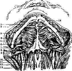
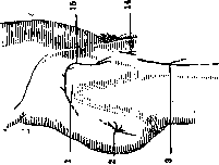
Figure 1. Muscles above the hyoid bone: 1-m. mylohyoideus; 2-mandible; 3-platysma; 4-m. masseter; 5-m. pterygoideus internus; 6-m. styloglossus; 7-hyoid bone; 8-m. stylohyoideus; 9-m. digastricus; 10-m. omohyoideus; 11-m. sternohyoideus; 12-thyroid cartilage; 13-m. sternocleidomastoideus; 14-attachment point of m. digastricus.
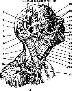
Figure 3. Superficial muscles of the head: 1-m. helicis major; 2-m. occipitalis; 3-antitragus; 4-m. auricularis posterior; 5-m. transversus nuchae (var.); 6-cauda helicis; 7-m. digastricus (venter posterior); 8-m. sternocleidomastoideus; 9-trigonum omotrapezoides; 10-m. trapezius; 11-trigonum omoclaviculare; 12 and 16-platysma; 13-m. omohyoideus; 14-thyroid cartilage; 15-apex of the carotid triangle (s. fossa carotica); 17-hyoid bone; 18-trigonum submaxillare; 19-m. digastricus (venter anterior); 20-m. transversus menti; 21-m. mentalis; 22-m. triangularis; 23-m. risorius; 24-m. quadratus labii inferioris; 25-m. orbicularis oris; 26-m. zygomaticus; 27-m. nasalis (pars transversa); 28-m. quadratus labii superioris; 29-m. orbicularis oculi (pars palpebralis); 30-m. procerus; 31-m. depressor supercilii; 32-m. corrugator supercilii; 33-m. orbicularis oculi (pars orbitalis); 34-m. frontalis; 35-parotidomasseteric fascia; 36-m. auriculofrontalis; 37-tragus; 38-m. auricularis superior et anterior; 39-galea aponeurotica.
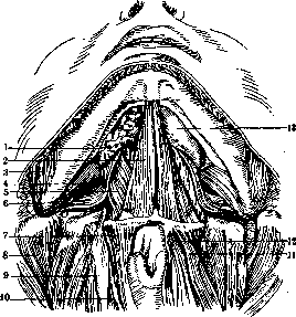
Figure 2. Muscles above* the hyoid bone: 1-sublingual gland; 2-m. geniohyoideus; 3-m. masseter; 4-m. pterygoideus internus; 5-m. hyoglossus; 6-m. styloglossus; 7-m. thyreohyoideus; 8-m. splenius capitis; 9-m. longus capitis; 10-m. sternothyreoideus; 11-m. omohyoideus; 12-m. sternohyoideus; 13-tongue.
;
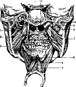
Figure 4. Masticatory muscles from behind (posterior part of the head removed by a saw cut passing through the temporomandibular joints; tongue, soft palate and pharynx with muscles removed): 1-lig. sphenomandibulare; 2-m. pterygoideus internus; 3-m. masseter; 4-m. geniohyoideus; 5-m. mylohyoideus; 6-m. genioglossus; 7-m. pterygoideus internus; 8-m. pterygoideus externus; 9-lig. pterygospinosum; 10-auditory tube; 11-choanae. 701
HUMAN MUSCLES
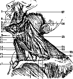
Figure 5. Muscles of the neck: 1-m. masseter; 2-m. digastricus; 3-m. styloglossus; 4 and 7-m. mylohyoideus; 5-m. stylohyoideus; 6-m. hypopharyngeus; 8-m. hyoglossus; 9-m. thyreohyoideus; 10-m. laryngopharyngeus; 11-m. longus capitis; 12 and 20-m. omohyoideus; 13-m. sternohyoideus; 14-m. sternothyreoideus; 15-thyroid gland; 16-m. scalenus anterior; 17 and 27-m. sternocleidomastoideus; 18-m. pectoralis major; 19-apex of the lung; 21-m. serratus anterior; 22-m. trapezius; 23-m. scalenus medius; 24-m. scalenus posterior; 25-m. levator scapulae; 26-m. splenius capitis.
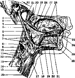
Fig. 7. Muscles of the tongue: 1-m. digastricus (venter posterior); 2-m. constrictor pharyngis superior; 3-m. styloglossus; 4-m. stylopharyngeus; 5-m. stylohyoideus; 6-m. constrictor pharyngis medius; 7-m. hyoglossus; 8-hyoid bone; 9 and 17-thyroid cartilage; 10-membrana hyothyreoidea; 11-m. constrictor pharyngis inferior; 12-m. thyreohyoideus; 13-thyroid gland; 14-m. omohyoideus; 15-m. sternothyreoideus; 16-m. sternohyoideus; 18-tendon of m. digastricus; 19-m. geniohyoideus; 20-m. longitudinalis superior; 21-m. mylohyoideus (raphe); 22-m. genioglossus; 23-m. longitudinalis inferior; 24-caruncula sublingualis; 25-vestibulum oris; 26-papillae fungiformes; 27-papilla foliata; 28-m. glossopharyngeus; 29-m. mylopharyngeus (cut); 30-arcus and m. glossopalatinus; 31-auditory tube; 32-zygomatic arch.
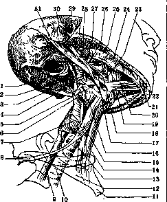
Figure 6. Deep muscles of the neck: 1-m. constrictor pharyngis superior; 2-raphe pharyngis; 3-m. stylopharyngeus; 4-m. constrictor pharyngis medius; 5-hyoid bone; 6-membrana hyothyreoidea; 7-m. constrictor pharyngis inferior; 8-m. omohyoideus; 9-esophagus; 10-trachea; 11-manubrium sterni; 12-1st rib; 13-m. sternothyreoideus; 14-m. sternohyoideus; 15-m. omohyoideus, venter superior; 16-m. thyreohyoideus; 17-m. stylohyoideus; 18-m. styloglossus; 19-m. digastricus, venter anterior; 20-m. mylohyoideus; 21-m. triangularis; 22-m. mentalis; 23-ductus submaxillaris (Warthoni); 24-platysma; 25-m. masseter; 26-m. pterygoideus internus; 27-parotid gland; 28-m. pterygoideus externus; 29-external acoustic meatus; 30-styloid process; 31-m. digastricus, venter posterior.
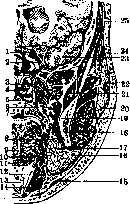
Figure 8. Frontal section of the head: 1-internal carotid artery; 2-sphenoidal sinus; 3-m. pterygoideus internus; 4-nasopharyngeal cavity; 5-m. levator uvulae; 6-m. tensor veli palatini; 7-soft palate; 8-m. styloglossus; 9-m. transversus linguae; 10-m. hyoglossus; 11-m. genioglossus; 12-m. thyreohyoideus; 13-lig. thyreohyoideum; 14-m. sternohyoideus; 15-platysma myoides; 16-submandibular ligament; 17-submandibular gland; 18-m. cephalopharyngeus; 19-mandibular ramus; 20-parotid gland; 21-temporal muscle; 22 and 24-galea aponeurotica; 23-temporal lobe of the brain; 25-pericranium. ____________.______ ______________ 703
HUMAN MUSCLES
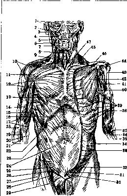
Figure 9.
Figure 9. Muscles of the trunk from the front: 1-m. orbicularis oculi; 2-m. quadratus labii sup.; 3-m. zygomaticus; 4-m. caninus; 5-m. masseter; 6-m. triangularis; 7-m. quadratus labii infer.; 8-m. sterno-hyoideus; 9-platysma; 10-m. deltoideus; 11-pars clavicularis m. pectoralis major; 12-pars sterno-costalis; 13-pars abdominalis; 14 and 15-m. biceps brachii: caput longum, caput breve; 16-costal margin; 17-m. brachialis; 18-m. brachio-radialis; 19-tendon of m. bicipitis; 20-lacertus fibrosus; 21-m. pronator teres; 22, 23 and 24-m. obliquus abdominis ext.; 25-annulus inguinalis subcutaneus; 26-m. ilio-psoas; 27-m. tensor fasciae latae; 28-m. pectineus; 29-m. sartorius; 30-lig. suspensorium penis; 31-lig. fundiforme penis; 32-m. cremaster; 33-aponeurosis m. obliqui abd. ext.; 34-m. obliquus abdominis externus; 35-radius; 36-attachment of m. brachialis; 37-capitulum radii; 38-m. brachialis; 39-m. serratus ant.; 40-m. latissimus dorsi; 41-m. coraco-brachialis; 42-m. pectoralis minor; 43-collum chirurgicum; 44-collum anatomicum; 45-m. subclavius; 46-m. trapezius; 47-m. sterno-cleido-mastoideus. Figure 10. Muscles of the human body from behind: 1 and 59-m. semispinalis capitis; 2-m. splenius capitis et cervicis; 3-m. levator scapulae; 4-m. serratus posterior superior; 5-m. serratus anterior; 6-m. rhomboideus; 7-m. supra-spinatus; 8-m. infraspinatus; 9-m. teres minor; 10-m. teres major; 11-m. coraco-brachialis; 12-m. brachialis; 13-epicondylus medialis; 14 and 45-olecranon; 15-m. glutaeus minimus; 16-m. piriformis; 17-m. gemellus superior; 18-bursa trochanterica m. glutaei medii; 19 and 22-m. quadratus femoris; 20 and 21-m. glutaeus maximus and bursa trochanterica; 23 and 25-bursae gluteo-femorales; 24-m. adductor minimus; 26-m. biceps femoris; 27-m. gastrocnemius; 28-m. plantaris; 29-planum popliteum femoris; 30-m. vastus medialis; 31-m. sartorius; 32-m. semimembranosus; 33-m. gracilis; 34-m. biceps femoris; 35-m. semitendinosus; 36-m. adductor magnus; 37-tractus ilio-tibialis (Maissiati); 38 and 39-m. glutaeus maximus; 40-m. glutaeus medius; 41-trigonum lumbale (Petiti); 42-m. obliquus abdom. ext.; 43-m. latissimus dorsi; 44-m. anconeus; 46-mm. extensores; 47, 48, 49 and 50-m. triceps (47 and 49-caput lat., 48-caput med., 50-caput long.); 51-m. rhomboideus; 52-m. teres major; 53-m. infraspinatus; 54-m. deltoideus; 55-spina scapulae; 56-m. trapezius; 57-vertebra prominens; 58-m. sterno-cleido-mastoideus.
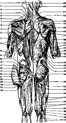
MUSCLES
HUMAN
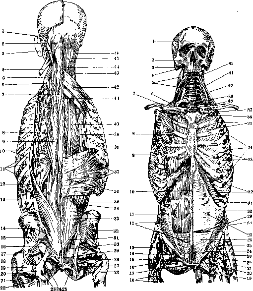
Figure 11.
Figure 11. Muscles of the back: 1-m. splenius capitis; 2-m. semispinalis capitis; 3-m. longissimus capitis; 4-m. splenius cervicis; 5-m. longissimus cervicis; 6-m. ilio-costalis cervicis; 7-m. semispinalis dorsi; 8-m. ilio-costalis dorsi; 9-m. longissimus dorsi; 10-mm. levatores costarum; 11-mm. intercostales externi; 12-m. ilio-costalis lumborum; 13-m. obliquus abd. ext.; 14-spina iliaca posterior superior; 15-m. glutaeus minimus; 16-foramen ischiadicum majus; 17-trochanter major; 18-bursa trochanterica; 19-m. quadratus femoris; 20-tuber ischiadicum; 21-m. glutaeus maximus; 22-m. adductor minimus; 23-bursa ischiadica; 24 and 25-lig. sacro-tuberosum; 26-m. obturator ext.; 27-m. obturator int.; 28-m. piriformis; 29-lig. sacro-coccygeum post, superficiale; 30 and 36-fascia lumbo-dorsalis; 31-origin of m. glutaei maximi; 32-m. glutaeus minimus; 33-tendon of m. longissimi and ilio-costalis; 34-m. ilio-costalis lumborum; 35-m. longissimus lumborum; 37-m. serratus post, inf.; 38-m. spinalis dorsi; 39-m. ilio-costalis dorsi; 40-m. longissimus dorsi; 41-m. semispinalis dorsi; 42-m. serratus posterior superior; 43-m. longissimus cervicis; 44-m. biventer; 45-m. complexus major; 46-m. longissimus capitis. Figure 12. Muscles of the trunk from the front: 1-frons; 2-arcus zygomaticus; 3-processus mastoideus; 4-maxilla; 5-m. levator scapulae; 6, 9, 33 and 34-m. serratus ant.; 7-clavicle; 8-m. pectoralis minor; 10-linea alba; 11 and 30-linea semilunaris (Spigeli); 12-m. transversus abdominis; 13-m. glutaeus medius; 14-lig. ilio-pectineum; 15-m. glutaeus minimus; 16-lacuna vasorum; 17-mm. vasti femoris; 18 and 19-m. obturator externus; 20-m. pectineus; 21-m. tensor fasciae latae; 22-lig. ilio-pectineum; 23-m. ilio-psoas; 24-hiatus inguinalis abdominalis; 25-lig. inguinale; 26 and 31-m. transversus abdominis; 27-posterior sheath of m. recti; 28-linea semicircularis; 29-m. rectus abdom.; 32-arcus costarum; 35-m. intercostalis externus; 36-sulcus venaesubclaviae; 37-tuberculum scaleni (Lisfranci); 38-m. scalenus post.; 39 and 40-m. scalenus medius; 41-m. scalenus ant.; 42-m. intertransversarius. B. M. E. m. XIX. 707
HUMAN MUSCLES
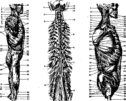
Figure 13.
Figure 14.
Figure 15.
Figure 13. Superficial muscles of the body: 1-m. semispinalis capitis; 2-m. sterno-cleido-mastoideus; 3-m. splenius; 4-m. levator scapulae; 5-m. scalenus medius; 6-m. trapezius; 7-acromion; 8-m. deltoideus; 9-m. infraspinatus; 10-m. teres major; 11-m. latissimus dorsi; 12-fascia lumbo-dorsalis; 13-trigonum lumbale (Petiti); 14-crista iliaca; 15-m. glutaeus maximus; 16-gluteal fold; 17-m. biceps femoris (caput longum); 18-m. semitendinosus; 19-m. semimembranosus; 20-m. biceps femoris (caput breve); 21-m. plantaris; 22-m. gastrocnemius; 23-capitulum fibulae; 24-condylus lateralis tibiae; 25-lig. patellae; 26-patella; 27-m. vastus lateralis; 28-tractus ilio-tibialis (Maissiati); 29-m. rectus femoris; 30-m. sartorius; 31-m. tensor fasciae latae; 32-spina iliaca ant. sup.; 33-aponeurosis; 34-point of Mac. Vigneau; 35-inscriptio II et III; 36-costal margin; 37-subcostal region; 38-inscriptio I; 39-sheath of m. recti; 40-pars abdominalis m. pectoralis majoris; 41-m. brachialis; 42-m. biceps brachii; 43-m. pectoralis major; 44-clavicula; 45-m. sterno-hyoideus; 46-m. omo-hyoideus; 47-m. digastricus; 48-m. masseter; 49-m. temporalis. Figure 14. Deep musculature of the back: 1-m. rectus capitis post, minor; 2-m. interspinalis; 3-m. rotator I; 4-m. rotator XII; 5-m. intertransversarius; 6 and 10-m. multifidus; 7-m. levator costae longus; 8 and 9-m. levator costae brevis; 11-m. obliquus capitis inf.; 12-m. rectus capitis post, major; 13-m. obliquus capitis sup. Figure 15. Muscles of the trunk from the side: 1-m. semispinalis capitis; 2-m. splenius capitis; 3-m. splenius cervicis; 4-m. levator scapulae; 5-m. serratus post, sup.; 6-m. rhomboideus minor; 7-m. serratus ant.; 8-fossa supraspinata; 9-spina scapulae; 10-margo vertebralis scapulae; 11-fossa infraspinata; 12-margo axillaris scapulae; 13-angulus inferior scapulae; 14 and 53-m. serratus anterior; 15-m. serratus post, inf.; 16-fascia lumbo-dorsalis; 17-mm. intercostales externi; 18-m. serratus post, inf.; 19-XII rib; 20-fascia lumbo-dorsalis; 21-m. ilio-costalis lumborum; 22-fascia lumbo-dorsalis; 23-foramen ischiad. majus; 24-m. piriformis; 25-lig. sacro-tuberosum; 26-lig. sacro-spinosum; 27-foramen ischiad. minus; 28-m. obturator internus cum gemellis; 29-m. quadratus femoris; 30-femur; 31-m. ilio-psoas; 32-trochanter major; 33-tuberculum pubicum; 34-m. glutaeus minimus; 35-m. cremaster; 36-canalis inguinalis; 37-lig. Pouparti; 38-spina iliaca ant. sup.; 39-crista iliaca (labium ext.); 40 and 41-m. obliquus abd. ext.; 42 and 44-m. obliquus abd. int.; 43-costal margin; 45-m. rectus abd.; 46-V rib; 47 and 49-m. serratus ant.; 48-m. sub-scapularis; 50-tuberositas infraglenoidalis; 51-collum scapulae; 52-incisura clavicularis sterni; 54-acromion; 55 and 56-m. scalenus ant.; 57 and 58-m. scalenus medius. 709
HUMAN MUSCLES
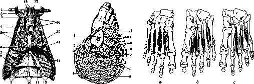
Figure 17.
Figure 18.
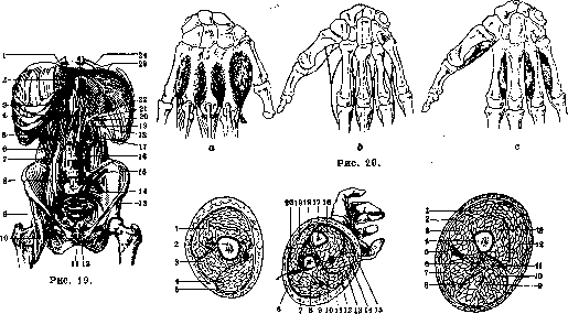
Figure 21.
Figure 16. Internal surface of the anterior thoracic wall: 1-clavicle; 2-I rib; 3-m. sterno-thyreoideus; 4-vasa mammaria int.; 5-m. transversus thoracis; 6-xiphoid process of sternum; 7-VII rib; 8-m. transversus abdominis; 9-m. obliquus abdominis internus; 10-mm. recti abdominis; 11-groove for a. and v. epigastrica superior; 12-m. transversus abdominis; 13-costal part of diaphragm; 14-trigonum sterno-costale; 15-sternal part of diaphragm; 16-mm. intercostales interni; 17-m. sterno-thyreoideus; 18-mm. sterno-hyoidei. Figure 17. Cross-section of the leg: 1-tibia; 2-m. flexor digitorum longus; 3-m. tibialis posterior; 4-heel; 5-m. gastrocnemius; 6-m. soleus; 7-fibula; 8-m. peronaeus longus; 9-m. extensor digitorum longus; 10-m. tibialis anterior; 11-fascia cruris. Figure 18. Interosseous muscles of the foot: a-mm. interossei dorsales; b-same on the plantar side; c-mm. interossei plantares. Figure 19. Posterior wall of the abdominal cavity with diaphragm: 1-foramen venae cavae; 2-hiatus oesophageus; 3-pleura; 4-hiatus aorticus; 5-XII rib; 6-m. quadratus lumborum; 7-m. psoas minor; 8-m. psoas major; 9 and 10-m. iliacus; 11-symphysis; 12-site of attachment of m. pyramidalis; 13-ligamentum Pouparti; 14-ligamentum sacro-iliacum anterius; 15-ligamentum ilio-lumbale; 16-ligamentum longitudinale anterius; 17-crus intermedium; 18-crus laterale; 19-crus mediale; 20 and 21-arcus lumbo-costalis medius et lateralis; 22-trigonum lumbo-costale; 23-centrum tendineum; 24-VIII rib. Figure 20. Interosseous muscles of the hand: a-mm. interossei dorsales; b-scheme of attachment of interosseous muscles; c-mm. interossei volares. Figure 21. Cross-section of the arm and forearm: 1-m. triceps; 2-humerus; 3-m. brachialis; 4-m. biceps; 5-fascia brachii; 6-m. supinator; 7-radius; 8-m. extensor carpi radialis longus et brevis; 9-m. brachio-radialis; 10-m. pronator teres; 11-fascia antebrachii; 12-m. flexor carpi radialis; 13-m. palmaris longus; 14-m. flexor digitorum sublimis; 15-m. flexor carpi ulnaris; 16-m. flexor digitorum profundus; 17-ulna; 18 and 19-m. extensor carpi ulnaris; 20-m. extensor digitorum communis. Figure 22. Cross-section of the middle of the right thigh: 1-m. rectus femoris; 2-fascia lata; 3-m. vastus medialis; 4-femur; 5-m. sartorius; 6-m. adductor magnus; 7-m. gracilis; 8-m. semimembranosus; 9-m. semitendinosus; 10 and 11-m. biceps, caput longum, caput breve; 12-m. vastus lateralis; 13-m. vastus intermedius. 23 Figure 23. Points and lines of irritation of muscles and nerves of the hand from the front: 1 and 2-m. deltoideus; 3-m. coraco-brachialis; 4-m. triceps, caput laterale; 5-n. radialis; 6-m. brachialis, portio lateralis; 7-m. brachio-radialis; 8-m. extensor carpi radialis longus; 9-m. flexor carpi radialis; 10-m. flexor sublimis digiti III, caput humerale; 11-m. extensor carpi radialis brevis; 12-m. flexor sublimis digiti III, caput radiale; 13-m. flexor pollicis longus; 14-m. pronator quadratus; 15-n. medianus; 16-m. abductor pollicis brevis; 17-ramus muscularis n. mediani; 18-m. interosseus dorsalis I; 19-mm. lumbricales I et II; 20-m. flexor pollicis brevis, caput superficiale; 21-m. flexor brevis digiti V et n. interosseus volaris IV; 22-m. abductor digiti V; 23-m. palmaris brevis; 24-n. ulnaris; 25-venter distalis m. flexoris sublimis digiti II; 26-m. flexor sublimis digiti V; 27-m. flexor sublimis digiti IV; 28-m. palmaris longus; 29-m. flexor carpi ulnaris; 30-m. pronator teres; 31-n. medianus; 32-n. ulnaris; 33-m. triceps, caput mediale; 34-m. biceps; 35-m. triceps, caput longum; 36-n. radialis. Figure 24. Muscles of the hand from the front: 1-m. trapezius; 2-m. deltoideus; 3-m. biceps; 4-m. triceps, caput laterale; 5-m. brachialis; 6-m. brachio-radialis; 7-m. extensor carpi radialis longus; 8-m. flexor digitorum sublimis, caput radiale pro digito III; 9-m. flexor pollicis longus; 10-m. pronator quadratus; 11-m. abductor pollicis brevis; 12-m. flexor pollicis brevis, caput superficiale; 13-aponeurosis palmaris; 14-m. abductor digiti V; 15-m. palmaris brevis; 16-m. flexor digitorum sublimis, digitus IV; 17-m. flexor carpi ulnaris; 18-m. palmaris longus; 19-m. flexor carpi radialis; 20-lacertus fibrosus m. bicipitis; 21-m. pronator teres; 22-m. brachialis; 23-m. triceps, caput mediale; 24-m. triceps, caput longum; 25-m. coraco-brachialis; 26-m. pectoralis major; 27-m. omo-hyoideus. Figure 25. Points and lines of irritation of muscles and nerves of the hand from behind: 1-m. infraspinatus; 2-m. teres minor; 3-m. teres major; 4-m. triceps, caput longum; 5-m. triceps, caput mediale, portio medialis; 6-n. ulnaris; 7-m. flexor carpi ulnaris; 8-m. supinator; 9-m. flexor digitorum profundus; 10-m. extensor carpi ulnaris; 11-m. pronator quadratus; 12-m. abductor digiti V; 13-mm. interossei dorsales I-IV; 14-m. extensor indicis proprius; 15-m. extensor pollicis longus; 16-m. extensor pollicis brevis; 17-m. abductor pollicis longus; 18-m. extensor digitorum communis et digiti V; 19-m. extensor carpi radialis brevis; 20-m. extensor carpi radialis longus; 21-m. brachio-radialis; 22-m. anconaeus; 23-n. radialis; 24-m. triceps, caput laterale; 25-m. deltoideus. Figure 26. Muscles of the hand from behind: 1-m. latissimus dorsi; 2-m. triceps, caput longum; 3-m. triceps, caput mediale; 4-epicondylus medialis; 5-m. flexor carpi ulnaris; 6-m. adductor pollicis; 7-m. interosseus dorsalis I; 8-m. extensor pollicis longus; 9-m. extensor pollicis brevis; 10-m. abductor pollicis longus; 11-m. extensor digitorum communis; 12-m. extensor digiti V; 13-m. extensor carpi radialis brevis; 14-m. extensor carpi ulnaris; 15-m. extensor carpi radialis longus; 16-m. anconaeus; 17-epicondylus lateralis; 18-m. brachio-radialis; 19-m. brachialis; 20-m. triceps, caput lateralis; 21-m. teres major; 22-m. teres minor; 23-m. infraspinatus; 24-m. deltoideus; 25-acromion; 26-m. trapezius. (For the illustration of the article Muscles of man.) / Sh&
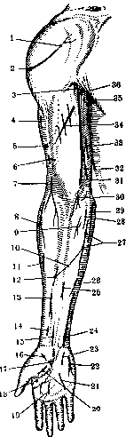
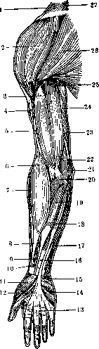
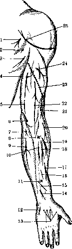
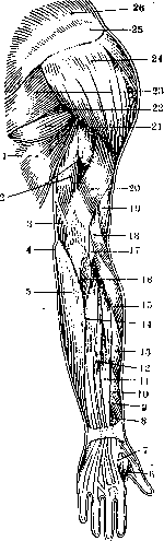
Figure 23.
Figure 24. Figure 25. Figure 26. Figure 27. Points and lines of irritation of muscles and nerves of the hand from within: 1-m. deltoideus; 2-m. coraco-brachialis; 3-m. biceps; 4-m. pronator teres; 5-m. brachio-radialis; 6-m. flexor carpi radialis; 7 and 23-m. flexor sublimis digiti IV; 8-m. flexor sublimis, caput radiale pro digito III; 9-venter distalis m. flexoris sublimis indicis; 10-n. medianus; 11-m. abductor pollicis brevis; 12-ramus muscularis n. mediani; 13-m. flexor pollicis brevis; 14 and 15-m. adductor pollicis; 16-mm. lumbricales I et II; 17-m. interosseus volaris IV; 18-m. palmaris brevis; 19-m. abductor digiti V; 20-n. ulnaris; 21-m. flexor sublimis digiti V; 22-m. flexor digitorum profundus; 24-m. palmaris longus; 25-m. flexor carpi ulnaris; 26-venter proximalis m. flexoris sublimis indicis; 27-n. medianus; 28-n. ulnaris; 29-m. triceps, caput mediale, portio medialis; 30-m. triceps, caput longum; 31-n. radialis. Figure 28. Muscles of the hand from within: 1-m. biceps; 2-aponeurosis palmaris; 3-m. abductor digiti V; 4-m. abductor pollicis brevis; 5-m. flexor carpi ulnaris; 6-m. flexor digitorum sublimis; 7-m. palmaris longus; 8-m. flexor carpi radialis; 9-m. brachio-radialis; 10-m. pronator teres; 11-m. brachialis; 12-m. triceps, caput mediale; 13-m. triceps, caput longum; 14-m. coraco-brachialis; 15-m. teres major; 16-m. pectoralis major; 17-m. deltoideus. Figure 29. Points and lines of irritation of muscles and nerves of the hand from outside: 1-m. infraspinatus; 2-m. teres minor; 3-m. teres major; 4-m. triceps, caput laterale; 5-n. radialis; 6-m. triceps, caput mediale, portio lateralis; 7-m. extensor carpi radialis longus; 7a-m. supinator; 8-m. extensor carpi ulnaris; 9-m. extensor digitorum communis; 10-m. adductor pollicis; 11-m. interosseus dorsalis I; 12-m. extensor pollicis longus; 13-m. pronator quadratus; 14-m. extensor pollicis brevis; 15-m. abductor pollicis longus; 16-m. extensor carpi radialis brevis; 17-m. brachio-radialis; 18-m. brachialis, portio lateralis; 19-m. biceps; 20-m. deltoideus. Figure 30. Muscles of the hand from outside: 1-m. trapezius; 2-m. deltoideus; 3-m. infraspinatus; 4-m. teres minor; 5 and 6-m. latissimus dorsi; 7-m. triceps, caput laterale; 8-m. brachio-radialis; 9-m. triceps, caput mediale; 10-m. anconaeus; 11-m. extensor carpi ulnaris; 12-m. extensor digitorum communis; 13-m. extensor pollicis brevis; 14-ligamentum carpi dorsale; 15-m. lumbricalis I; 16-m. adductor pollicis; 17-m. interosseus dorsalis I; 18-m. extensor pollicis longus; 19-m. abductor pollicis longus; 20-m. extensor carpi radialis brevis; 21-m. extensor carpi radialis longus; 22-m. brachialis; 23-m. biceps; 24-m. serratus anterior; 25-m. pectoralis major; 26-m. omo-hyoideus; 27-m. sternocleidomastoideus. (For the illustration of the article Muscles of man.)
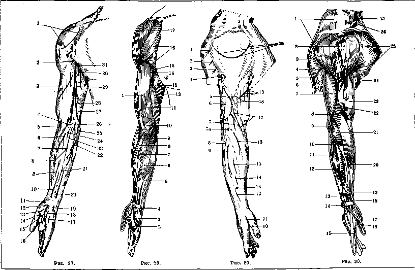
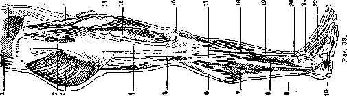
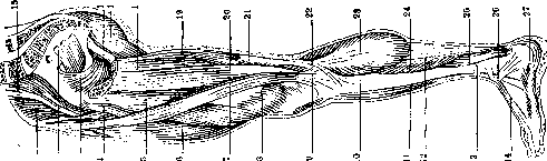
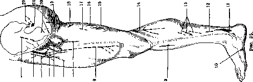
Figure 31. Lines of irritation of muscles and nerves of the leg from the front: 1-m. tensor fasciae latae; 2-m. triceps femoris; 3-m. rectus femoris; 4-m. vastus lateralis; 5-m. peronaeus longus; 6-m. extensor digitorum longus; 7-m. extensor hallucis longus; 8-m. extensor digitorum et hallucis longus; 9-m. abductor digiti quinti; 10-m. interossei; 11-m. abductor hallucis; 12-m. flexor digitorum longus; 13-m. soleus, portio tibialis; 14-m. tibialis anterior; 15-m. gastrocnemius, caput med.; 16-m. vastus medialis; 17-m. adductor magnus; 18-m. gracilis; 19-m. adductor longus; 20-m. pectineus; 21-m. sartorius; 22-n. femoralis; 23-m. ilio-psoas. Figure 32. Muscles of the leg from the front: 1-m. iliacus; 2-m. tensor fasciae latae; 3-m. sartorius; 4-m. rectus femoris; 5-tractus ilio-tibialis; 6-m. vastus lateralis; 7-patella; 8-tuberositas tibiae; 9-m. peronaeus longus; 10-m. extensor digitorum longus; 11-lig. transversum cruris; 12-lig. cruciatum pedis; 13-m. extensor hallucis brevis; 14-m. psoas minor; 15-m. psoas major; 16-m. pectineus; 17-m. adductor longus; 18-m. adductor magnus; 19-m. gracilis; 20-m. vastus medialis; 21-retinaculum patellae mediale; 22-pes anserinus (patte d'oie); 23-m. gastrocnemius, caput mediale; 24-m. tibialis anterior; 25-m. soleus; 26-m. flexor digitorum longus; 27-m. extensor hallucis longus; 28-m. abductor hallucis. Figure 33. Lines of irritation of muscles and nerves of the leg from behind: 1-m. glutaeus medius; 2-m. glutaeus maximus; 3-m. semitendinosus, venter superior; 4-m. adductor magnus; 5-m. semitendinosus, venter inferior; 6-n. tibialis; 7-m. gastrocnemius medialis; 8-m. soleus, portio tibialis; 9-n. tibialis; 10-m. abductor digiti quinti; 11-m. peronaeus brevis; 12-m. soleus, portio fibularis; 13-m. gastrocnemius lateralis; 14-n. peronaeus communis; 15-m. biceps, caput breve; 16-m. semimembranosus; 17-m. vastus lateralis; 18-m. biceps, caput longus; 19-n. ischiadicus; 20-m. tensor fasciae latae. Figure 34. Muscles of the leg from behind: 1-m. obliquus abdominis externus; 2-spina iliaca posterior superior; 3-m. glutaeus maximus; 4-apex ossis coccygis; 5-m. adductor magnus; 6-m. biceps, caput longum; 7-m. semitendinosus, tendo intermedius; 8-m. gracilis; 9-m. semimembranosus; 10-m. plantaris; 11-m. gastrocnemius, caput mediale; 12-m. gastrocnemius, caput laterale; 13-m. soleus; 14-mm. peronaei; 15-m. flexor digitorum longus; 16-lig. laciniatum. (For the illustration of the article Human Muscles.) 12 Figure 31. Figure 32. Fig. 33. X>л Figure 34. Figure 35. Lines of irritation of muscles and nerves of the leg from within: 1-n. obturatorius; 2-n. femoralis; 3-m. sartorius; 4-m. pectineus; 5-m. adductor longus; 6-m. rectus femoris; 7-m. gracilis; 8-m. vastus medialis; 9-m. tibialis ant.; 10-m. abductor hallucis; 11-n. tibialis; 12-m. flexor digit, longus; 13-m. soleus, portio tibialis; 14-m. gastrocnemius, caput mediale; 15-m. semimembranosus; 16-m. semitendinosus, venter inferior; 17-m. abductor magnus; 18-m. semitendinosus, venter superior; 19-m. glutaeus maximus и n. ischiadicus; 20-m. obturator int. и n. pudendus; 21-n. ischiadicus; 22-m. piriformis. Figure 36. Muscles of the leg from within: 1-m. iliacus; 2-m. piriformis; 3-m. obturator internus; 4-m. pectineus; 5-m. adductor longus; 6-m. rectus femoris; 7-m. sartorius; 8-m. vastus medialis; 9-pes anserinus (patte d'oie); 10-tibia; 11-m. tibialis anterior; 12-m. plantaris; 13-lig. transversum cruris; 14-m. extensor hallucis longus; 15-m. psoas major; 16-lig. sacro-tuberr°sum; 17-m. glutaeus maximus; 18-m. adductor magnus; 19-m. semitendinosus; 20-m. gracilis; 21-m. semimembranosus; 22-m. semitendinosus; 23-m. gastrocnemius; 24-m. soleus; 25-m. flexor digitorum longus; 26-lig. cruciatum pedis; 27-m. abductor hallucis. Figure 37. Lines of irritation of muscles and nerves of the leg from outside: 1-m. glutaeus medius; 2-m. glutaeus maximus; 3-m. vastus lateralis; 4-m. biceps femoris, caput breve; 5-n. peronaeus communis; 6-m. gastrocnemius, caput later.; 7-m. peronaeus longus; 8-m. soleus, portio fibularis; 9-m. peronaeus brevis; 10-m. extensor digitorum brevis; 11-m. abductor digiti quinti; 12-m. extensor digitorum longus; 13-m. tibialis anterior; 14-m. rectus femoris; 15-m. tensor fasciae latae. Figure 38. Muscles of the leg from outside: 1-m. latissimus dorsi; 2-tractus supratrochantericus; 3-m. glutaeus maximus; 4-tractus ilio-tibialis; 5-m. biceps femoris; 6-m. gastrocnemius, caput laterale; 7-m. soleus, portio fibularis; 8-m. peronaeus brevis; 9-m. peronaeus tertius; 10-m. abductor digiti quinti; 11-m. obliquus abdominis externus; 12-m. tensor fasciae latae; 13-m. sartorius; 14-m. rectus femoris; 15-m. vastus lateralis; 16-retinaculum patellae laterale; 17-m. peronaeus longus; 18-m. tibialis anterior; 19-m. extensor digitorum longus; 20-lig. transversum cruris; 21-lig. cruciatum pedis; 22-m. extensor digitorum brevis. (For the illustration of the article Human Muscles.) 25 <м
«
Ю
~ (M CO
P) И *й
CO Г~
Figure 39. Deep muscles of the forearm from the palmar side: 1, 3 and 39-m. brachialis; 2-tendon of m. bicipitis; 4-lig. collateral laterale; 5-lig. annulare radii; 6-entrance to the canal for the radial nerve; 7-bursa bicipito-radialis; 8-tendon of m. bicipitis; 9-m. supinator; 10-m. flexor digit.sublimis; 11 and 41-m. pronator teres; 12-m. flexor digitorum sublimis; 13-radius; 14-m. flexor carpi radialis; 15-m. pronator quadratus; 16-lig.radio-carpeum volare; 17-m. abductor pollicis longus; 18-m. flexor pollicis brevis; 19-m. flexor pollicis longus; 20-m. interosseus ext. I; 21-m. lumbricalis I; 22-m. flexor digitorum sublimis; 23-m. flexor digitorum profundus; 24-crossing of fibers of the deep tendon; 25-ligamenta capitulorum transversa; 26-vagina tendinum; 27-m. flexor digiti V; 28-m. opponens digiti V; 29-lig. pisometacarpeum; 30-lig. carpi transversum; 31-lig. pisohamatum; 32-os pisiforme; 33-tendon of m. flexoris carpi ulnaris; 34-m. pronator quadratus; 35-ulna; 36, 37 and 38-m. flexor digitorum sublimis; 40-epicondylus medialis humeri; 42-lacertus fibrosus; 43-septum intermusculare mediale. Figure 40. Deep muscles of the forearm from the palmar side: 1-m. brachialis; 2-tendinous part of the canal for entrance of n. radialis; 3-bursa bicipito-radialis; 4-m. supinator; 5-m. flexor pollicis longus; 6-tendon of m. extensoris carpi radialis longus; 7-m. pronator quadratus; 8-m. abductor pollicis longus; 9-tendon of m. brachioradialis; 10-lig. carpi dorsale; 11-m. extensor pollicis brevis; 12-m. abductor pollicis brevis; 13-m. flexor pollicis brevis; 14-tendon of m. flexoris pollicis longi; 15-m. adductor pollicis; 16-m. lumbricalis I; 17-tendon of m. flexoris digitorum sublimis; 18-tendinous sheath; 19-ligamenta capitulorum transversa; 20-m. flexor digiti V; 21-m. opponens digiti V; 22-lig. carpi transversum; 23-m. abductor digiti V; 24-os pisiforme; 25-tendon of m. flexoris carpi ulnaris; 26-m. pronator quadratus; 27-m. flexor digitorum profundus; 28 and 29-m. flexor pollicis longus; 30-epicondylus medialis humeri. Figure 41. Deep muscles of the forearm and hand from the dorsal side: 1-olecranon; 2-m. anconaeus; 3, 29 and 30-canal for n. radialis; 4-ulna; 5-m. extensor carpi ulnaris; 6-m. extensor digitorum communis; 7-septum intermusculare; 8-m. extensor pollicis longus; 9-m. extensor carpi ulnaris; 10-m. extensor indicis proprius; 11-entrance for tendon of m. extensoris indicis proprii; 12-lig. carpi dorsale; 13-tendon of m. extensoris carpi ulnaris; 14-tendon of extensor digiti quinti; 15-aponeurosis; 16-m. interosseus ext. I; 17-m. adductor pollicis; 18-tendon of m. extensoris pollicis longi; 19-tendon of m. extensoris pollicis brevis; 20-canal for tendon; 21-m. extensor carpi radialis longus et brevis; 22-m. extensor pollicis brevis; 23-m. abductor pollicis longus; 24-m. extensor carpi radialis brevis; 25-m. extensor carpi radialis longus; 26-radius; 27-septum intermusculare; 28-m. supinator; 31-lig. annulare radii; 32-epicondylus lateralis humeri; 33-m. brachialis. Figure 42. Attachment of muscles of the forearm and hand from behind: 1 and 42-m. triceps; 2, 12, 27 and 33-line separating flexors from extensors; 3-m. flexor digitorum sublimis; 4-m. flexor carpi ulnaris; 5-m. flexor digitorum profundus; 6-ulna; 7-m. extensor carpi ulnaris; 8-m. extensor pollicis longus; 9-membrana interossea; 10-m. extensor indicis proprius; 11-m. extensor digitorum communis; 13-m. extensor digiti V proprius; 14-m. extensor carpi ulnaris; 15, 16, 19 and 22-mm. interossei externi III, IV, II and I; 17-m. lumbricalis III; 18-dorsal aponeurosis; 20-m. extensor pollicis longus; 21-m. adductor pollicis; 23 and 29-m. extensor pollicis brevis; 24 and 40-m. extensor carpi radialis brevis; 25 and 41-m. extensor carpi radialis longus; 26 and 31-m. abductor pollicis longus; 28-m. brachioradialis; 30-mm. extensores carpi radiales; 32-m. pronator teres; 34-radius; 35 and 37-m. supinator; 36-m. anconaeus; 38-m. extensor carpi ulnaris; 39-m. extensor digitorum communis. (For the illustration of the article Human Muscles.)
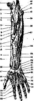
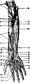
Figure 40.
Figure 39.
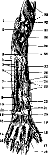
Figure 41.
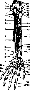
Figure 42.
Figure 43. Attachment of muscles on the anterior surface of the forearm: 1 and 14-m. brachioradialis; 2-m. extensor carpi radialis longus; 3-m. extensor carpi radialis brevis; 4-m. extensor digitorum communis; 5-m. supinator; 6-m. biceps; 7-radius; 8-m. flexor digitorum sublimis (caput radiale); 9 and 47-line dividing flexors from extensors; 10-m. pronator teres; 11-membrana interossea; 12-m. flexor pollicis longus; 13-m. pronator quadratus; 15-m. abductor pollicis brevis; 16-m. flexor pollicis brevis, caput profundum; 17-m. opponens pollicis; 18-m. abductor pollicis longus; 19-m. flexor carpi radialis; 20-m. adductor pollicis; 21-m. flexor pollicis brevis, caput profundum; 22-m. abductor pollicis brevis; 23-m. flexor pollicis longus; 24-m. lumbricalis I and m. interosseus internus I; 25-m. lumbricalis II; 26-m. flexor digitorum sublimis; 27-m. flexor digitorum profundus; 28-m. interosseus internus II and m. lumbricalis III; 29-m. interosseus internus III and m. lumbricalis IV; 30-m. abductor digiti V; 31-m. flexor digiti V; 32-m. opponens digiti V; 33-m. extensor carpi ulnaris; 34-m. flexor digiti V brevis; 35-m. abductor digiti V; 36-m. flexor carpi ulnaris; 37-ulna; 38 and 39-m. flexor digitorum profundus; 40-m. pronator teres (caput ulnare); 41-m. flexor pollicis longus (caput ulnare); 42-m. flexor digitorum sublimis (caput ulnare); 43-m. brachialis; 44-m. flexor digitorum sublimis, m. palmaris, m. flexor pollicis longus; 45-m. flexor carpi radialis; 46-m. pronator teres (caput humerale); 48-m. brachialis. Figure 44. Muscles of the thigh from within: 1-spina iliaca anterior superior; 2 and 10-tendon of m. rectus femoris; 3-bursa ilio-pectinea; 4-trochanter major; 5-ligamentum ilio-femorale; 6-m. adductor minimus; 7-trochanter minor; 8-m. vastus lateralis; 9-m. vastus intermedius; 11-m. sartorius; 12-m. vastus medialis; 13-m. semimembranosus; 14-beginning of Hunter's canal; 15 and 16-a. and v. femoralis; 17-m. adductor magnus; 18-m. adductor brevis; 19-m. obturator externus; 20-m. pectineus; 21-m. piriformis; 22-plexus sacralis; 23-promontorium; 24-ligamentum ilio-lumbale. Figure 45. Group of adductor muscles: 1-bursa ilio-pectinea; 2-capsula articularis; 3-linea intertrochanterica; 4-m. adductor minimus; 5-m. articularis genu; 6-bursa infrapatellaris; 7-ligamentum collaterale tibiale; 8-retinaculum; 9-epicondylus medialis; 10-bursa suprapatellaris; 11-hiatus adductorius (Hunteri); 12-membrana vasto-adductoria; 13-m. adductor magnus; 14-tuber ischiadicum; 15-angulus pubicus; 16 and 18-os pubis. Figure 46. Muscles of the pelvis and thigh from the front: 1-tendon of m. psoas minor; 2-m. quadratus lumborum; 3-m. iliacus; 4-spina iliaca anterior superior; 5-m. glutaeus minimus; 6-capsula articularis; 7-caput externum m. iliaci; 8-connection of m. vastus lateralis and intermedius; 9-m. vastus lateralis; 10-tendon incision; 11 and 17-retinaculum; 12-ligamentum collaterale fibulare; 13-ligamentum capitulae fibulae; 14-tuberositas tibiae; 15-ligamentum patellae; 16-tendon of m. gracilis; 18-ligamentum collaterale tibiale; 19-m. vastus medialis; 20-m. articularis genu; 21-m. gracilis; 22-m. adductor magnus; 23-m. adductor minimus; 24-m. pectineus; 25-cartilage; 26-m. sacro-coccygeus anterior; 27-m. piriformis; 28-m. psoas major. Figure 47. Deep muscles of the posterior surface of the leg: 1-tendon of m. semimembranosus; 2, 3 and 4-tendons of m. sartorii, m. gracilis, m. semitendinosi; 5-bursa of m. semitendinosi; 6-m. flexor digitorum longus; 7-m. tibialis posterior; 8-tendonous membrane; 9-tibia; 10-groove for a. tibialis posterior and n. tibialis; 11-tendon of m. tibialis posterior; 12-fascia cruris; 13-malleolus medialis; 14-processus posterior tali; 15-ligamentum laciniatum; 16-m. tibialis anterior; 17-junctura tendinum; 18-tendon of m. flexoris digitorum longus; 19-tendon of m. flexor hallucis longus; 20-m. abductor hallucis; 21-m. flexor hallucis brevis; 22-mm. lumbricales; 23-m. flexor digiti V; 24-m. interosseus internus III; 25-m. interosseus externus IV; 26-m. opponens digiti V; 27-tuberositas ossis metatarsi V; 28-tendon of m. peronaei brevis; 29-incision of ligamentum plantare longum; 30-tendon of m. peronaei longi; 31-m. quadratus plantae; 32-beginning of m. flexor digitorum brevis; 33-beginning of m. abductor hallucis; 34-tuber calcanei; 35-tendo calcaneus; 36-tendon of m. plantaris; 37-m. peronaeus brevis; 38-m. flexor hallucis longus; 39-m. peronaeus longus; 40-groove for a. peronaea; 41-septum intermusculare; 42-canal for a. tibialis anterior; 43-fibula; 44-beginning of arcus solei; 45-m. popliteus; 46-m. soleus; 47-tendon of m. bicipitis. (For the illustration of the article Human Muscles.)
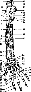
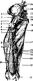
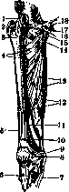
Figure 43.
Figure 44. Figure 45.
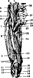
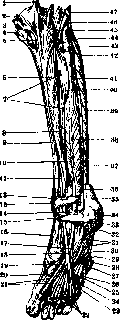
Figure 46.
Figure 47. Figure 48. Muscles of the hip joint and thigh from within: 1-bursa ilio-pectinea; 2-spina iliaca anterior; 3-m. glutaeus minimus; 4 and 8-m. iliacus; 5-spina iliaca anterior superior; 6-m. tensor fasciae latae; 7-m. sartorius; 9 and 11-m. rectus femoris; 10-ligamentum ilio-femorale; 12-m. vastus lateralis; 13-m. vastus medialis; 14-patella; 15-m. sartorius; 16-tendon of m. adductoris magnus; 17-m. semimembranosus; 18-entrance to Hunter's canal; 19 and 20-a. and v. femoralis; 21-m. gracilis; 22-m. adductor magnus; 23-m. adductor brevis; 24-m. adductor longus; 25-m. pectineus; 26-m. piriformis; 27-m. obturator internus; 28-plexus sacralis; 29-promontorium; 30-m. psoas. Figure 49. Attachment of muscles in the area of the hip joint from the front: 1-m. quadratus lumborum; 2-m. transversus abdominis; 3-m. obliquus abdominis internus; 4-m. obliquus abdominis externus; 5-m. sartorius; 6-m. iliacus; 7-m. tensor fasciae latae; 8-m. rectus femoris; 9-m. glutaeus minimus; 10-m. glutaeus medius; 11-m. quadratus femoris; 12-m. pectineus; 13-m. vastus lateralis; 14-m. adductor brevis; 15-m. vastus intermedius; 16-m. adductor longus; 17-m. articularis genu; 18-m. vastus lateralis; 19-m. quadriceps femoris; 20-tractus ilio-tibialis; 21-m. biceps femoris; 22-m. sartorius; 23-m. semitendinosus; 24-m. gracilis; 25-m. adductor magnus; 26-m. adductor minimus; 27-m. obturator externus; 28-m. pyramidalis; 29-m. rectus abdominis; 30-m. piriformis; 31-m. obturator internus; 32-m. psoas major. Figure 50. Attachment of muscles in the area of the hip joint from behind: 1-flexors of the thigh; 2-m. obturator internus; 3-m. adductor minimus; 4-m. adductor brevis; 5 and 14-m. adductor magnus; 6-m. vastus medialis; 7-m. gracilis; 8-m. semimembranosus; 9 and 11-m. popliteus; 10-m. biceps femoris; 12-m. gastrocnemius; 13-m. plantaris; 15-m. biceps (caput breve); 16-m. vastus lateralis; 17-m. adductor longus; 18-m. glutaeus maximus; 19-m. iliacus; 20-m. pectineus; 21-m. psoas major; 22-m. quadratus femoris; 23-m. obturator externus; 24-m. obturator internus cum gemelli; 25-m. piriformis; 26-m. rectus femoris; 27-m. glutaeus minimus; 28-m. glutaeus medius; 29-m. tensor fasciae latae; 30-m. glutaeus maximus; 31-m. transversus abdominis; 32-m. obliquus abdominis internus; 33-m. quadratus lumborum; 34-m. latissimus dorsi; 35-m. iliocostalis. Figure 51. Attachment of muscles on the antero-lateral surface of the leg: 1-m. tensor fasciae latae; 2-m. biceps femoris; 3, 7 and 12-m. peronaeus longus; 4, 6 and 10-m. extensor digitorum longus; 5-membrana interossea; 6-m. peronaeus brevis; 9-m. peronaeus tertius; 11-tuber calcanei; 13-m. flexor hallucis longus; 14 and 15-m. tibialis posterior; 16 and 18-m. tibialis anterior; 17-m. extensor hallucis longus; 19-m. semitendinosus; 20-m. gracilis; 21-m. sartorius; 22-tuberositas tibiae. Figure 52. Attachment of muscles on the posterior surface of the leg: 1-m. semimembranosus; 2-m. popliteus; 3-m. flexor digitorum longus; 4-membrana interossea; 5-m. triceps surae; 6-tuber calcanei; 7-m. peronaeus brevis; 8 and 11-m. tibialis posterior; 9-m. flexor hallucis longus; 10 and 13-m. peronaeus longus; 12-m. soleus; 14-m. biceps femoris. Figure 53. Muscles of the posterior surface of the leg: 1-m. semimembranosus; 2-m. sartorius; 3-tendon of m. gracilis; 4-caput mediale m. gastrocnemii; 5-tendon of m. semitendinosi; 6-m. popliteus; 7-pes anserinus profundus; 8-contour of m. gastrocnemii; 9-tendon of m. plantaris; 10-m. gastrocnemius; 11-tendo calcaneus; 12-canal for vessels and nerves; 13-heel; 14-lamina superficialis fasciae cruris; 15 and 16-m. soleus; 16-lamina profunda fasciae cruris; 18-m. plantaris; 19-caput laterale m. gastrocnemii; 20-m. biceps femoris. (For the illustration of the article Human Muscles.)
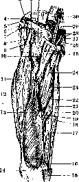
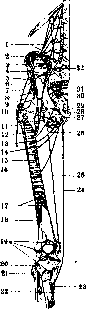
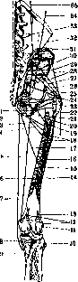
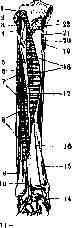
Figure 48.
Figure 49. Figure 50. -.§9$ -1Я Figure
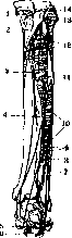
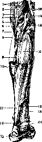
Figure 52.
Figure 53.
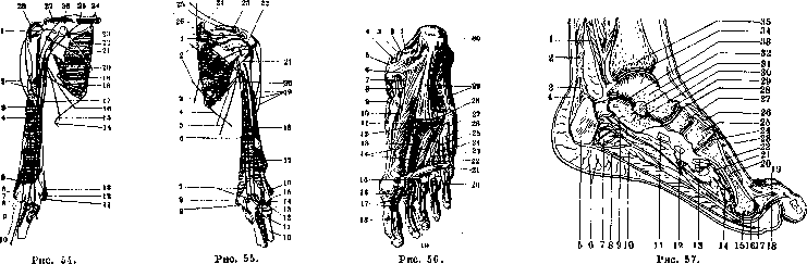
Figure 54. Attachment of muscles of the arm and forearm (front view): 1-m. supraspinatus; 2, 27 and 28-m. deltoideus; 3-m. biceps; 4-m. brachialis; 5-m. extensor carpi radialis longus; 6-m. extensor carpi radialis brevis; 7-m. extensor digitorum communis; 8-m. supinator; 9-m. biceps; 10-m. brachioradialis; 11-m. flexor digitorum sublimis; 12-m. flexor carpi radialis; 13-m. pronator teres; 14-m. serratus anterior; 15-m. latissimus dorsi; 16-m. pectoralis major; 17-m. coracobrachialis; 18-m. teres major; 19, 22 and 24-m. serratus anterior; 20-m. pectoralis minor; 21-m. subscapularis; 23-m. omohyoideus; 25-m. pectoralis major; 26-coracoid process. Figure 55. Attachment of muscles of the arm and forearm (rear view): 1 and 3-m. trapezius; 2-m. rhomboideus; 4-m. serratus anterior; 5-m. latissimus dorsi; 6-m. triceps (caput longum); 7-m. triceps (caput mediale); 8-m. flexor digitorum sublimis; 9-m. flexor carpi ulnaris; 10-radius; 11-m. anconaeus; 12-m. supinator; 13-m. extensor carpi ulnaris; 14-m. extensor digitorum communis; 15-m. extensor carpi radialis brevis; 16-m. extensor carpi radialis longus; 17-m. brachioradialis; 18-m. triceps (caput laterale); 19-m. deltoideus; 20-m. teres major; 21-m. teres minor; 22-m. infraspinatus; 23-m. supraspinatus; 24-m. serratus anterior; 25-m. trapezius; 26-m. levator scapulae. Figure 56. Deep muscles of the foot: 1-m. flexor hallucis longus; 2-posterior process of talus; 3-tendon of m. flexor digitorum longus; 4-m. tibialis posterior; 5-lig. laciniatum; 6-sustentaculum tali; 7-m. flexor digitorum longus; 8-m. tibialis posterior; 9-m. tibialis anterior; 10-m. abductor hallucis; 11-m. flexor digitorum longus; 12-m. flexor hallucis longus; 13-m. adductor hallucis; 14-m. flexor hallucis brevis; 15-m. interosseus extensor II; 16-m. flexor digitorum longus; 17-lig. vaginale; 18-m. flexor hallucis longus; 19-m. flexor digitorum brevis; 20-crossing of tendons of m. flexor digitorum longus and brevis; 21-m. adductor hallucis; 22-m. interosseus extensor III; 23-m. interosseus internus II; 24-radial interosseus extensor IV; 25-m. interosseus internus III; 26-m. flexor digiti V; 27-radial abductor digiti V; 28-m. flexor digitorum brevis; 29-plantar aponeurosis; 30-tuber calcanei. Figure 57. Sagittal section of the foot: 1-lamina profunda fasciae cruris; 2-lam. superficialis fasciae cruris; 3-tendo calcaneus; 4-bursa tendinis calcanei; 5-tuber calcanei; 6-heel; 7-m. abductor hallucis; 8-plantar aponeurosis; 9-m. quadratus plantae; 10-m. flexor digitorum brevis; 11-cuboid bone; 12-third cuneiform bone; 13-tendon of peroneus longus; 14-m. adductor hallucis; 15-capitulum; 16-metatarsophalangeal joint; 17-tendon of m. flexor digitorum brevis; 18-tendon of m. flexor digitorum longus; 19-dorsal aponeurosis; 20-second metatarsal bone; 21-plantar arch (artery); 22-m. interosseus extensor; 23-arcuate artery; 24-tarsometatarsal joint; 25-second cuneiform bone; 26-m. extensor hallucis brevis; 26-navicular bone; 27-talocalcaneonavicular joint; 28-head of talus; 29-body of calcaneus; 30-interosseous talocalcaneal ligament; 31-talus; 32-talocalcanean joint; 33-talocrural joint; 34-tibia. 7 42 IV. Pathological anatomy of muscles. Since smooth muscles do not constitute a separate system of significance but are everywhere a component part of various organs (for example, blood vessels, digestive tube, respiratory tract, uterus, etc.), pathological changes in smooth muscles are rarely independent: in most cases they are either general with changes in parenchymal elements as such (for example atrophy in cachexia, fatty degeneration in anemia, intoxications, etc.) or are closely connected with changes in the organ in which they are located. In connection with this, it is very difficult and even impractical to consider the pathological anatomy of smooth muscles as a separate chapter: those pathological changes characteristic of smooth muscles that are general with changes in parenchymal elements are described in the presentation of individual types of changes (e.g. atrophy, fatty degeneration, etc.), while those that are closely connected with pathological changes in one or another organ are indicated in the presentation of the pathological anatomy of each of the corresponding organs. Only tumors originating from smooth muscles, called leiomyomas [see separate table (Volume XV, pp. 431-432), Figure 5], have the significance of independent changes, which rarely occur in pure form (i.e. as tumors consisting only of smooth muscle fibers) (see also Myoma). The main postmortem change in muscle is rigor mortis, which is expressed in the fact that after death muscles undergo hardening, fixing the parts of the body in the position in which they are seized by rigor (see Corpse). Rigor can also occur in a fetus that died in utero. Rigor also affects the muscles of the heart, the muscular tissue of arteries, and the musculature of the stomach. Rigor of the arrector pili muscles gives gooseflesh on the corpse. Postmortem rigor of muscles has great importance in forensic medicine, as it can help clarify the time elapsed from the moment of death to the time of examination of the corpse. Developmental defects of muscles mainly concern defects in the formation of one or another muscle or part of a muscle. Among such malformations, developmental defects of the diaphragm are of greatest importance. Among developmental defects of other muscles, mention should be made of the absence of m. pectoralis major and minor, observed more frequently in men (5:1) and on the right side (4:3); sometimes there is simultaneously underdevelopment of the corresponding (2, 3, 4) ribs and abnormal development of the skin (its thinness, weak development of hair, more rarely excessive hairiness). Defects of m. deltoideus, supraspinatus and infraspinatus, latissimus dorsi, trapezius are less common; together with underdevelopment of the latter, there is also underdevelopment of m. sternocleidomastoideus. Some forms of congenital torticollis (see) are based on underdevelopment of one of the sternocleidomastoid muscles or intrauterine change (sclerosis) of it on the basis of abnormalities in the position of the fetus. Developmental defects of various muscles of the abdomen, limbs, face, and orbit are described very rarely. Among circulatory disorders in muscles, anemia may be of significance. [It goes without saying that those cases in which muscles, depending on general anemia or local causes (compression), turn out to be bloodless and consequently pale and dry, do not represent special practical interest.] If complete cessation of arterial blood flow to a certain group of muscles occurs, this can result in complete exsanguination, ischemia of muscular tissue, and its necrosis; the muscle becomes clay-like, dry, and brittle. The causes of muscle ischemia are embolism of the artery or more often traumatic damage to the arterial trunk; it should be noted that in general the arterial network in muscles is so rich in anastomoses that even with complete closure of an artery, ischemia is not observed in most cases; only with closure of the lumen of a large artery or in connection with weakening of blood flow from collateral causes (shock) do ischemia and necrosis develop. Moreover, in some areas (e.g. in the area of forearm flexors) ischemia and necrosis easily form due to the weaker development of the anastomotic network here. Ischemia and necrosis of a group of muscles form the basis of so-called ischemic contractures (see). Hyperemia of muscles, arterial and venous, changing the shade of the color of muscular tissue, has no special significance; according to observations by Berger and Bier (Berger, Bier), in congestive hyperemia of muscles, hypertrophy of muscle fibers develops, which is later replaced by some atrophy of them with proliferation of interstitial connective tissue. Hemorrhage into muscles occurs with injuries, with ruptures of muscles due to strong contractions (tetanus), as well as with various hemorrhagic diatheses (blood diseases, infectious diseases, intoxications, scurvy, etc.); in some cases, the cause of hemorrhage is a change in muscular tissue (waxy degeneration, for example in typhoid fever, various inflammatory conditions of muscles). With hemorrhage into muscles, the spilled blood either impregnates, infiltrates the muscular tissue, located between the muscle fibers and their bundles, or forms a hematoma in the muscle (for example in the rectus abdominis muscle). Edema in muscles manifests as watery infiltration of the interstitial connective tissue with displacement of muscle fibers, which themselves do not show particular changes. Often with muscle edema, a particularly pronounced manifestation of edema is noted in the muscle spindles. Necrosis of muscles can be a consequence of cessation of arterial blood flow (see above), is observed with traumatic damage to muscles, with inflammations, with the growth of tumors into muscles. The origin of muscle necroses developing as a result of concussions and contusions without violation of the integrity of the covering is not entirely clear; here too, the main significance may belong to circulatory disorders. Dead muscular tissue has a yellow, clay-like appearance, is dry, and lacks luster; necrosis of individual muscle fibers may be invisible to the naked eye. Under the microscope, necrotized muscle fibers are found to be devoid of nuclei, while the cross-striation is well preserved at first.
Subsequently, the breakdown of muscle fibers occurs, which initially may take the form of disc-shaped splitting (see below), and later assumes the appearance of a continuous granular disintegration. Through the inflammatory reaction in the surrounding area, the dead mass is absorbed and replaced by scar tissue. Atrophy of M. is expressed in the fact that muscle fibers gradually become thinner and thinner: the diameter of the fiber decreases to 20-15-10 μ (normally approx. 20-50 μ), and in severe degrees of atrophy to 5-3 μ and finally to the complete disappearance of the contractile substance. The cross-striation is preserved for a very long time, however, in severe degrees of atrophy it disappears; the longitudinal striation persists somewhat longer, which can sometimes still be seen in fibers even 3 μ thick. In some cases of atrophy, with a certain degree of thinning of muscle fibers, dystrophic changes occur in the form of coarse-grained disintegration of the protoplasm, resembling wax-like degeneration; sometimes longitudinal splits of atrophying fibers can be seen. Often in atrophy, a granular brown pigment appears in the muscle fibers, belonging to lipofuscin, and sometimes a fine-grained pigment can be observed, giving a reaction to iron and apparently being a product of the metamorphosis of hemoglobin of muscle fibers. Glycogen from muscle fibers begins to disappear only in severe degrees of atrophy. A very constant change in atrophy of M. is the amitotic multiplication of muscle nuclei (so-called atrophic multiplication of nuclei), which tends to increase as the muscle fibers thin; in severe degrees of atrophy, the thinned muscle fibers are completely devoid of contractile substance, and the narrow tubes of their sarcolemma contain rows of multiplied muscle nuclei (German Muskelkernschlauche). Often the multiplied muscle nuclei are very pyknotic, are arranged in groups in which they fuse with each other and form bizarre figures of 'nuclear plates' (German Kernplatten), which are sharply stained by nuclear dyes. The essence of atrophic multiplication of muscle nuclei is still not clear. Some look upon this phenomenon as a special kind of degeneration of nuclei, others attribute it to regenerative manifestations, a third connect it with the fact that with a decrease in the volume of contractile substance, the nuclei of muscle fibers find themselves in better conditions of nutrition; finally, some see in the multiplication of nuclei a phenomenon of the formation of special cells ('myoclasts') that absorb the contractile substance. The connective tissue in places of atrophy of M. as a rule proliferates; sometimes, however, there is proliferation of fatty tissue, which can be so significant that the volume of the atrophied M. can exceed the norm (atrophia lipomatosa, pseudohypertrophia). The spread of atrophic changes in individual muscles is not the same; sometimes atrophy uniformly affects all muscle fibers of a given muscle, in other cases atrophy is spread throughout the muscle, but the change in muscle fibers is not expressed to the same degree in all fibers; sometimes there is a bundle-like distribution of atrophy, expressed in the fact that in some bundles atrophy affects the muscle fibers completely, while other bundles are completely unchanged; finally, the preservation of individual muscle fibers in the midst of atrophying muscle tissue can be observed. Very often such preserved individual muscle fibers, groups of them, and entire bundles are in a state of hypertrophy, being thickened to 250 μ or more in the diameter of the fiber. Muscle spindles usually do not participate in the atrophic process. Macroscopically, the atrophied M. is flaccid, paler than normal, and in severe degrees of atrophy begins to resemble fish meat; a yellow tint indicates proliferation of fatty tissue, a brown tint indicates the presence of brown pigment in the atrophying fibers. The volume of atrophying muscles can be reduced to a very significant degree, while in other cases, due to proliferation of fatty tissue in the atrophied muscle, it is preserved or even increased. The causes of muscle atrophy are diverse. Some atrophies develop as a consequence of diseases of the nervous system (peripheral nerves, spinal or brain), others depend on joint lesions, a third represent the result of inactivity or a manifestation of senile or marantic atrophy; sometimes atrophy of M. depends on one or another pressure on it (bandages, tumor, hematoma, etc.). In an attempt to divide the various types of muscle atrophy according to their causes, numerous classifications have been proposed. From a patho-anatomical point of view, the following scheme of Meyenburg appears most convenient: I. Neurogenic muscle atrophies: a) neural muscle atrophies, b) spinal muscle atrophies, c) cerebral muscle atrophies. II. Non-neurogenic muscle atrophies: a) atrophies from inactivity, b) artrogenic muscle atrophies, c) myogenous and tenogenous muscle atrophies, developing from 1) external causes, 2) internal (unexplained) causes (dystrophies). Regarding the distribution of atrophy of M. in the body, it can be noted that many of them have only local significance, others have the character of general diseases of the entire muscular system. To the first of neurogenic forms belong those atrophies that develop in connection with lesions of individual nerves and limited lesions of the spinal cord or brain; from non-neurogenic forms—artrogenic atrophies developing in accordance with the affected joint, atrophies from inactivity, depending on the absence of function, usually in one limb or another, myogenous and tenogenous atrophies occurring in case of damage to M. itself and its tendon, in inflammation of M., in pressure and other purely local causes. To muscle atrophies of a general character from neurogenic forms belong atrophies in multiple nerve lesions, progressive spinal muscular atrophy and the myatonia adjacent to it; and from neurogenic forms—progressive muscular dystrophy and partly myasthenia. (For details of patho-anatomical changes in these diseases—see Muscle atrophy in diseases of the nervous system, Dystrophy, Myatonia, Myasthenia.) From degeneration in M., wax-like degeneration is very common. Cloudy swelling, or granular degeneration, is also observed, in which muscle fibers swell and become cloudy due to the appearance of a mass of small protein granules. The cross-striation disappears in this case. This type of degeneration is encountered as a general change in severe infections and intoxications and as a local change in circulatory disorders and near foci of inflammation. Vacuolar degeneration in the form of the appearance of droplets of moisture inside muscle fibers occurs in inflammatory processes, in edema, and in various dystrophies. Amyloidosis of M. in the form of local deposition of amyloid in the perimysium internum and in the sarcolemma is extremely rare. Disorders of fat metabolism in M. can manifest in the form of lipomatosis, i.e., proliferation of fatty tissue among muscle tissue, and degenerative obesity (fatty degeneration), expressed in the appearance inside muscle fibers of a mass of small fat droplets. Lipomatosis can occur in general obesity, but is most often observed in various muscle atrophies (see above). In degenerative obesity of muscles, which occurs in infections (diphtheria), intoxications (phosphorus), severe anemias and cachexias (tuberculosis) and often affects mainly the diaphragm M., small droplets of fat may initially be arranged in rows along the course of the transverse striation and longitudinal fibrils; in severe degrees of obesity, the droplets completely fill the entire muscle fiber and merge into larger droplets, and the transverse and longitudinal striation disappear. All these phenomena often mainly affect those muscles that lie close to the main infectious focus (e.g., neck muscles in scarlet fever, diphtheria, abdominal muscles in typhoid fever). The degree of tension and activity (diaphragm, mm. stapedius, tensor tympani) is also of importance, which explains the significant frequency of hearing abnormalities after infections. In relation to degenerative obesity, it should be borne in mind that in normal muscle fibers, especially the diaphragm, as a physiological phenomenon, small droplets of fat are encountered in varying amounts. A special degenerative change of muscle fibers is their fibrillar and disc-shaped splitting; in the first, the muscle fiber begins to split into primitive fibrils, in the second, rarer one—into transverse discs corresponding to the cross-striation. These changes are found in various circulatory disorders, inflammations, edema of M., also in their mechanical injuries.
It is interesting to note that Grawitz, in the fibrillar splitting of muscle fibers, saw signs of the transition of muscle fibrils into connective tissue (formation of myogenic connective tissue).-All the above-mentioned degenerative changes in M. are reversible; even with significant degeneration of the contractile substance, the latter can be restored from the remnants of sarcoplasm that usually remain near the muscle nuclei; only strongly expressed degeneration leads to complete disintegration and death of the fibers.-Petrification in M. can manifest as the deposition of lime in the interstitial connective tissue of M. (perimysium internum et externum) and in muscle fibers. Cases of the first category either refer to local deposition of lime in scar-modified connective tissue or to a general disease type of lime metastases (see Lime deposits, metastases), in which sometimes widespread deposition of lime (calcinosis universalis) occurs, in particular involving the intermuscular connective tissue. Petrification of muscle fibers sometimes occurs in areas of muscle tissue necrosis, in areas of former inflammatory processes, adjacent to chronic abscesses (e.g. tuberculous); it must be assumed that in all these cases petrification affects dead muscle fibers; deposition of lime in wax-like degenerated muscle fibers has also been described. Hypertrophy of M., in most cases regarding its genesis as working hypertrophies, represents true hypertrophy and usually has only thickening of muscle fibers as its basis; most morphological and experimental studies establish that this thickening of muscle fibers is explained by an increase in the amount of sarcoplasm and the number of fibrils; thickening of fibrils is rejected by the majority. Nuclei increase in volume, but the number of nuclei does not increase, so that compared to the volume of the muscle, the number of nuclei is relatively smaller than in normal conditions; if an increase in the number of nuclei is observed, this indicates existing atrophic changes in the hypertrophied M. Some describe in hypertrophy, in addition to thickening, also an increase in the number of muscle fibers by splitting the previous ones; however, these data are not entirely convincing. It is interesting that muscle spindles do not participate in hypertrophy. Hypertrophy of M., besides cases of increased work load of certain muscle groups and besides compensatory hypertrophy of preserved individual muscle fibers and their groups in atrophies and in the death of part of the muscle tissue, is also observed as one of the manifestations of some special diseases of the muscle tissue (e.g. myotonia, myopathie hypertrophiante). Regeneration of M. can lead to complete restoration of M. damage if the damage is small. When a significant defect is formed in M., incomplete regeneration occurs, expressed in the development of a connective tissue scar. Regeneration in M. can proceed in two ways depending on the nature of the muscle tissue damage. 1. In those cases where mainly the contractile substance is damaged, while sarcoplasm and muscle nuclei are preserved to one degree or another, restoration of M. occurs by a method called the embryonal type of regeneration. It is observed in waxy and other degenerations of M., after frostbite, crushing, and after non-sharp injuries to muscle tissue. In these cases, the contractile substance of the muscle fiber dies, and the sarcoplasm disintegrates into lumps, some containing nuclei (sarcolytes); at the same time, multiplication of muscle nuclei (amitotic and mitotic) occurs. From these nuclei, surrounded by sarcoplasm, cellular elements of round and spindle-shaped form with fine-grained protoplasm, called sarcoblasts, are formed; they are elements analogous to embryonal myoblasts. If the sarcolemma is intact, the formation of these elements occurs inside the sarcolemma tube, in which the sarcoblasts are arranged in rows, partially fuse with each other, forming multinucleated syncytial masses. If the integrity of the sarcolemma is violated, the sarcoblasts (myoblasts) are found lying freely, and in such cases they appear as rich in fine-grained protoplasm epithelioid cells; later they take on an elongated form and upon multiplication of nuclei they also form multinucleated masses of heavy protoplasm (myoblasts), around which a sarcolemma shell differentiates. In such protoplasmic multinucleated masses, lying either in the tubes of the former sarcolemma or outside them, fibrils differentiate, giving longitudinal striation; then transverse striation appears, which gives restoration of muscle fibers. Sometimes due to the unfavorable influence of the environment in which regeneration takes place (inflammation, proliferation of connective tissue), formation of muscle fibers from sarcoblasts does not occur, and the latter either degenerate and disappear or, in the form of multinucleated (giant) cells, remain for a long time among the connective tissue. 2. Another type of restoration of M. is called regeneration by budding; it occurs in cases where there is significant damage to muscle tissue with disruption of the continuity of a series of muscle fibers. The defect formed due to the violation of the integrity of M. is filled with young connective tissue, while at the ends of the torn muscle fibers, club-shaped bulging of sarcoplasm containing a large number of multiplying muscle nuclei occurs; these bulging formations from the ends of muscle fibers, called muscle buds, grow into the connective tissue; in cases where the separated ends of muscle fibers are not far from each other, the growing muscle buds moving towards each other can fuse, which restores the continuity of the muscle fiber. In the sarcoplasm of the fused muscle buds, fibrils and transverse striation differentiate. In many cases, however, when either the distance between the torn ends of muscle fibers is considerable or the connective tissue in the area of the defect has undergone scarring, such fusion of muscle buds does not occur; a scar is formed, around which club-shaped thickenings of muscle fibers with numerous nuclei are still visible for a long time; on cross-sections they appear as multinucleated giant cells. Regeneration according to the embryonal type can be combined with regeneration by budding; in such cases both processes proceed side by side, and young muscle fibers formed from sarcoblasts can connect with muscle buds.-Formation of bone in M.- see Myositis, myositis ossificans. Inflammation of M.- see Myositis. Gas phlegmon of M.- see Gas phlegmon. Among infectious granulomas, tbc in M. is not often encountered, and usually it is a matter of the transition of the tuberculous process to M. from bones, joints, pleura, skin, mucous membranes (for example in the tongue). Under these conditions, tuberculous nodes, caseating, and sometimes suppurating and giving tuberculous abscesses, form among M.; the latter can rupture and leave fistulas or descend and form cold abscesses. In other cases, tbc in M. has a more productive character and is expressed in the development of fibrous tissue in M., among which caseous nodules are found. Rare cases of hematogenous tbc of the muscle, representing hematogenous metastases (usually from the lungs and lymph glands), but often incorrectly called primary muscular tbc, are characterized by the development of conglomerates of nodules and tuberculous infiltrates, among which disintegration of muscle fibers occurs; hemorrhages around these foci are not uncommon. According to Saltikov, the epithelioid cells of nodules may have a muscular origin.-Syphilis affects M. in the late period of the disease and manifests as the formation of gummas in M. or the development of fibrous myositis. Gummas in M. 74» can reach a significant volume and often appear as large tumors that bulge under the skin; they tend to break down and in these cases form deep crater-like ulcers, which subsequently heal with strongly retracted scars. Syphilitic fibrous myositis gives a picture that often coincides with banal, non-specific fibrous myositis; sometimes in the connective tissue miliary gummas and obliterating vasculitis are found. Often fibrous myositis and gumma are combined with each other; in such cases, in the field of fibrous connective tissue, a dry yellowish-gray caseous focus with potato-like edges is visible. Most often syphilis affects the M. of the arm, especially the biceps M., masticatory M., tongue, M. of the neck, M. of the back.--On M. can pass from the skin a granuloma in leprosy. Tumors in M. are relatively rare; in this regard, for neoplasms primarily developing in M., it should be borne in mind that tumors originating from the interstitial connective tissue, i.e. from the perimysium, are more common here, while tumors whose source is the muscle fibers themselves are very rare.
The first group includes fibromas, which most often develop as dense nodes from the fascia of the rectus abdominis muscle; intramuscular lipomas and the rarer intermuscular ones, which most often occur in the muscles of the back and lower extremities; myxomas, hemangiomas, and lymphangiomas; chondromas are rarer. Sarcomas of the muscle occur more frequently than the benign tumors listed above and can arise from intermuscular and intramuscular connective tissue; most often they are round-cell, spindle-cell, and pleomorphic-cell sarcomas; in addition, liposarcomas, myxosarcomas, and angiosarcomas are encountered. All these tumors, in their growth, push apart and atrophy muscle tissue; sarcomas, in addition, through their infiltrating growth, destroy and eliminate it. Tumors arising from striated muscle tissue, i.e., those having striated muscle fibers as the source of their development, can have various characteristics. First of all, it can be pointed out that neoplasms consisting of striated muscle fibers (of embryonic type) or having these fibers as one of the components of the tumor most often occur outside muscle tissue, i.e., where there are no voluntary striated muscles. These benign rhabdomyomas and malignant rhabdomyosarcomas (see Myomas), usually found along the urinary tract and explained as a consequence of tissue maldevelopment, are not related to tumors developing from voluntary muscles. From the muscle elements of voluntary muscles, tumors of two kinds can possibly develop. 1. Mioblastomiomas, i.e., myomas from mioblasts, tumors consisting of large round cells with fine-grained, slightly basophilic protoplasm; in 1925 Abricosov established that these cells are mioblasts, on the basis of which these tumors were given the above-mentioned name, which is now generally accepted. Sometimes the cells of the tumors have slightly elongated oval shapes and in them differentiation of fibrillarity and cross-striation is visible. The formation of multinucleated strands with the same differentiation can also be observed. Mioblastomiomas are benign tumors, most often developing in the tongue muscle, less often in other muscles; it can be thought that they may originate from mioblasts appearing in the course of regenerative processes in muscles. Similar tumors developing in the skin may be a product of embryonic irregularities in the development of the skin-muscle plate. - 2. Rhabdomyosarcomas, or malignant rhabdomyomas, tumors of polymorphic, predominantly spindle-shaped cells; in some of them fibrillarity and cross-striation are visible. The course of rhabdomyosarcomas is malignant, similar to the course of sarcomas. Abricosov's idea that these tumors can also originate from mioblasts, i.e., represent mioblastosarcomas, has been confirmed in our time by the case of Meyenburg, in which in a tumor of the tongue there were transitions from mioblastomioma to rhabdomyosarcoma. It should be noted that in these tumors it is often not possible to find cells with fibrillarity and cross-striation; in such cases it can easily happen that this muscle tumor will be determined as a sarcoma. Morphologically, rhabdomyosarcomas originating from voluntary muscles are very similar to the same tumors developing extramuscularly. - Treatment is surgical. Of the tumors secondarily affecting muscles, the most important is cancer, which often spreads to muscles; e.g., cancer of the breast to the pectoral muscle, cancer of the uterus, cancer of the rectum to the muscles of the pelvis and perineum; similarly, other malignant tumors can also spread to muscles. Metastases of tumors (cancer, sarcomas, melanomas) in muscles are relatively rare. A tumor secondarily growing in muscles causes degeneration, atrophy, and destruction of muscle tissue, and the cells of the tumor (most often of cancer that has grown into the muscle) can penetrate inside the sarcolemma tubes and grow within them, destroying the contractile substance.
A. Abricosov. Parasites of Muscles. Parasites can be localized in the thickness of the muscle fibers themselves or be located in the connective tissue layers. The first group includes sarcosporidia, forming Miescher's sacs (Miescher) or Rainey's bodies (Rainey). More than 25 species of sarcosporidia of mammals are known (e.g., Sarcocystis Lindemannani of man, S. tenella of sheep); sarcosporidia parasitize also in birds and reptiles. In muscle fibers of fish, myxosporidia can be found (e.g., Myxobolus Pfeifferi). Leishmania-like forms of Trypanosoma Cruzi are also found in muscle fibers. The larvae of Trichinella spiralis become encapsulated in muscle fibers. In the localization sites of parasites, muscle fibers undergo strong changes—they increase in volume, are replaced by parasites, and degenerate. In the connective tissue layers of muscles, the larvae of Taenia solium and T. saginata, echinococcus, larvae (Sparganum) of Diphyllobothrium mansoni, Sparganum prolifer, Coenurus glomeratus and others (various mammals) are localized. Muscles can undergo deep destruction by the larvae of parasitic (e.g., Wohlfart's fly) and non-parasitic flies in cases of tissue myiasis (see). Lit.—see lit. to the article Muscular system.
Related articles
Mentioned in
Cite this page
“Human Muscles.” Soviet Medical Encyclopedia. English translation of Bolshaya Meditsinskaya Entsiklopediya, 1st ed. (Moscow, 1928–1936), ed. N. A. Semashko. https://sovietmedicalencyclopedia.pages.dev/article/human-muscles/