Epiglottis
Historical document, translated for reference. It reflects medical knowledge of the 1920s–30s and is not medical advice.
Summary
The epiglottis is the upper cartilage of the larynx, extremely diverse in form and size. It has laryngeal and lingual surfaces, and is connected to various ligaments including thyro-epiglottic, hyo-epiglottic, ary-epiglottic, and glosso-epiglottic ligaments.
Encyclopedia article (1928–1936)
Epiglottis, epiglottis, supraglottic cartilage. Anatomy. E.-upper cartilage of the laryngeal skeleton, extremely diverse in form and size (see Larynx). The larger part (upper) of the epiglottis overhangs the entrance to the larynx (pharyngeal). The remaining (lower) part forms the anterior wall of the upper part of the larynx (laryngeal). The laryngeal and lingual surfaces of the epiglottis are distinguished. The base of the epiglottis ends in a sharp process (petiolus), on the inner surface of which there is a noticeable thickening (tuberculum), formed by the accumulation of fatty tissue and numerous glands in this place.-Ligaments of the epiglottis. 1. Thyro-epiglottic (lig. thyreo-epiglotticum)-a short elastic ligament (about 1 cm in length), connecting the apex of the epiglottis with the upper notch of the thyroid cartilage. Individual fibers of this ligament descend lower and fuse with the surface of the thyroid cartilage. 2. Hyo-epiglottic (lig. hyo-epiglotticum)-a wide ligament extending from the body and greater horns of the hyoid bone and attaching to the lingual surface of the epiglottis. Between the laryngeal part of the epiglottis and the hyo-thyroid ligament, a wedge-shaped space is formed, in which elastic fatty tissue is located, the so-called fat body (corpus adiposum laryngis), which contributes to the involuntary movements of the epiglottis. 3. Ary-epiglottic ligaments (lig. ary-epiglotticum), stretched between the lateral surfaces of the epiglottis and the arytenoid cartilages. 4. Glosso-epiglottic (lig. glosso-epiglotticum)-a median ligament stretched between the root of the tongue and the epiglottis. The mucous membrane is raised above the ligament and forms the so-called frenulum of the epiglottis (frenulum); on both sides of the frenulum there are two oval depressions (valleculae). 5. Between the lateral surface of the root of the tongue and the lateral surface of the epiglottis, two thin glosso-epiglottic lateral ligaments are located.-Blood vessels consist of a pair of epiglottic arteries (a. epiglottis), originating from the superior laryngeal arteries (a. laryng. sup.), and supply the laryngeal surface; the lingual surface is supplied by branches of the lingual artery (arteria lingualis). Veins of the same name accompany these arteries and form a dense superficial venous network in the area of valleculae.-Lymphatic network of the epiglottis is in direct connection with the lymphatic network of the thyroid cartilage and the false vocal cords. Regional glands of this network are located between the hyoid bone and the thyroid cartilage.-Innervation of the epiglottis is carried out by a separate branch arising from the superior laryngeal nerve (nervus laryngeus superior). Histological structure of the epiglottis. The basis of the epiglottis is reticular cartilage, penetrated in many places by ducts of glands. The lingual surface of the epiglottis is lined with cylindrical epithelium, which extends here from the tongue. The laryngeal surface is covered with stratified squamous epithelium. Loose submucosal tissue is present only in the thickness of the ary-epiglottic ligaments and on the lingual surface of the epiglottis, especially near the root of the tongue. Glands, predominantly of the acinar type, are especially strongly developed
^
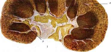
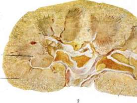
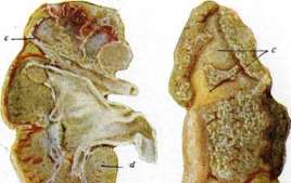
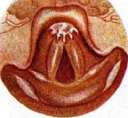
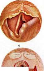
Fig. 1. Anatomical preparations: a - thickened cartilage with epiglottic folds; b - median section. Fig. 2. Lymphatic follicles: a - swollen lymph follicle (*Peyer's patches*); b - collection of lymph; c - lymphatic follicle. Fig. 3. Epiglottis. Fig. 4. Defect in the epiglottis cartilage. Fig. 5. Epiglottis carcinoma. Fig. 6. Epiglottis after syphilitic perichondritis. Fig. 7. Epiglottis tuberculosis. Epiglottis. Adrenal glands. Nephritis. Nephrosclerosis. On the epiglottis tubercle and on the lower part of the lingual surface of the epiglottis, in addition, individual lymphatic follicles and taste buds are always found. In the area of valleculae, there are a significant number of glands, lymphatic elements, and taste buds. Comparative anatomy and embryology - see Larynx. Physiology. The function of the epiglottis is not yet fully understood. Its role during the act of swallowing is to better protect the larynx from the penetration of food masses. This role recedes into the background compared to the role of the pharyngeal and lingual muscles (see Swallowing). The movements of the epiglottis are passive, and its own muscles participate little in this movement. As shown by Eijkman, the fatty body has special importance during the act of swallowing; the compression of this fat in the longitudinal direction, due to the resistance of this fat, helps to push the epiglottis downward. In the absence of the epiglottis and even with the tongue protruded forward, food does not enter the larynx (Beck). The function of the epiglottis in phonation is unclear. Apparently, it does not participate in phonation. Pathology. Inflammatory diseases of the epiglottis are rarely isolated; they are most often of the exudative type. Depending on the nature of the affected tissue, catarrhal, phlegmonous inflammations and perichondrites develop. Acute catarrhal inflammation of the epiglottis occurs in isolation and is described under the name "epiglottitis." Chronic catarrhal inflammation, compared with that of the entire larynx, has no peculiarities. Phlegmonous inflammation most often develops with acute phlegmon of the fauces, with erysipelas of the upper respiratory tract and (in isolation) with injuries to the epiglottis. Perichondrites of the epiglottis most often develop with secondary infection of tuberculous and syphilitic ulcers. A special place is occupied by edema of the epiglottis, which is usually described separately, since this symptom in diseases of the larynx acquires special clinical significance. Edema of the epiglottis is rarely stagnant, primary; more often it is secondary, inflammatory. The first develop with general circulatory stagnation in the small circle of blood circulation, with tumors of the neck and angioneurotic paralyses of the vessels. Close to these edemas are edemas caused by the action of some chemical substances, for example, bromine, iodine and chlorine, as well as insect bites. Secondary inflammatory edema of the epiglottis is sharply expressed with inflammation of the submucosa and especially with secondary infection of tuberculous, syphilitic, cancerous and typhoid ulcers. Perichondrites almost always represent a further stage of the phlegmonous process or occur with secondary infection of tuberculous and syphilitic ulcers of the epiglottis. Clinical symptomatology. Subjective complaints in acute inflammations of the epiglottis are especially sharply expressed with significant edema. Characteristic is difficulty in swallowing and sometimes breathing; in ulcerative phlegmonous processes and perichondrites (especially tuberculous) - severe pain when swallowing, radiating to the ear; in simple catarrhal inflammations - burning, smarting, sensation of a foreign body in the larynx. The voice rarely changes. In chronic inflammations, subjective complaints are expressed very weakly. Objectively, in catarrhal processes, there is a sharp hyperemia of the entire epiglottis and slight swelling of the mucosa; in inflammations accompanied by edema, the lingual surface of the epiglottis and the aryepiglottic ligaments swell especially strongly; the entire epiglottis has the appearance of an amorphous gelatinous mass of pale yellow color. In phlegmon of the epiglottis, it is swollen, bright red in color, sometimes on the mucosa the transparency of an abscess is noticeable. In perichondrites - a bumpy thickening of the epiglottis of bright red color and a sharp infiltration of the aryepiglottic ligaments. With cartilage decay, purulent fistulas are easily detected, sometimes of considerable size. Treatment is anti-inflammatory and analgesic or surgical in the form of scarification of the mucosa in edematous and phlegmonous processes; for severe edema accompanied by severe suffocation, tracheotomy is indicated. (For detailed treatment methods, see Larynx.) Among infectious diseases, the epiglottis is often affected in tuberculosis and syphilis and very rarely in lupus, scleroma, abdominal and typhus fever, diphtheria, actinomycosis. Tuberculosis of the epiglottis in the productive form is almost never observed. Much more often there are tuberculous ulcers on the epiglottis (see separate table, fig. 7), usually located on its free surface in the form of flat lenticular or even deeper ulcers, with infiltrated, uneven, undermined edges and a granulating surface. Perichondrites due to secondary infection of tuberculous ulcers are especially common and are accompanied by sharp edema. Characteristic for tuberculous lesions of the epiglottis is dysphagia, reaching exceptional strength. Treatment is mainly general. Locally - lubrication, injections and inhalations of anesthetic substances; for ulcers, in addition, cauterization with lactic and trichloroacetic acid, orthoform, hydrogen peroxide, etc. For severe dysphagias, prolonged anesthesia is achieved by injecting 85% ethyl alcohol into the area of the superior laryngeal nerve. In isolated ulcerative processes of the epiglottis, complete removal of the epiglottis down to the petiolus is sometimes used. Syphilis of the epiglottis is not encountered in the primary period. Secondary manifestations are found in the form of erythema and plaque muqueuse. In the tertiary period, gummas of the epiglottis are observed, initially in the form of a more or less delimited infiltrate the size of a hazelnut, more often on the lingual and free surface and on the corresponding aryepiglottic ligament. These gummas quickly undergo decay, and then their tumor-like nature recedes into the background, as a deep syphilitic ulcer is discovered. Extremely characteristic for syphilis of the epiglottis is slight pain even with ulcerations and perichondritis. The differential diagnosis between syphilis and tuberculosis is sometimes extremely difficult and can be resolved after a series of laboratory analyses. After syphilitic perichondritis, significant defects of the epiglottis and its scarred wrinkling are often observed (see separate table, fig. 6). Often syphilitic scars form mainly at the base of the anterior surface of the epiglottis, which results in its anteflexion. It is always necessary to keep in mind the possibility of a combination of syphilis and tuberculosis. Treatment is general antispecific. Tumors. Among benign tumors of the epiglottis, cysts are often found and rarely angiomas. The rest are described as rare diseases. Cysts are most often located at the root of the tongue in the area of valleculae and on the lingual surface of the epiglottis. In the vast majority, these are retention cysts of mucous glands. Among malignant tumors, cancer is most often found (see separate table, fig. 5), developing in the form of a bumpy soft infiltrate, prone to ulceration. Often, cancers of the root of the tongue spread to the epiglottis. In cancers of the epiglottis, metastases in the lymphatic glands located under the digastric muscle are characteristic. Cancers of the epiglottis ulcerate quite quickly and lead to edema and perichondritis. If there are doubts in the diagnosis, the best means is deep biopsy. Treatment of isolated cancer is reduced to the removal of the entire epiglottis by intra- and extralaryngeal routes, radio- and roentgenotherapy and diathermy.
Related articles
Mentioned in
Cite this page
“Epiglottis.” Soviet Medical Encyclopedia. English translation of Bolshaya Meditsinskaya Entsiklopediya, 1st ed. (Moscow, 1928–1936), ed. N. A. Semashko. https://sovietmedicalencyclopedia.pages.dev/article/epiglottis/