Larynx
Historical document, translated for reference. It reflects medical knowledge of the 1920s–30s and is not medical advice.
Summary
A comprehensive historical overview of the larynx, detailing its anatomy, physiology, and pathology as understood in the early 20th century. This section focuses on the cartilaginous framework, ligaments, and internal structure of the vocal apparatus.
Encyclopedia article (1928–1936)
LARYNX. Contents: Anatomy, physiology, and embryology .... 770 Pathology of the larynx...............777 Methods of treating diseases of the larynx.......788 The larynx, excised from a cadaver and freed from its surrounding soft tissues, represents a cartilaginous, irregularly shaped box, the lower and upper walls of which are absent, as the larynx opens into the pharynx from above and transitions into the trachea from below. Anatomy, physiology, and embryology. Three unpaired cartilages—the cricoid, thyroid, and epiglottis—and three paired cartilages—the arytenoid, Santorini's (corniculate), and Wrisberg's (cuneiform)—participate in the formation of the laryngeal skeleton (see Figure 1). The primary cartilage upon which all others rest is the cricoid (cartilago cricoidea). The narrower part of the cricoid cartilage faces anteriorly. Its posterior surface is a wide plate resembling the signet of a ring; it faces the lumen of the esophageal entrance. The largest of the unpaired cartilages is the thyroid (cartilago thyreoidea). It is formed by two quadrangular plates joined together at one edge at almost a right angle, and constitutes that part of the larynx which, protruding noticeably on the anterior surface of the neck, especially in thin men, is called the Adam's apple (see). The upper edge of the thyroid cartilage has a notch in the front along the midline, easily palpable through the skin (incisura thyreoidea superior), and at the back, each of the lateral plates bears a process or horn (superior horns), which are connected by ligaments to the horns of the hyoid bone. Since ligaments stretch from the anterior notch and from the entire free edge of the thyroid cartilage to the median and lateral bodies of the hyoid bone (ligamentum hyo-thyreoideum mediale et laterale), the impression is created that the entire larynx is suspended from this bone. On the lower edge of the lateral plates of the thyroid cartilage at the back, there are also horns (inferior), by which it articulates with the cricoid cartilage. Anteriorly and laterally between the two cartilages, a fairly wide gap is formed, which is filled by a ligament stretched here (ligamentum crico-thyreoideum or conicum). On the upper posterior edge of the signet of the cricoid cartilage are articular facets for articulation with the base of the paired arytenoid cartilages. The latter have the appearance of flat, slightly inwardly curved pyramids with a backward-tilted apex and a base slightly cut inward. The very complex curvature of the articular surfaces and the extensibility of the ligamentous apparatus allow the arytenoids to perform
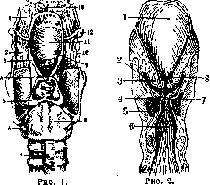
Figure 1. Larynx with cartilages and ligaments (posterior view): 1—vallecula; 2—membrana hyo-thyreoidea; 3—cartilago cuneiformis; 4—membrana mucosa laryngis; 5—cartilago arytaenoidea; 6—ligamentum crico-arytaenoideum posterius; 7—trachea; 8—cartilago cricoidea; 9—plica aryepiglottica; 10—ligamentum hyo-thyreoideum; 11—epiglottis; 12—os hyoideum; 13—plica glosso-epiglottica mediana. (From a specimen of the Anatomical Institute of the 2nd Moscow State University.) Figure 2. Frontal section through the larynx. View of the glottis from behind: 1—epiglottis; 2—ventriculus laryngis (Morgagni); 3—plica ventricularis; 4—plica vocalis; 5—musculus thyreo-arytaenoideus (externus); 6—cavum laryngis; 7—rima glottidis; 8—rima vestibuli. (After Toldt.)
rotational movements around a vertical axis and along a horizontal plane (lateral displacements). At the base of the arytenoids are two processes—one muscular, directed outward and backward, and the other vocal, directed forward. To the first are attached the posterior and lateral muscles (musculus posticus and musculus lateralis), and to the second—the fibers of the vocal cords. On the apex of the arytenoid cartilages sit small cartilages—Santorini's, connected to the arytenoids by connective tissue or joints. Between the inner surfaces of both arytenoid cartilages remains a space filled with muscles and covered with mucous membrane. It is called the interarytenoid space (incisura interarytaenoidea). The epiglottis is a thin, flexible cartilage of various shapes; its free edge is directed upward into the laryngeal part of the pharynx, and its thick apex (petiolus) is attached to the thyroid cartilage at the bottom of its superior notch. The middle of the anterior surface of the epiglottis is connected by a wide ligament to the body and horns of the hyoid bone (ligamentum hyo-epiglotticum). Anterior to the epiglottis, at the root of the tongue, are pits (valleculae), which are separated from each other in the midline by a frenulum and bounded laterally by the lateral glosso-epiglottic ligaments. In children, the epiglottis is shaped like a narrow, laterally compressed groove; in some people, this grooved appearance remains for life (canine epiglottis). We find the next stage of epiglottis development in women, in whom its structure shows flexibility and delicacy. In men, the epiglottis is generally wider and taller. From the lateral edges of the epiglottis, aryepiglottic ligaments extend posteriorly toward the arytenoid cartilages. Within these ligaments are the paired small Wrisberg's cartilages. On both sides of the larynx, between the inner surfaces of the thyroid cartilage plates, the outer side of the arytenoids, and the aryepiglottic ligament, are the so-called piriform fossae (sinus piriformis or recessus laryngo-pharyngeus), down which food rolls during swallowing. The lower edge of the cricoid cartilage is connected to the first cartilage of the trachea by the cricotracheal ligament. All laryngeal cartilages, except the epiglottis, Wrisberg's, and Santorini's cartilages, which have a reticular structure, belong to the hyaline type. In accordance with the skeleton, the lumen of the larynx, lined with mucous membrane, has an irregular shape and resembles an hourglass (see Figure 2). In a sagittal section on the lateral surface of the larynx, the true vocal cords (chordae vocales) are noted, protruding as a fold. In the whole larynx, both opposing cords begin anteriorly from the inner surface of the angle of the thyroid cartilage at the middle of its height, next to each other (anterior commissure); posteriorly they attach to the vocal process of the arytenoid cartilages. The length of the vocal cords varies on average: in men 20–24 mm, in women 18–20 mm. They consist of strong fibro-elastic fibers on the inner free edge and of muscle bundles embedded within (internal thyro-arytenoid muscle). In cross-section, the cord represents a triangle, the upper side of which looks into the pharynx, the outer side is attached to the lateral wall of the larynx, and the inner side faces its lumen. Immediately above the cords are elongated pocket-like depressions—slits (Morgagnian ventricles) of varying depth and shape, sometimes reaching giant sizes and palpable even from the side of the neck, as noted in some humans and monkeys (howlers). Rounded ridges of mucosa—false cords—hang over the ventricles. Their thickness contains glands, loose and elastic fibers, and muscle bundles (according to Simanovsky, inherent only to humans). The latter assist in the secretion of fluid from the glands of the false cord mucosa to moisten the true cords. The cavity of the larynx is divided into three sections: the upper—from the root of the epiglottis to the level of the false cords (vestibulum laryngis), the middle—extending from the false to
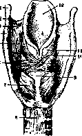
of the true vocal cords, and the lower—the subglottic space—extends to the lower edge of the cricoid cartilage. The space between both true vocal cords is called the glottis (rima glottidis). During breathing, this slit takes the form of a triangle, with the apex directed anteriorly and the base being the interarytenoid space; during phonation, it closes until the lumen is completely eliminated [see separate table (cols. 783–784), Figures 1 and 2].—The mucous membrane of the larynx is similar in its structure to the mucous membrane of the fauces, but contains elastic fibers in many places, especially in the lower (respiratory) part of the larynx and on the true vocal cords. On the false vocal cords and the aryepiglottic folds, the submucosal layer is abundantly developed; it is less pronounced in the interarytenoid space and on the epiglottis. The epithelium of the mucosa in the region of the true vocal cords, the interarytenoid space, the aryepiglottic folds, and on the laryngeal surface of the arytenoids consists of stratified squamous (pavement) epithelium with abundant formation of papillae. In all other places, the mucosa is covered with stratified columnar (ciliated) epithelium with goblet cells. Mucous glands of the acinous type are most commonly found on the laryngeal surface of the epiglottis, on the false vocal cords, and on the posterior wall of the larynx. On the free edge of the true vocal cords, there are almost no glands. Simanovsky, Hering, and others found them at the boundary of the elastic tissue with the muscular layer.—The laryngeal mucosa contains areas of adenoid tissue with follicles, which, accumulating in large numbers in the ventricles of Morgagni, can form the laryngeal tonsil (tonsilla laryngea), found in humans in approximately 12% of cases (Dobrovolsky); plasma cells are also scattered here and there. The muscular apparatus of the larynx, insofar as it connects this entire organ with neighboring ones, serves for its fixation, elevation, and depression. These processes occur with the help of the extrinsic muscles, which attach to the hyoid bone, the thyroid cartilage, and the sternum. To perform the respiratory and sound-forming functions, the intrinsic muscles of the larynx serve, which do not extend beyond its limits and are attached partly to its outer and partly to its inner surface on both sides.—The most important function of the larynx—respiratory—
Figure 3. Muscles of the larynx (posterior view): 1—cornu majus ossis hyoidei; 2—lig. hyo-thyreoideum lat.; 3—membrana hyo-thyreoidea; 4—cornu sup. cartil. thyreoideae; 5—m. aryepiglotticus; 6—m. arytaenoideus transv.; 7—m. crico-arytaenoideus post.; 8—trachea; 9—lamina cartilaginis cricoideae; 10—m. arytaenoideus obliq.; 11—cart. corniculata (Santorini); 12—epiglottis. (After Spalteholz.) (see Respiration)—can be carried out because the vocal cords diverge and form a wide opening for the passage of air. This work is performed by the paired posterior cricoarytenoid muscle (m. posticus)—the abductor muscle (abductors; see Figure 3). Paralysis of both muscles leads to suffocation, as there are no auxiliary muscles for opening the glottis. With its wide belly, the muscle attaches to the posterior surface of the cricoid, and with its tendinous end to the muscular process of the base of the arytenoids. The activity of all other intrinsic muscles is directed toward closing the glottis (adductors) and toward the phonatory function. The antagonist of the posterior muscle is the paired lateral cricoarytenoid muscle (m. lateralis). Attaching to the lateral surface of the cricoid at one end and to the vocal process of the arytenoids at the other (see Figure 4), the lateral cricoarytenoid muscle brings the vocal cords together to varying degrees and thus participates in both the act of breathing and the process of phonation. However, it cannot create the conditions for expressing the two basic properties of sound—pitch and timbre: to change the tone of the sound, the vocal cords must be stretched. This can be achieved either by moving the system of arytenoids backward or by sliding the thyroid cartilage forward. The dispute on this matter has been resolved in favor of the latter. This function is performed by the paired anterior cricothyroid muscle (m. anticus s. crico-thyr.), which, with fan-shaped diverging fibers, spans from the cricoid to the lower edge of the thyroid cartilage (see Figure 5). This muscle is very highly
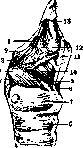
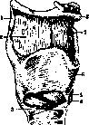
Figure 4.
Figure 5. Figure 4. Muscles of the larynx. The right lamina of the thyroid cartilage is removed (right side view): 1—m. aryepiglotticus; 2—cartil. corniculata; 3—m. arytaenoideus obliq.; 4—muscular process; 5—m. crico-arytaenoideus post.; 6—trachea; 7—cricoid cartilage; 8—lig. cricothyreoideum med.; 9—m. crico-arytaenoideus lat.; 10—m. thyreo-arytaenoid. ext.; 11—right lamina of the thyroid cartilage; 12—m. thyreo-epiglotticus; 13—epiglottis. (After Spalteholz.) Figure 5. View of the larynx from the right and front: 1—lig. hyo-thyreoideum lat.; 2—membrana hyo-thyreoidea; 3—lig. crico-thyreoideum ant.; 4—m. crico-thyreoideus; 5—lig. crico-thyreoideum med.; 6—cartil. thyreoidea; 7—lig. hyo-thyreoideum med.; 8—os hyoideum. (After Spalteholz.) developed in singers. Various nuances of the voice (see) depend on the fact that the vocal cords can change their shape, elasticity, and tension independently of their stretching. The blood vessels of the larynx branch off from the superior thyroid artery (a. thyreoidea sup.),
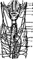
Figure 6.
Nerves of the pharynx and larynx: 1—n. glossopharyngeus; 2—ganglion nodosum; 3—rami pharyngei n. vagi; 4—n. laryngeus sup.: from which the superior laryngeal artery (a. laryngea sup.) separates, penetrating together with the nerve and vein the thyrohyoid membrane anterior to the greater horns. From it also branches the cricothyroid artery, which penetrates through the cricothyroid ligament into the lower part of the larynx.
part of the larynx. The inferior laryngeal artery (a. laryngea inf.) branches off from the inferior thyroid artery and, anastomosing with the superior laryngeal artery, spreads in the region of the posterior crico-arytenoid muscle. All arteries of the larynx are not terminal, as they are widely connected by their anastomoses (as are the veins). On the mucous membrane, the vessels are distributed unevenly. Thus, on the true vocal cords they are much less pronounced than on the false ones.—The lymphatic vessels of the larynx were studied by Most. According to their abundance, the larynx can be divided into upper and lower sections. The greatest number of vessels is in the region of the arytenoids and the ventricles of Morgagni; the true vocal cords, however, contain a negligible amount of them. The lymphatic vessels of the upper part of the larynx are directed toward the deep cervical glands along the neurovascular bundle, while the vessels of the lower part go anteriorly to the prelaryngeal glands and posteriorly to the glands along the course of the vagus and recurrent nerves, as well as between the trachea and esophagus.—The nerves of the larynx branch off from the n. vagus under the names of the superior and inferior, or recurrent, nerves. The first is mixed: its motor branch goes outside the larynx only for the crico-thyroid muscle (m. crico-thyreoideus), while the sensory branch supplies the mucous membrane of the laryngeal inlet and is distinguished by great sensitivity. Having entered the thoracic cavity, the vagus nerve gives off purely motor branches on both sides (n. laryngeus inf. s. recurrens; see Figure 6). The right branch loops from front to back around the subclavian artery, while the left one goes around the aortic arch; then both of them ascend and supply all the muscles of the larynx with the exception of the m. crico-thyreoidei, giving off small branches to the trachea and esophagus along the way. Position of the larynx. The larynx occupies the anterior part of the neck, along its midline, and for a large extent can be palpated through the skin and muscles; posteriorly it borders on the esophagus, and on the sides on the large vessels and nerves of the neck. In a state of rest (vagus; b—n. sympathicus; 7—gangl. cervicale med.; 8—n. recurrens; 9—rami cardiaci; 10—ansa Vieussenii; 11—ganglion stellatum; 12—plexus caroticus; 13—rami pharyngei n. glossopharyngei. According to L. K. Müller), the upper edge of the larynx is at the level of the III cervical vertebra, and the lower edge at the lower edge of the VI. The middle of the thyroid cartilage corresponds to the body of the V vertebra. During swallowing and phonation, the larynx rises and falls within wide limits. The thyroid gland covers the lateral parts of the larynx, reaching the lower edge of the thyroid cartilage, and its isthmus often gives a process upward along the midline of the larynx.—The question of the brain centers for the larynx has not yet been clarified. According to Krause, Munk, Bekhterev, and others, the cerebral cortex center for the adductors is located in the gyrus praefrontalis. Upon stimulation of this area, closure of the glottis and contraction of the muscles of the pharynx and soft palate occur. According to Russell, the center for the abductors lies anterior to and below the center of the adductors. Comparative anatomy of the larynx. The rudiments of the larynx in a primitive form are observed in some tailed amphibians. Its cartilaginous skeleton in them is represented by one pair of so-called lateral cartilages, which were transformed from the VII branchial visceral arch of lower animals. In tailless amphibians, the cricoid and thyroid cartilages (from the IV and V branchial arches) already participate in the formation of the skeleton, and a hint of vocal cords is noted, while in reptiles there is a rudiment of the epiglottis. In birds, there are 2 larynges: the upper one, homologous to the larynx of reptiles, consists of the arytenoid and cricoid cartilages; the lower one is located at the lower end of the trachea; in it, external and internal vocal membranes are noted, between which are 2 glottises. In mammals, the skeleton of the larynx consists of the same cartilages as in humans. The axis of the laryngeal lumen in animals lies in the direction of the nasal openings. Therefore, the epiglottis protrudes into the nasopharyngeal cavity and is located behind the soft palate, in connection with which some animals (the horse) cannot breathe through the mouth at all; the paths for air and food do not cross in them. In animals, the mobility of the larynx is very limited due to its strong connection with the hyoid bone, which in turn is fused with the bones of the skull. In humans, movements of the larynx are possible within wide limits. In all mammals, the glottis cannot close tightly in its posterior part when producing sound, since a more or less wide opening always remains between both arytenoid cartilages—the hiatus intervocalis, reaching the diameter of the index finger in some animals (in the pig and horse); humans represent an exception in this regard. In many breeds of animals, there are special resonator sacs or very developed ventricles of Morgagni in the larynx. Development of the larynx, associated with the development of the respiratory apparatus, begins in the first month of intrauterine life in the posterior section of the pharyngeal gut (see), which is compressed from the sides in this place. On its ventral surface, a horseshoe-shaped fold (furcula) is formed, with its convexity facing forward, between the branches of which is a longitudinal depression—the rudiment of the respiratory organs. At the beginning of the second month, two tubercles—tubercula arytaenoidea—develop on the sides of the depression, and the fold forms the epiglottis and plicae aryepiglotticae. The longitudinal depression at this time is divided into two sections: the anterior one, stretched in width between the epiglottis and plicae aryepiglotticae—the laryngeal inlet, and the posterior slit-like one—incisura interarytaenoidea. In the third month, all the component parts of the larynx are formed; the cartilages of the larynx in all probability develop from the cartilages of the IV and V pharyngeal arches. The lumen of the larynx closes for a short time during development due to the growth of the epithelium. The laryngeal inlet in the fetus and newborn is located high and almost protrudes into the nasal part of the pharynx, which facilitates breathing during sucking. Typical signs of the male and female larynx are formed by the period of puberty, when its rapid growth occurs. In castrates, the larynx remains small and resembles the female type. Transection of the laryngeal nerves has almost no effect on the development of the larynx. No racial difference in the larynx is noted.—Examination of the larynx—see Laryngoscopy. Pathology of the larynx. The larynx, compared to other sections of the upper respiratory tract, is less frequently subject to diseases. Almost always, inflammatory processes involve the mucous membrane, even if the primary disease began with the cartilages and joints. A special section is occupied by afflictions of the muscles and nerves, as they can proceed without inflammatory local changes.—Most often one has to deal with inflammations of the larynx (see Laryngitis).—Closely related to laryngitis is a group of edemas, which are either of congestive or inflammatory origin. In the first case, serous infiltration of the mucous membrane and submucous tissue is encountered in those diseases accompanied by general edema (nephritis, heart defects), as well as during compression of the lymphatic vessels by neoplasms, swollen glands, and scars. Closely related to these forms are edemas from insect bites, burns, trauma, and poisoning. Inflammatory acute or chronic edema occurs in all forms of inflammation of the mucosa and cartilages, in tuberculous, cancerous, syphilitic, and typhoid ulcers, as well as in inflammations of neighboring organs (anginas). The most important symptom of laryngeal edema is shortness of breath, which can lead to phenomena of suffocation, while the voice is often almost not disturbed, since the parts with a looser submucous layer—the epiglottis, arytenoids, aryepiglottic fold, and false cords—are in an edematous state. The edematous parts appear as soft rounded tumors having a seemingly transparent surface of a grayish or red color. On the cadaver, the tension of the edematous tissues disappears, and they look flabby and wrinkled. In acute edema, death from suffocation may occur, while in chronic edema, even in the case of subglottic localization, due to the habit of inhaling small amounts of air, the danger of suffocation threatens only in extreme degrees of stenosis. Treatment consists in eliminating the causative factors and prescribing ice; at the critical moment, intubation or tracheotomy is performed (see).—In addition to inflammations, the following pathological processes are noted on the laryngeal mucosa. Anemia of the larynx—occurs as a partial manifestation of general anemia and tuberculosis and is accompanied by weakness of the voice.—Hyperemia of the larynx—in the form of physiological redness is observed in some individuals either as a constant phenomenon or as a partial manifestation of general hyperemia of the skin, e.g., when the skin reddens from shame. Congestive hyperemia occurs in general plethora, heart and liver diseases, and emphysema (cyanosis).—Hemorrhages in the larynx can be intratissue and open. They are encountered in hemorrhagic laryngitis and hemorrhagic diathesis (scurvy), in trauma, and septic diseases; not infrequently, a limited hemorrhage results from too loud a cry, as well as during inflammatory and ulcerative processes (cancer), and in severe venous congestion, sometimes accompanied by phlebectasia. Cases of vicarious bleeding instead of menstruation (Schech) and from vascular tumors have been described. Tuberculosis of the larynx is expressed in a specific inflammation caused by the Koch bacillus; the mucous and submucous tissues participate almost exclusively in the process.
The disease usually does not proceed as an independent form but is a complication of the primary affliction—pulmonary tuberculosis (in approximately 30% of the latter). Infection of the larynx occurs either through sputum, and consequently through the epithelial covering, or via the lymphatic and blood vessels from the lungs and tuberculous glands. Hering, Esch, and others have pointed out the path of infection into the mucosa through the excretory ducts of the mucous glands. According to various statistics, men fall ill twice as often as women, mostly in the prime of life; in children under 10 years of age, tuberculosis of the larynx is a great rarity. In the early stages of the disease, hyperemia of individual parts of the larynx is observed, most often the true and false vocal cords, the arytenoids, and the epiglottis (catarrhal form); then an increase in volume is added, depending on the development of infiltrates. The tubercles of these infiltrates initially lie under the epithelium, then penetrate deeper into the submucous tissue and even between the muscles. Over time, the direct consequence of infiltration is the formation of ulcers. Due to the desquamation or breach of the integrity of the epithelium, a superficial abrasion first forms, which, with further breakdown of deeper layers, turns into an ulcer with undermined edges. Deepening into the underlying tissue, such an ulcer forms a funnel, the edges of which undergo necrosis, and the floor may reach the perichondrium; thus, the ulcer serves as a cause of perichondritis, which, with secondary infection, turns into purulent inflammation of the cartilage. In other cases, the infiltrate grows in the form of so-called tuberculomas and papillomas spreading throughout the larynx. The clinical picture of tuberculosis of the larynx presents great variety, which, however, fits into a certain regular sequence determined by immuno-biological laws (Ranke, Petruschky). The earliest manifestation of tuberculosis of the larynx is most often found in the form of catarrhal inflammation of the true vocal cords and the interarytenoid space, where, with the help of optical magnification, the presence of many small tubercles (subepithelial miliary nodules) can be noted. This limited redness against the paleness of the rest of the laryngeal and pharyngeal mucosa, especially when it is located on one cord, represents the most reliable early sign of beginning tuberculosis of the larynx (see separate table, Fig. 5). Developing thickenings—infiltrates—can be pale or bright red, with a smooth or bumpy surface. The true vocal cords often appear uneven, spindle-shaped, thickened, with rounded edges (see separate table, Fig. 4). The false cords become infiltrated and cover the true ones, causing their low mobility and hoarseness. On the epiglottis, infiltration more often begins on its laryngeal surface and then is located along its edges, turning it into a rounded, turban-like tumor. On the arytenoids, this infiltrative stage is expressed either as diffuse hyperemia or in the form of a spherical, pale, and edematous tumor, which makes the movement of the cords difficult, sometimes to the point of complete immobility. From here, the thickenings often pass to the aryepiglottic folds.
These manifestations of laryngeal tuberculosis are accompanied by a series of subjective and objective signs: a hoarse voice, pain, tingling, a sensation of dryness, coughing, etc. When infiltrates break down, flat lenticular or crater-like ulcers with overgrown edges are produced (see separate table, Fig. 6). In these cases, pain during swallowing is of purely mechanical origin. It can reach an unbearable degree (dysphagia) and be accompanied by pain in the ear. Ulcers arise mainly in places subjected to friction; therefore, their favorite localization is the true cords. The fate of the ulcers varies: they may heal by scarring with the ingrowth of fibrous tissue or break down further and serve as the cause of perichondritis. The latter, extending into the subglottic region, are often accompanied by stenosis of the larynx, which may necessitate tracheotomy. The course of laryngeal tuberculosis is chronic; the morbid process may alternately improve or worsen depending on the state of the pulmonary affliction. In general miliary tuberculosis, all stages of the disease in the larynx proceed extremely rapidly, the entire mucosa turns into a continuous ulcer, and in a short time, the death of the patient occurs, who is forced to refuse food and drink (galloping throat consumption). In view of the fact that the course of laryngeal tuberculosis changes depending on the ratio of the immuno-biological forces of the organism, many authors strive to fit all symptoms of the clinical manifestations of laryngeal tuberculosis into a scheme that would give a clear idea of what is happening at a given moment. These classifications are based on both pathological-anatomical (Hering, Schmidt, Manasse) and clinical (Basenko, Melnikov) signs. A. N. Voznesensky, speaking of the nomenclature of clinical laryngeal tuberculosis, believes that two main forms emerge in its course: destructive (exudative and parenchymatous) and productive (fibrous). At the same time, laryngeal tuberculosis can be active or inactive; under conditions of activity, like pulmonary tuberculosis, it can be acute, subacute, and chronic. The recognition of laryngeal tuberculosis in the presence of characteristic phenomena and indications from the lungs presents no difficulties; in other cases, the differential diagnosis wavers between tuberculosis, syphilis, and malignant tumors. Treatment of laryngeal tuberculosis is reduced to general and local. The former is conducted according to the rules of phthisiotherapy, among which the organization of dispensary and hospital care is especially important; in the latter, the action is directed against the disease itself or its symptoms. Attempts to treat locally have resulted in a large list of the most diverse medications acting as disinfectants, cauterants, or analgesics. Surgical techniques in the form of excising infiltrates and ulcers with special curettes are extremely useful in many cases; furthermore, galvanocautery, diathermy, radium, X-rays, and phototherapy are used; finally, laryngofissure and tracheotomy are sometimes resorted to in order to provide the larynx with complete rest. Against dysphagia, lubrication with cocaine, menthol, and other anesthetic agents is used. In cases of severe dysphagia, 80% alcohol is injected into the superior laryngeal nerve or it is transected. In pregnancy, artificial miscarriage is indicated. Scleroma of the larynx—see Rhinoscleroma. Syphilis of the larynx usually manifests both in the secondary stage in the form of catarrh, papules, and condylomas, and in the tertiary stage in the form of gummas, infiltrates, ulcerations, and perichondritis. Syphilitic erythema is almost indistinguishable from acute catarrhal laryngitis and is diagnosed only on the basis of coincidence with the appearance of other syphilitic signs. Papules are localized mainly on the epiglottis, on the vocal cords, and on the posterior wall of the larynx. In the tertiary period, the course of laryngeal syphilis is distinguished by a great variety of the external picture, since gummy infiltrates are prone to breakdown and transformation into ulcers, causing secondary perichondritis and the formation of scars. The gummy process most often affects the epiglottis, less frequently the interarytenoid space and the false cords. Gummy ulcers are deep, with sharply defined undermined edges, with a fatty floor and an edematous circumference. The further fate of such ulcers is either spreading in width and depth or scarring, leading to the formation of bands, adhesions, membranes, diaphragms, to ankylosis of the joints, and to disfigurement of the laryngeal lumen up to complete stenosis. Subjective sensations in all forms of laryngeal syphilis are insignificant unless phlegmons and perichondritis are added due to secondary infection. Despite the fact that the course of the destructive process in laryngeal syphilis proceeds very quickly, it is still difficult in many cases to distinguish it from tuberculosis, and it is also easy to confuse it with cancer, especially since histological examination often does not provide a convincing result.
results; not infrequently one must resort to the Wassermann reaction. Treatment is mostly limited to general energetic anti-syphilitic treatment. When using salvarsan, in view of the possibility of rapid stenosis, a follow-up examination with a mirror is required. Local treatment is reduced to the removal of overgrown persistent tissue and to the excision of scars followed by bougienage or laryngofissure. Perichondritis of the Larynx is rarely a primary disease (after trauma, wounds); it mostly develops as a secondary affliction in acute and chronic infectious diseases, in malignant neoplasms, as well as in various types of typhus, diphtheria, smallpox, syphilis, and tuberculosis; scarlet fever and measles are of particular importance. A coccal infection, having penetrated deep, leads to an accumulation of pus between the cartilage and the perichondrium. Such an abscess can develop on both the outer and inner surfaces of the laryngeal cartilages; its outcome is usually necrosis of the cartilages and the formation of fistulas. Of the cartilages, the arytenoid and the epiglottis are most often affected; the cricoid and thyroid are less frequently inflamed. A. F. Ivanov indicates that in typhus, the arytenoids are more often affected, while in tuberculosis and syphilis, the cricoid and thyroid cartilages are involved. The course of acute perichondritis begins with minor general symptoms and slight pain during swallowing, often radiating to the ear. The inflamed area and its surroundings swell due to collateral edema of the tissues, whereby the lumen of the Larynx narrows, and difficulty in breathing and suffocation appear, sometimes requiring tracheotomy. In diseases of the arytenoids, the swelling also spreads to the aryepiglottic folds, and a restriction of their mobility during phonation is observed. In perichondritis of the cricoid cartilage, subglottic swelling appears. If the abscess does not open quickly, then after prolonged suppuration, the necrotic cartilage is sloughed off with the subsequent formation of fistulous tracts and cicatricial contractions. With prolonged inflammation, the cartilages of the Larynx have a tendency to ossify even at a young age. Treatment for acute perichondritis is directed against the underlying disease. Rest for the larynx and the application of ice are recommended; abscesses are opened with a Tobold knife. In chronic cases, surgical treatment is used with dissection of the Larynx and excision of scars and hypertrophic formations, followed by expansion of the Larynx lumen using special cannulas.

Inflammation of the joints of the Larynx—acute and chronic—relates almost exclusively to the crico-arytenoid joint; it often accompanies perichondritis during infectious diseases. Such inflammation is described in articular rheumatism and gout. Objective symptoms are reduced to redness and swelling of the arytenoid and to the restriction of its mobility. Subjectively, hoarseness and slight pain during swallowing and phonation are observed. Treatment: rest, warming compresses, salicylate preparations.—Diseases of the nerves and muscles of the Larynx. Disorders of the innervation of the Larynx can occur both on the part of the motor and sensory nerves. Disturbances in the movements of the laryngeal muscles are caused either by an affliction of the nervous system or by changes in the muscle tissue itself (hyperfunction, hypofunction). Increased excitability of the neuromuscular system manifests in the form of spasms, which are either caused reflexively or are of central origin and belong to the category of neuroses.—Spasm of the glottis in children (laryngismus stridulus) at the age of 1/2–2 years occurs on the basis of rachitic disorders or as a partial phenomenon in general spasmophilia. The disease is expressed in spasmodic inspiratory movements, in twitching in the limbs with constricted pupils, in cyanosis of the face, and attacks of suffocation. In treatment, cold rubdowns, mechanical irritation of the nasal mucosa, pressure on the root of the tongue with a finger are used, and the general state of health is also influenced by strengthening agents.—Spasm of the glottis in adults (laryngospasmus)—a reflex spasmodic closure of the glottis, occurs from the action of local irritations (inhalation of gases, foreign bodies, etc.), and is also found in hysteria, epilepsy, tetanus, and tabes. Here the act of inspiration is accompanied by short intermittent expiratory movements. Treatment is directed against the underlying disease.
Phonatory spasm of the glottis (dysphonia spastica) is related to stuttering and represents a professional neurosis in persons who excessively strain the voice, and in neurasthenics. The sound of the voice appears squeezed, syllables and words seem to split, speech becomes difficult due to spastic closure of the glottis and can turn into complete aphonia.—Excessive tension of the vocal cords often occurs during mutation of the voice or during its breaking in the period of transition to puberty (in boys), when rapid growth of the Larynx occurs—here the matter is reduced to a disorder of innervation.—Of much greater importance is the disturbance of movement of the laryngeal muscles in the sense of weakening their function in paresis and paralysis, both in terms of frequency and clinical importance, since these disorders are an early sign of disease in other, often distantly located organs. This includes changes in the movement of the Larynx musculature of purely mechanical origin, e.g., due to scars, tumors, edema, etc.; changes in the muscle tissue itself in various debilitating diseases, as well as paresis of the Larynx muscles in singers and speakers due to overstrain.
In addition to these myopathic pareses, it is necessary to dwell on neuropathic paralyses, which can be of cerebral, bulbar, and peripheral origin. A distinction is made between organic diseases of the nerves and functional ones. A characteristic sign of organic diseases of the laryngeal nerves is considered to be the fact that the nerves going to the abductor muscles (m. posticus) are affected first, which is why the vocal cords cannot leave the median position and do not open the glottis (Rosenbach-Semon law). In functional (hysteria) diseases of the nerves, on the contrary, the adductor nerves suffer, which is why the glottis cannot close. In an affliction of the motor branch of the superior laryngeal nerve (after diphtheria), the anterior crico-thyroid muscle (m. anticus), which stretches and tenses the vocal cords, is paralyzed in isolation, whereby the voice loses high tones and tires quickly. Much more often, muscle paralyses are caused by a disease of the recurrent nerve, which is explained by the length of this nerve and its poorly protected position.
Figure 7. a—bilateral paralysis of m. posticus (during breathing); b—paralysis of the left recurrent nerve (during breathing).
In complete paralysis, usually unilateral, complete immobility of the corresponding half of the larynx results (see Figure 7, b). Clinical symptoms consist of a disorder of phonation, coughing, and expiratory straining. In fresh paralysis, the vocal cord assumes the so-called cadaveric position. In bilateral paralysis at the beginning, while only the m. posticus is affected, breathing is sharply difficult, and then, when the tone of other muscles also weakens, the glottis gapes in the form of an oval, rather narrow slit, sufficient however for breathing, and the matter may proceed without tracheotomy (see Figure 7, a). The cause of the affliction sometimes lies in a disease of the central nervous system, but mainly it is of peripheral origin—compression of the inferior laryngeal nerve; thus, the etiological factor is aortic aneurysm, esophageal cancer, tuberculosis of the bronchial glands, tabes, syphilis, and other brain diseases. In partial paralysis of the recurrent nerve, which can be from mechanical injury, in infectious diseases and chronic poisonings (nicotine, alcohol), individual groups of adductor and abductor muscles suffer.

Figure 8. Paralysis of m. interni (phonation): a—left-sided; b—bilateral; c—bilateral paralysis of m. lateralis.
The m. internus can be affected in isolation (see Figure 8, a and b), most often on the basis of voice overstrain, in chronic laryngitis, hysteria, and cachexia: the timbre and pitch of the voice are lost. If the m. lateralis is affected (see Figure 8, c), during phonation the glottis assumes a rhomboid shape with the largest diameter between the tips of the vocal processes of the arytenoids—resulting in aphonia.—Paralysis of the m. transversus (see Figure 9, a) causes hoarseness;

Figure 9. a—paralysis of m. transversus (phonation); b—paralysis of m. transversus and m. interni (phonation).
the glottis cannot fully close during phonation because a triangular opening forms in its posterior section between the arytenoid cartilages; this is often accompanied by paralysis of the musculus internus (see Figure 9, b). Sensory disorders of the larynx. The degree of sensitivity of the laryngeal mucosa in healthy individuals fluctuates within wide physiological limits; therefore, pathological disturbances can be spoken of with certainty only in cases of complete anesthesia. In the elderly, a significant decrease in reflex excitability is sometimes observed, which can also weaken in young people due to habituation to a stimulus. Hyperesthesia appears during inflammation of the mucosa, as well as in neurasthenics and hysterics (neuralgia laryngis). Sensitivity can be impaired either completely or partially; furthermore, it may change regarding tactile, pain, and temperature sensations. In certain diseases, such as syringomyelia and syringobulbia, all types of sensation may be disturbed, and temperature sensation especially often. Motor paralyses are usually not accompanied by changes in sensitivity, which argues for the independence of the respective nerve fibers of the larynx. Tumors of the larynx. The cause of the development of laryngeal tumors mostly remains unknown. Undoubtedly, various types of inflammatory processes, maintained by external irritation (dust, tobacco, alcohol), play a significant role in their origin. However, in a whole series of cases, they occur in a completely healthy larynx. Mechanical factors should also be included among the causative reasons. The complex histological structure of the larynx leads to a great variety of anatomical and clinical forms of tumors, among which benign ones, according to large statistics, occur 12 times more often than malignant neoplasms. Among benign growths, connective tissue polypoid formations—fibromas—rank first in frequency, followed by papillomas, cysts, lipomas, myxomas, enchondromas, and pachydermia. Fibromas, or true polyps of the larynx, range in size from a millet seed to a pea or a hazelnut (see separate plate, Fig. 3). Fibromas grow slowly, most often on the anterior or middle third of the true vocal cords, preferring their free edge, and sometimes they also begin below the vocal cord; in exceptional cases, they originate from the false cords, the epiglottis, or the posterior wall of the larynx. They appear as tumors sitting on a pedicle or a broad base, of a fairly soft or firm consistency, grayish-white, pink, or red in color depending on the number of vessels, with a smooth surface covered with epithelium. Not infrequently, hemorrhages can be observed in them
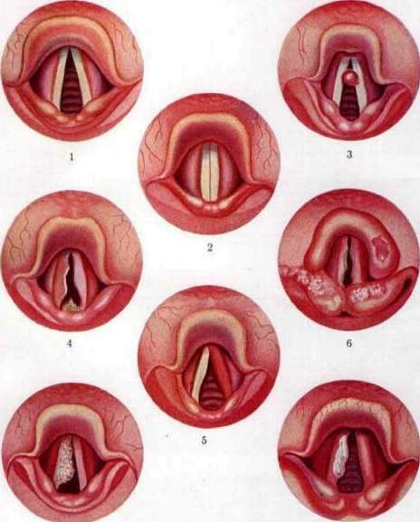
Figure 1. Normal male larynx during respiration. Figure 2. Normal male larynx during phonation. Figure 3. Fibroma of the left vocal cord. Figure 4. Tuberculosis of the larynx (ulceration of the right vocal cord and infiltration of the interarytenoid space). Figure 5. Tuberculosis of the larynx (unilateral ulceration of the vocal cord (left)). Figure 6. Tuberculosis of the larynx (ulcers on the vocal cords; perichondritis of the epiglottis, arytenoid cartilage, and aryepiglottic folds). Figure 7. Papilloma of the right vocal cord. Figure 8. Carcinoma of the right vocal cord. To the article Larynx. 788 influence due to constant contusion during the closure of the vocal cords, causing the color to change to dark or brownish-red. Glands are almost never found in them; in other cases, polyps serve as the cause for the development of retention cysts lined with cylindrical epithelium. The first disturbance produced by a polyp of the larynx consists of a gradually increasing change in the voice in the form of hoarseness and huskiness, which may reach aphonia. At first, patients can still overcome the obstacle to the closure of the cords by strong muscle tension, but then the latter weaken, causing a narrow slit to remain between the cords even during phonation. Tumors situated above and below the cords may not affect the voice for a long time. When a polyp is localized in the middle part of the glottis during phonation, two sounds may be formed—diphthonia. In other cases, a sudden transition of the voice to falsetto or a trill is noted. Often patients complain of paresthesia and a sensation of a foreign body, which they try to remove by expectoration. Respiratory function in laryngeal polyps usually does not suffer. Only in rare cases is there sudden death from suffocation. A polyp is easily diagnosed by laryngoscopy if the cords are normal, but during inflammation, it is sometimes overlooked. The only therapeutic measure is removal with a snare, Krause forceps, or a Cordes curette. In rare cases, the polyp tears off and spontaneous cure occurs. — Angiomas, being tuberous tumors of spongy soft consistency, bluish-red to black in color, reach the size of a cherry and are located on the true and false cords. They sometimes grow together with similar tumors in the mouth and fauces. — Lipomas and myxomas of the larynx are very rare. — Chondromas, consisting of hyaline cartilage, originate from the cricoid and thyroid cartilage; growing into the larynx, they cause its stenosis. — Cysts originating from glands are found on the epiglottis. A polypoid tumor of the larynx is sometimes simulated by local amyloid, manifesting in the deposition of amyloid clumps (see Amyloid degeneration) in the submucosal connective tissue of the larynx. — Among tumors of epithelial origin, papillomas (synonyms: papillary fibroma or epithelioma, condyloma acuminatum) are the most common phenomenon. They are of three kinds: 1) in the form of nodules the size of a hemp seed to a pea, 2) in the form of tumors shaped like a cockscomb, 3) in the fine-grained form of cauliflower or raspberry (see separate plate, Fig. 7). The cause of their development is unknown, but some link it to tuberculosis. Histologically, they consist of a framework of papillae made of connective tissue with thin-walled capillaries; the papillae are covered with proliferations of stratified epithelium, which is why an inexperienced researcher can easily confuse the microscopic picture with cancer. Papillomas occur at all ages, but especially often in children, sometimes shortly after birth or during the first and second year. It can be said that prolonged huskiness in a child is almost always caused by this tumor. In view of the tendency toward dissemination, papilloma can scatter throughout the entire mucosa, but it is more common on the true and false cords, epiglottis, arytenoids, and even in the trachea. The color of the papilloma is sometimes shiny white, sometimes pink and red. When they proliferate, papillomas can cause symptoms of suffocation. — The prognosis is generally good, but their complete removal is always very difficult, as they have a tendency to recur and grow vigorously; on the other hand, by the period of puberty, they may disappear on their own, and in adults, they may resolve after severe infectious diseases. Some point to their property of being transferred by inoculation to healthy tissue during surgical interventions. — Papillomas are removed endolaryngeally or after splitting the larynx along the midline, following a preliminary tracheotomy. Recently, the Seifert laryngoscope, built on the model of a vaginal speculum and convenient for direct endolaryngeal operations, has been used with success. For conservative treatment, radio-roentgenotherapy and cauterization are used, and arsenic and prolonged doses of magnesia are prescribed. Of the malignant tumors in the larynx, cancers and sarcomas are found. Among the various forms of cancer, squamous cell carcinoma with abundant keratinization and localization on the true cord is observed more often, in approximately 2/3 of cases; it is followed by adenocarcinoma of the medullary type, and scirrhus is the rarest. Usually, these tumors appear primarily in the larynx and only rarely grow into it from neighboring organs. By localization, internal and external cancers are distinguished. The former, being within the limits of the laryngeal cavity itself, in the region of the vocal cords, can for a long time be considered a purely local disease, as they give neither metastases nor swelling of the neighboring cervical lymph glands, whereas external ones, originating from the aryepiglottic folds, the posterior wall of the larynx, the pyriform sinuses, and the epiglottis, soon produce involvement of the regional lymph glands and themselves grow into the width and depth of the tissues. Cancer has no definite shape or size, but at the beginning of the disease, it may resemble a fibroma, papilloma, or pachydermia on the vocal cords (see separate plate, Fig. 8) or look like tuberculosis and syphilitic infiltrate. With its further growth, the tumor has a tendency to ulcerate, whereby reactive inflammation in the form of edema, infiltrates, and perichondritis may appear in the vicinity; finally, signs of general disease appear—cachexia and metastases in distant organs. Death usually occurs in 1–3 years, often from a secondary infection. The disease affects men more often at the age of 40–70 years, rarely at an early age (cancer has been described even in a 16-year-old). Statistics show that among cases of carcinoma of various organs, cancer of the larynx accounts for about 1.5% (Kolb, Marschik). According to the data of Moscow autopsy rooms for 1923–27, cancer of the larynx was observed in 3.47% of all cancers. — Course. Cancer of the larynx in many cases proceeds completely latently or its symptoms are similar to the signs of benign tumors. In this initial period, patients amidst complete health note a slight but persistent huskiness or discomfort, to which they pay little attention; upon examination with a mirror at this time, in internal cancer, one can notice a small nodule or wart on a slightly reddened vocal cord that seems firmly grown into it. In external cancer, even these signs are not visible; the disease reveals itself by a distressing urge to cough and mild pains radiating to the ear during talking and swallowing. Glandular carcinomas originating from the cavity of the ventricle of Morgagni already at this time give characteristic rounded swellings, very similar to syphilitic or tuberculous infiltrates. In a later stage, all subjective and objective signs appear more clearly: pains and hoarseness intensify, and the first symptoms of approaching stenosis and suffocation are added. Due to the growth of the tumor into the depths, a firm infiltration and sharp restriction of movement or even complete immobility of half of the larynx and edema of the surrounding parts are clearly noticeable. Fraenkel believes that immobility of the cords occurs if the tumor is located near the arytenoid cartilages. In the final stage, simultaneously with disintegration and the formation of ulcers, the cancer grows through the framework of the larynx and fuses the cervical glands into a dense, shapeless mass; the patient experiences unspeakable agony from dysphagia, suffocation, choking, and an exhausting cough and often perishes from rapid or slow suffocation if a tracheotomy was not performed in time. Often, an unbearable odor from the mouth is added to everything else. — The diagnosis is made on the basis of the symptom complex, taking into account the patient's age, anamnesis, course, and general examination. However, it is not always easy to differentiate cancer from tuberculosis and syphilis, especially since a trial excision of a piece does not in all cases reliably confirm the clinical diagnosis and since cases of combinations of cancer with lupus or tuberculosis are occasionally observed. It should be noted that biopsy often gives an impetus to more rapid growth of the tumor. — Treatment is conservative and surgical. The former is used in the initial stages or in inoperable cases in the form of radium, X-rays, and other destructive agents. Morphine is used against pain. Surgical intervention may vary depending on the localization and spread of the tumor.
If the tumor is on the edge of the vocal cord or the epiglottis, endolaryngeal removal or laryngofissure is recommended; if the entire half of the larynx is affected, partial excision is performed, and with wider spread, the entire larynx should be removed completely, sometimes with part of the esophagus, pharynx, and tongue. Tracheotomy is performed at the first signs of respiratory distress. Sarcoma of the larynx is encountered less frequently than cancer, in the form of primary tumors from spindle cells, chondro-, fibro-, melano-, and lymphosarcomas, at all ages, more often in men. Affecting predominantly the vocal cords or the lower part of the larynx, it has the appearance of a clearly circumscribed, smooth tumor, while its color and consistency vary depending on the histological structure. Clinical phenomena are less pronounced than in cancer and are reduced to the mechanical action of the tumor. Sarcoma has no tendency toward ulcerative destruction but affects the cervical glands and is often a sign of general sarcomatosis. In treatment, besides surgical intervention, X-ray and radiotherapy are successfully used, and arsenic is prescribed. Methods of treating diseases of the larynx. Many diseases of the larynx are only local manifestations of a general disease of the organism; therefore, in treating them, it is always necessary to pay primary attention to the underlying disease, the course of which, in turn, is often significantly eased by the elimination of local symptoms. Prophylaxis is of great importance for the prevention of diseases of the larynx, which amounts to the elimination of all factors acting harmfully on the mucous membrane of the upper respiratory tract (e.g., inhalation of contaminated and dry air in certain industries, vocal strain, etc.), as well as those associated with the phenomena of the so-called common cold, the essence of which lies in pathological changes in vasomotor reflexes depending on the cooling of the body. From this point of view, the hardening of the peripheral neurovascular apparatus to increase the reactive capacity and adaptability of the vessels (air, water, and sun baths with simultaneous body movements, balneological and climatic treatment) is of considerable importance for combating diseases of the larynx. Local treatment is applied either in the form of direct application of a medicinal substance to the laryngeal mucosa or in the form of action upon it from the skin by means of heat, cold, massage, electricity, radium and X-rays, congestive hyperemia, or special surgical techniques. Endolaryngeal local treatment can be medicinal, mechanical-physical, and operative. Medicines are applied to the laryngeal mucosa in liquid, solid, and powder, vapor, and gaseous forms. Swabbing is performed under the control of a laryngoscopic mirror using a laryngeal probe with a cotton brush attached to it. In small children, the larynx is swabbed blindly, pushing the tongue downward with the left index finger. For swabbing, solutions of various substances are used: silver nitrate in 1%–5%, tannin, alum, iodine-glycerin, phenosalyl, lactic acid, cocaine, etc. These agents can be instilled into the larynx from a syringe with a long curved tip adapted for this purpose. With this method, the gag reflex and spasms of the vocal cords are usually weakly expressed; instillation is tolerated by the patient more easily than swabbing. For a deeper influence on the affected tissue, it is often necessary to resort to chemical cauterization with acids in substantia; lactic, chromic, and trichloroacetic acids are most commonly used. Insufflation of powders from special devices fulfills its purpose with more precise localization of the painful process (ulcers); potassium bromide, anesthesin, orthoform, morphine, cocaine mixed with sugar, and other soothing, disinfecting, and narcotic agents are used. Mechanical-physical methods of treatment. 1. Galvano-cautery. For cauterization, an arcuately curved cautery is used, the sharp platinum tip of which is plunged into the tissue in a red-hot state under mirror control. After cauterization, a more or less strong reaction appears, and the site of the operation is covered with a fibrinous white crust that lasts for several days. Subsequent scarring is a good aid for the treatment of tuberculosis, infiltrates, and tumors. Current is conducted to the cautery from a source through a Kuttner or Schech handle. 2. Electrolysis. Platinum-iridium needles of a curved laryngeal electrode connected to the cathode are stuck into the tumor, and the anode in the form of a plate is placed on the skin. The strength of the direct current is 5–40 mA. The appearing gas bubbles serve as proof of tissue decomposition. 3. Electrocoagulation and diathermy serve to destroy tissue by the heat of high-voltage currents (Kaltkaustik); the method for the larynx is little developed, requires great caution, and apparently has no particular advantage over galvano-cautery; it is used in tuberculosis and malignant tumors. 4. Light therapy, in the form of sun rays directed at the site affected by tuberculosis using reflecting mirrors, often serves as an addition to general treatment in the south. Recently, treatment using the so-called Sollux lamp up to 2,400 volts with tungsten wire and a nitrogen medium has become widespread. For endolaryngeal surgical intervention, which can be performed under the control of a laryngeal mirror or by the direct method via autoscopy, deep anesthesia is required not only for pain relief but also to suppress local reflexes. The best agent for anesthesia, despite the large number of proposed agents, is cocaine in a 10–20% aqueous solution, with an admixture of adrenaline (2–3 drops of a 1:1,000 adrenaline solution per 1 g of cocaine solution); this solution is applied to the mucosa by swabbing in an amount of 2–3 g. Anesthesia lasts 5–20 minutes. Complete anesthesia can be obtained by injecting 1/3–1% cocaine through the skin into the superior laryngeal nerve; in dysphagia, 80% alcohol is injected into it. Endolaryngeal operations include: curettage of ulcers, incisions, punctures, and removal of pieces of tumors and polyps using curettes, knives, forceps, double spoons, guillotines, snares, and other instruments by various authors. All these instruments for indirect operations have a curved shape, the curvature of which can be changed by bending. Instruments with tips that turn in all directions, adapted to the Krause universal handle, enjoy the greatest popularity. For direct operations in the larynx, autoscopic devices are used—the laryngoscope (Seifert) or bronchoscope (Brünings)—through the tubes of which special instruments are inserted. The advantage of this method lies in easier access to the posterior wall of the larynx, in the calm confidence of natural direct movements, and in the possibility of operating under general anesthesia in children. Endolaryngeal operations also include the method of gradual expansion of laryngeal strictures using hollow, curved bougies made of hard rubber according to Schrötter, or metal ones according to Brünings, of various diameters. External operations through the skin take place in cases where access to the interior of the larynx through natural passages is difficult or impossible, as well as for partial or complete extirpation of the larynx. 1. Laryngofissure (see)—splitting of the larynx—is often performed for the removal of infiltrates, tumors, and in laryngeal stenoses. 2. Laryngostomy. In persistent laryngeal stenoses after perichondritis, when there is no hope that laryngofissure will immediately restore breathing, the laryngeal cavity is tamponed for a long time through a skin incision with gauze or special rubber T-shaped tubes for dilation, so that an opening remains on the anterior surface of the larynx, which is subsequently, after removal of the tracheotomy tube, closed by a plastic operation.
L. Rabotnov. 3. Extirpation of the Larynx (total) was first performed experimentally on dogs by Albers (1829). Watson (1886) removed the Larynx in a human for syphilis of the Larynx, and Billroth (1873) for cancer. In Russia, the first removal of the Larynx was performed by Multanovsky in 1875. Among Russian surgeons, the greatest number of Larynx removals were performed by P. I. Dyakonov, N. M. Volkovich, and V. M. Mintz. In 1894, 199 cases were recorded in the world literature (Sęndziak), and by 1907, there were already 397 cases of total removal of the Larynx (Terebinsky). Indications for the removal of the Larynx are almost exclusively malignant neoplasms. Careful preliminary preparation of the oral cavity is necessary—removal or filling of carious teeth, thorough daily brushing, and systematic rinsing of the mouth and pharynx with disinfectant solutions. It is necessary to eliminate or alleviate as much as possible diseases of the respiratory tract and lungs, prepare the heart in weakened patients, and also impress upon the patient the need for endurance and patience in the initial period after the operation, until they can speak and eat independently. Removal of the Larynx is performed under general anesthesia with chloroform (ether irritates the respiratory tract), intravenous hedonal, or local, infiltration or regional anesthesia, guided by general rules. In cases of pronounced, threatening respiratory distress, the operation begins with a tracheotomy. There are principled supporters of preliminary tracheotomy, but many surgeons consider it unnecessary, not being deterred by the cyanosis and increased venous bleeding present before the throat is opened, which must be carefully stopped. If a preliminary tracheotomy has been performed, anesthesia is, of course, administered through the cannula. The patient lies on their back, with a bolster under the shoulders and the head end of the torso slightly lowered. A T-shaped incision is most often used. Its horizontal upper part is made at the level of the hyoid bone or slightly lower, between the medial edges of the sternocleidomastoid muscles; the vertical part runs from the middle of the hyoid bone to the jugular notch along the midline. If necessary, a lower transverse incision can be added, thus obtaining a flap incision recommended by Gluck. A rectangular flap incision with a lateral base or an oval flap incision with a superior base have also been proposed. In the transverse
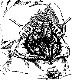
Figure 10. Removal of the larynx. The trachea is sutured into the
skin wound. The larynx is pulled upward and separated from behind. In the transverse part of the T-shaped incision, the sternohyoid and thyrohyoid muscles are cut; along the midline, they and the sternothyroid muscles are bluntly pushed to the sides, with the sternothyroid muscles being cut at their attachment to the thyroid cartilage. By blunt and sharp dissection, the Larynx is exposed anteriorly and laterally, cutting the pharyngeal constrictor at its attachment, which can also be done later. Simultaneously, the cricothyroid arteries are ligated. To expose the upper part of the trachea, the isthmus of the thyroid gland is dissected between ligatures, or sometimes part of it is excised, but one can also limit oneself to pushing the isthmus downward (Gluck). Exposing an excessive section of the trachea leads to a disruption of its nutrition and the possibility of necrosis. Once freed, the trachea is cut transversely between the cricoid cartilage and the first tracheal ring (lower in cases of extensive tumors) and immediately taken up on ligatures, placed if possible without damaging the mucosa. Lifting it on the ligatures, the separation of the Larynx from the trachea is quickly completed, and the mucosa is immediately swabbed with 10% cocaine to prevent coughing from the usual entry of tiny amounts of blood. Some recommend cutting the trachea after first separating it from the esophagus; this may cause unnecessary trauma to the recurrent nerve. It is better to immediately sew the freed end of the trachea into the edges of the skin incision with several sutures, again avoiding piercing the mucosa if possible. Above the trachea, the edges of the skin incision are sutured, also capturing the anterior wall of the esophagus without the mucosa. The opening of the Larynx is filled with gauze, its edge is grasped with strong forceps, pulled upward and forward, and the anterior wall of the esophagus is separated from the posterior surface of the Larynx (see Figure 10). After the trachea is cut, cyanosis and minor venous bleeding disappear. If the operation is performed under chloroform, a tube of suitable diameter is inserted into the cocaine-treated trachea, equipped at the other end with a funnel covered with flannel or gauze, onto which the chloroform is dripped. After this, the isolation of the Larynx is completed, moving from bottom to top, cutting the pharyngeal constrictor if this was not done earlier, and ligating the inferior and superior laryngeal arteries along the way. In the region of the arytenoid cartilages, the anterior wall of the pharynx is opened, and isolating gauze is introduced into its cavity. The superior horns of the thyroid cartilage are cut or isolated, and the mucosa lining the piriform sinuses is dissected away if it does not cause concern. The thyrohyoid membrane and the hyo-epiglottic ligament are cut, the epiglottis is separated from the root of the tongue, and the entire Larynx is removed. Gluck recommends starting the isolation of the Larynx from above and cutting the trachea toward the end of the operation. The pharyngeal opening is closed with sutures in one or two layers at the edges of the mucosal defect, without piercing it and protecting the wound at all times from the entry of mucus from the mouth. The cut sternohyoid muscles are sutured, which reduces the tension on the sutures of the pharyngeal defect; sutures are placed on the skin. Drains are placed toward the pharynx at the ends of the transverse incision. Suturing the pharyngeal opening protects the wound from infection from the mouth and from deglutition bronchopneumonia. The sutures do not always hold, but isolating the wound even for the first 3–4 days improves subsequent healing. If it is impossible to suture the pharyngeal opening, it is left open, and it is recommended not to suture the entire wound for better drainage. Part of the wound is also left open in cases of partial or total excision of the adjacent part of the pharynx and esophagus, which is sutured into the skin incision behind the trachea. To feed the patient for the first 6–7 days, a gastric tube is inserted into the esophagus a demeure, most conveniently through the nose. A tracheotomy tube is placed in the trachea. A standard dressing is applied, not compressing the veins of the neck. During the operation, at the beginning or end, the regional lymph nodes and cellular tissue are inspected and removed. Total removal of the Larynx for malignant neoplasms yields over 15% recoveries lasting longer than one year. One of the patients operated on by P. I. Dyakonov was in good condition after 18 years. Mortality after total removal of the Larynx among various surgeons ranges from 3% (Moure and Portmann) to 40%. Death is less often directly related to the operation itself (shock) and occurs in the first days after it with increasing decline in cardiac activity. To prevent shock, it is necessary to traumatize the nerves of the Larynx, which are closely connected with the nerves of the heart, as little as possible. More often, death is caused by postoperative diseases—aspiration bronchopneumonia and other pulmonary complications (about 47% of all deaths), purulent mediastinitis, sometimes phlegmons, emboli, septicopyemia, etc. To prevent lung diseases, it is necessary to carefully protect the trachea from the entry of blood during the operation, and saliva, wound discharge, and swallowed food in the postoperative period. It is also necessary to prevent phenomena of tracheitis and bronchitis, which easily arise due to the direct entry of atmospheric air into the trachea. It is necessary to humidify the inhaled air by hanging a moist apron of 2–3 layers of gauze in front of the tube opening, performing repeated inhalations with some alkaline water, and humidifying the air in the room. It is important to carefully clean the tracheotomy tube as it becomes clogged with mucus. If phlegmonous inflammation develops, the wound must be opened wide and drained to prevent the spread of inflammation to the mediastinal tissue. Some perform the removal of the Larynx in two stages so that the developed scars act as a barrier to the spread of infection into the mediastinum. In all methods of Larynx removal, careful care of the wound and the patient is the most important condition for the success of the operation. One must monitor every detail, the slightest change in the condition of the patient, who is unable to speak and sometimes cannot sufficiently clearly state their sensations and wishes in writing. If the pharyngeal sutures hold, then after 8–12 days the patient can be allowed to swallow small portions of liquid and semi-liquid food. If the sutures have diverged or the pharynx was not sutured, a gastric tube must be inserted at each feeding until the opening in the pharynx is formed enough that a suitable dressing can sufficiently prevent swallowed food from leaking out. After the wound has healed, it is necessary to give the patient the opportunity to speak. Occasionally, patients with a sutured pharyngeal opening learn to speak without an external
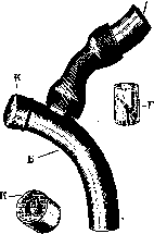
Figure 11. A—pharyngeal part, B—tracheal part of the larynx; K—cap placed on the end of A during eating; Г—voice apparatus inserted into A. At this time, it is necessary to remove the cap K from the outer opening of the tube and put in its place the cap K with a metal door Д, which opens upon inhalation.
establishing a connection between the pharynx and the throat, the so-called "false voice"—a loud whisper produced with the help of a supply of air accumulated in the pharynx and the upper part of the esophagus. Usually, one must resort to an artificial larynx, first constructed by Gussenbauer and subsequently subjected to various modifications. At the present time, in patients with a sutured pharyngeal opening, Gluck's vocal apparatus is used, with air pumped through the nose into the pharynx or with an external communication between the pharynx and the throat by means of a tube through the nose or mouth (Gottstein type). Less bulky and noticeable is the artificial larynx of the Gussenbauer type, which directly connects the throat with the pharynx through an opening made additionally in the sutured pharynx or left unsutured. The most simple and convenient is the Dyakonov larynx (see Figures 11, 12, and 13). With the help of its soundless device, patients breathe easily

Figure 12. Diagram of a sagittal section after removal of the larynx and installation of the Dyakonov larynx.

Figure 13. Appearance of a patient with the Dyakonov larynx inserted. through the mouth and learn to speak in such a loud whisper that they can converse over the telephone. The sound-producing device gives a clear, loud, but monotonous sound, is quite easily damaged, and furthermore
its use somewhat restricts
breathing. Even more simple,
and capable of being made from rubber tubes at home, is the artificial larynx of A. F. Ivanov (see Figure 14). Figure 14. a—outer end with valve b, which lets air in from the outside during inhalation and closes during exhalation; г—pharyngeal end with a valve that closes the entrance to the larynx during swallowing but lets air through during exhalation; д—throat end. In initial forms of malignant tumors occupying a limited area of the larynx poor in lymphatic vessels, but already involving the cartilage, many surgeons perform partial, most often hemilaryngeal, removal of the larynx with preservation of the epiglottis. The operation begins with splitting and inspection of the larynx, often with a preliminary tracheotomy. After removal of the affected part, careful cessation of bleeding follows. Depending on the defect, there is either a primary partial suturing of the wound with tamponade and a tracheotomy tube, or open treatment of the wound with subsequent plastic closure. Removal of regional lymph nodes is mandatory. Gluck, when removing half of the larynx, recommends suturing the edges of a hinged skin flap to the posterior, upper, and lower edges of the laryngeal defect. The operation yields a lower mortality rate compared to total removal, but recurrences are somewhat more frequent. Sometimes a fairly loud voice is restored, in which case the role of the missing vocal cord is played by scars or the remaining aryepiglottic folds.
Related articles
Cite this page
“Larynx.” Soviet Medical Encyclopedia. English translation of Bolshaya Meditsinskaya Entsiklopediya, 1st ed. (Moscow, 1928–1936), ed. N. A. Semashko. https://sovietmedicalencyclopedia.pages.dev/article/larynx/