Laryngotomy
Historical document, translated for reference. It reflects medical knowledge of the 1920s–30s and is not medical advice.
Summary
This article from the 1928–1936 Soviet medical encyclopedia details the surgical procedure of laryngotomy, including its types, indications, and technique. It explains the distinction between partial and complete laryngotomy, the use of local anesthesia, and the management of complications like hemorrhage and airway obstruction.
Encyclopedia article (1928–1936)
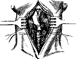
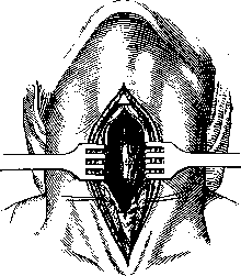
ЛЯРИНГОФИССУРА (from Greek larynx-larynx and Latin fissura-slit), incision of the larynx; this operation is more correctly called laryngotomy, since a laryngofissure is the result of the operation. Complete and partial laryngotomies are distinguished; in the first, the thyroid and cricoid cartilages, the cricothyroid ligament, and the upper tracheal rings are incised along the midline, while in the second, the cricothyroid ligament, cricoid cartilage, and upper tracheal rings are incised. (Partial laryngotomy is preferable, as the vocal cords are not injured and the voice is not impaired.) A transverse laryngotomy according to Gersuny is also used, in which a transverse incision of the larynx is made above the region of the vocal cords, and a longitudinal incision along the midline is added above it. For small lesions of the larynx, it is sometimes sufficient to incise only one thyroid cartilage. Indications for laryngotomy include foreign bodies, neoplasms, cicatricial stenosis, injuries to the larynx, and tuberculosis. The operation is performed under local anesthesia with the head hanging down or in the Trendelenburg position, bringing the patient into this position only at the moment of tracheal incision to avoid aggravation of suffocation due to venous stasis. Technique of the operation. In complete laryngotomy, a skin incision is made along the midline from the hyoid bone down to the jugular fossa; after the skin, the fascia is incised, and several small arteries running along the cricothyroid membrane and at the isthmus of the thyroid gland have to be ligated and cut. Pushing the muscles aside exposes the hyoid-thyroid ligament, thyroid cartilage, cricothyroid ligament, and cricothyroid cartilage; to expose the trachea, the isthmus of the thyroid gland must be bluntly separated, ligated in two places, and cut (Fig. 1); it is even better to expose the trachea according to Bose: the deep fascia of the gland attached to the cricoid cartilage is separated from the cartilage by a transverse incision and bluntly pushed down together with the gland. Then the trachea is opened, the edges of the incision are spread with blunt hooks, and through the resulting opening the inner surface of the trachea and larynx is lubricated with 20% cocaine-adrenaline. A tracheotomy tube is inserted and 5–10 minutes are waited for complete anesthesia to set in, after which the cricoid, and then the thyroid cartilages are cut with strong scissors (and with Lis-trovet scissors if the cartilage is ossified), trying to stay strictly on the midline so as not to damage the vocal cords. Spreading the hooks, the halves of the thyroid cartilage are separated (Fig. 2), the interior of the larynx is inspected, the changes found there are determined, and action is taken accordingly. Upon completion of the intralaryngeal operation, the wound is sutured: the perichondrium and muscles with catgut, the skin with silk; if there is no bleeding and breathing is free, the trachea is also sutured, inserting a strip of gauze into the lower corner of the wound; if, however, there is suspicion of the possibility of stenosis, it is better to insert a cannula for several days. If the intralaryngeal intervention was quite extensive and there is a danger of bleeding, it is safer to tamponade the laryngeal cavity and withdraw the tampon above the tracheotomy tube; after the tampon and cannula are removed, the opening in the trachea closes with granulations. Where the lesions of the larynx are deeper and the postoperative wound requires prolonged observation and treatment, the split larynx is not sutured, and treatment of the open larynx is carried out; laryngotomy is thus converted into laryngostomy (see). In partial laryngotomy, the skin incision begins somewhat above the lower edge of the thyroid cartilage, and the cricothyroid membrane, cricoid cartilage, and upper tracheal rings are incised. Various laryngotracheal cannulas constructed on the principle of T-shaped tubes (Dupuis, Schimmelbusch, Killian) have been proposed instead of an ordinary tracheal cannula for laryngotomy. When a tumor is located closer to the entrance to the larynx, it is necessary to combine laryngotomy with pharyngotomy (median subhyoid pharyngolaryngotomy, according to Kocher); in this case, the larynx is split in the middle, the sternohyoid and geniohyoid muscles are cut at the hyoid bone, then the geniohyoid membrane is incised, the hyoid bone is strongly pulled upward with a hook, and the mucous membrane of the pharynx is opened; thus a wide access to the pharyngeal space and the entrance to the larynx is obtained.
Figure 1. The first moment of the operation.
Then the trachea is opened, the edges of the incision are spread with blunt hooks, and through the resulting opening the inner surface of the trachea and larynx is lubricated with 20% cocaine-adrenaline. A tracheotomy tube is inserted and 5–10 minutes are waited for complete anesthesia to set in, after which the cricoid, and then the thyroid cartilages are cut with strong scissors (and with Lis-trovet scissors if the cartilage is ossified), trying to stay strictly on the midline so as not to damage the vocal cords. Spreading the hooks, the halves of the thyroid cartilage are separated (Fig. 2), the interior of the larynx is inspected, the changes found there are determined, and action is taken accordingly. Upon completion of the intralaryngeal operation, the wound is sutured: the perichondrium and muscles with catgut, the skin with silk; if there is no bleeding and breathing is free, the trachea is also sutured, inserting a strip of gauze into the lower corner of the wound; if, however, there is suspicion of the possibility of stenosis, it is better to insert a cannula for several days. If the intralaryngeal intervention was quite extensive and there is a danger of bleeding, it is safer to tamponade the laryngeal cavity and withdraw the tampon above the tracheotomy tube; after the tampon and cannula are removed, the opening in the trachea closes with granulations. Where the lesions of the larynx are deeper and the postoperative wound requires prolonged observation and treatment, the split larynx is not sutured, and treatment of the open larynx is carried out; laryngotomy is thus converted into laryngostomy (see). In partial laryngotomy, the skin incision begins somewhat above the lower edge of the thyroid cartilage, and the cricothyroid membrane, cricoid cartilage, and upper tracheal rings are incised. Various laryngotracheal cannulas constructed on the principle of T-shaped tubes (Dupuis, Schimmelbusch, Killian) have been proposed instead of an ordinary tracheal cannula for laryngotomy. When a tumor is located closer to the entrance to the larynx, it is necessary to combine laryngotomy with pharyngotomy (median subhyoid pharyngolaryngotomy, according to Kocher); in this case, the larynx is split in the middle, the sternohyoid and geniohyoid muscles are cut at the hyoid bone, then the geniohyoid membrane is incised, the hyoid bone is strongly pulled upward with a hook, and the mucous membrane of the pharynx is opened; thus a wide access to the pharyngeal space and the entrance to the larynx is obtained.
Figure 2. The second moment of the operation.
Then the trachea is opened, the edges of the incision are spread with blunt hooks, and through the resulting opening the inner surface of the trachea and larynx is lubricated with 20% cocaine-adrenaline. A tracheotomy tube is inserted and 5–10 minutes are waited for complete anesthesia to set in, after which the cricoid, and then the thyroid cartilages are cut with strong scissors (and with Lis-trovet scissors if the cartilage is ossified), trying to stay strictly on the midline so as not to damage the vocal cords. Spreading the hooks, the halves of the thyroid cartilage are separated (Fig. 2), the interior of the larynx is inspected, the changes found there are determined, and action is taken accordingly. Upon completion of the intralaryngeal operation, the wound is sutured: the perichondrium and muscles with catgut, the skin with silk; if there is no bleeding and breathing is free, the trachea is also sutured, inserting a strip of gauze into the lower corner of the wound; if, however, there is suspicion of the possibility of stenosis, it is better to insert a cannula for several days. If the intralaryngeal intervention was quite extensive and there is a danger of bleeding, it is safer to tamponade the laryngeal cavity and withdraw the tampon above the tracheotomy tube; after the tampon and cannula are removed, the opening in the trachea closes with granulations. Where the lesions of the larynx are deeper and the postoperative wound requires prolonged observation and treatment, the split larynx is not sutured, and treatment of the open larynx is carried out; laryngotomy is thus converted into laryngostomy (see). In partial laryngotomy, the skin incision begins somewhat above the lower edge of the thyroid cartilage, and the cricothyroid membrane, cricoid cartilage, and upper tracheal rings are incised. Various laryngotracheal cannulas constructed on the principle of T-shaped tubes (Dupuis, Schimmelbusch, Killian) have been proposed instead of an ordinary tracheal cannula for laryngotomy. When a tumor is located closer to the entrance to the larynx, it is necessary to combine laryngotomy with pharyngotomy (median subhyoid pharyngolaryngotomy, according to Kocher); in this case, the larynx is split in the middle, the sternohyoid and geniohyoid muscles are cut at the hyoid bone, then the geniohyoid membrane is incised, the hyoid bone is strongly pulled upward with a hook, and the mucous membrane of the pharynx is opened; thus a wide access to the pharyngeal space and the entrance to the larynx is obtained.
Then the trachea is opened, the edges of the incision are spread with blunt hooks, and through the resulting opening the inner surface of the trachea and larynx is lubricated with 20% cocaine-adrenaline. A tracheotomy tube is inserted and 5–10 minutes are waited for complete anesthesia to set in, after which the cricoid, and then the thyroid cartilages are cut with strong scissors (and with Lis-trovet scissors if the cartilage is ossified), trying to stay strictly on the midline so as not to damage the vocal cords. Spreading the hooks, the halves of the thyroid cartilage are separated (Fig. 2), the interior of the larynx is inspected, the changes found there are determined, and action is taken accordingly. Upon completion of the intralaryngeal operation, the wound is sutured: the perichondrium and muscles with catgut, the skin with silk; if there is no bleeding and breathing is free, the trachea is also sutured, inserting a strip of gauze into the lower corner of the wound; if, however, there is suspicion of the possibility of stenosis, it is better to insert a cannula for several days. If the intralaryngeal intervention was quite extensive and there is a danger of bleeding, it is safer to tamponade the laryngeal cavity and withdraw the tampon above the tracheotomy tube; after the tampon and cannula are removed, the opening in the trachea closes with granulations. Where the lesions of the larynx are deeper and the postoperative wound requires prolonged observation and treatment, the split larynx is not sutured, and treatment of the open larynx is carried out; laryngotomy is thus converted into laryngostomy (see). In partial laryngotomy, the skin incision begins somewhat above the lower edge of the thyroid cartilage, and the cricothyroid membrane, cricoid cartilage, and upper tracheal rings are incised. Various laryngotracheal cannulas constructed on the principle of T-shaped tubes (Dupuis, Schimmelbusch, Killian) have been proposed instead of an ordinary tracheal cannula for laryngotomy. When a tumor is located closer to the entrance to the larynx, it is necessary to combine laryngotomy with pharyngotomy (median subhyoid pharyngolaryngotomy, according to Kocher); in this case, the larynx is split in the middle, the sternohyoid and geniohyoid muscles are cut at the hyoid bone, then the geniohyoid membrane is incised, the hyoid bone is strongly pulled upward with a hook, and the mucous membrane of the pharynx is opened; thus a wide access to the pharyngeal space and the entrance to the larynx is obtained.
Related articles
Mentioned in
Cite this page
“Laryngotomy.” Soviet Medical Encyclopedia. English translation of Bolshaya Meditsinskaya Entsiklopediya, 1st ed. (Moscow, 1928–1936), ed. N. A. Semashko. https://sovietmedicalencyclopedia.pages.dev/article/laryngotomy/