Local Anesthesia
Historical document, translated for reference. It reflects medical knowledge of the 1920s–30s and is not medical advice.
Summary
Local anesthesia refers to techniques that use safe, minimally toxic substances to eliminate pain sensitivity locally while maintaining consciousness. This article covers the history, pharmacology, and various methods of local anesthesia as practiced in the 1920s-1930s.
Encyclopedia article (1928–1936)
LOCAL ANESTHESIA (from the Greek a- neg. part, and aisthesis - sensation), or local pain relief, are methods that aim to destroy pain sensitivity (mainly during surgical operations) locally with the help of safe, minimally toxic substances, while preserving consciousness completely. Local anesthesia, being significantly less dangerous than general anesthesia, has attracted increasing attention from surgeons over the last 20-30 years. Attempts at local pain relief were made long ago; even in ancient times, cooling was used, which dulled sensitivity, and they tried to smear the skin with various medicinal substances, and compressed and tied off limbs to reduce pain. With the introduction of cocaine as an anesthetic substance by the German ophthalmologist Koller (1884), local anesthesia became firmly established and was able to develop further in a systematic and rational manner. Local anesthesia with cocaine was significantly improved by Reclus (1889) in France, using 1-1½% solutions, and by Schleich (1891) in Germany, who recommended for infiltrative anesthesia the safe dilution of cocaine 0.01-0.1% in combination with a 0.2% solution of NaCl. But only with the discovery of novocaine, a substance significantly safer than cocaine, by Einhorn (1905), and the proposal by Braun to add adrenalin to the anesthetic substance to prolong its effect, did local anesthesia begin to be used more widely. A large number of pharmacological substances have been proposed for pain relief. The following requirements are made for a substance suitable for local anesthesia: 1) weak toxicity and strong selective action on nervous tissue; 2) absence of local damage and irritation of tissues; 3) stability of the substance during sterilization and solubility in water. The most important of these substances are: cocaine (Cocaine C17H21NO4), colorless crystals; with hydrochloric acid it forms Cocainum hydrochloricum, the most commonly used preparation; for injection in local anesthesia, 0.1-1% solution is used (maximum dose - 0.03 pro dosi). In otorhinolaryngology, cocaine is widely used for anesthetizing the mucous membranes of the nose, pharynx, and larynx; for this purpose, a 10% solution of cocaine is used. Tropococaine, its hydrochloride salt (Tropococain muriaticum) is used in 1-5% solutions, mainly for spinal anesthesia. Eucain, a derivative of cocaine, is used in a 1/10% solution, doses: up to 0.1-0.15 eucain. Stovain is 2-3 times less toxic than cocaine, ½-1% solutions are used, dose 0.2-0.3. Novocain (Novocain C18H21O2N2HCl), a white crystalline powder, readily soluble in alcohol and water, the most commonly used and best preparation for all types of local anesthesia. In surgery, novocaine is currently used extremely widely. Usually for infiltrative anesthesia, ½-1% solutions are used in amounts up to 200-250 cubic cm without side (harmful) effects or poisoning phenomena. To enhance the effect of novocaine, adrenalin is added to the solution (Sol. Adrenalini-1:1000); to 10.0 of the anesthetic solution, 1 drop of adrenalin. When using novocaine in this way, the duration of anesthesia is 1½-2 hours. Very recently in Germany, new preparations have also been released, such as tutocaine and psicaine; they have all the advantages of novocaine but act much more strongly, therefore these preparations for local anesthesia are used in greater dilution, which further reduces the possibility of poisoning. Tutocaine and psicaine are used in 1-1½% solutions, also with the addition of adrenalin. The results, as with novocaine, are very good. The latest preparations also include borocaine, dolantin (see also Anesthetic substances). Methods of Local Anesthesia Anesthetic substances can be applied in various ways. They can be applied to superficial tissues or mucous membranes. Using substances such as chloroethyl and ether, and spraying them on the surface, causes cooling and thus achieves insensitivity of the given area. But the main method, which has primary importance in local anesthesia, is the injection of anesthetic substances. This injection can be done by infiltrating all tissues (infiltrative anesthesia), or the injection is made directly into one nerve or another, thereby interrupting its conductivity (conductive or regional anesthesia). Then the anesthetic substance can also be introduced into the circulatory system, into a vein or artery of the limb (venous or arterial anesthesia) in order to cause insensitivity of the entire limb or part of it. These methods have very limited application, are rarely used, and have no practical value. Anesthesia by smearing superficial tissues and mucous membranes. In diseases of the ear, nose, and throat, this simplest and oldest method of pain relief is widely used. Insensitivity of the larynx is achieved by smearing its mucous membrane with a 20% solution of cocaine; for the mucous membrane of the mouth and pharynx, a 10% solution is sufficient. In ophthalmic practice, 1-2% solutions of cocaine are used. When anesthetizing mucous membranes, it must be remembered that cocaine is rapidly absorbed and can cause poisoning phenomena. The danger of the toxic effect of cocaine depends on the concentration of the solution and the size of the area of mucous membrane being treated. Special caution is required when introducing cocaine for local anesthesia of the mucous membrane of the bladder and urethra. A. by cooling. The most commonly used substances for causing anesthesia by cooling are ether and chloroethyl. Ether-Aether sulf. (C2H5)2O, with a boiling point of +35°, is poured into a Richardson atomizer, consisting of a bottle with a fitted stopper, through which two metal tubes pass; one of them is connected to a double rubber bulb for pumping air. The ether is caught by the air stream and atomized. The stream, directed onto the skin, makes it white and freezes it to complete A.; the temperature at this time drops to 15-20°. The same A. is achieved with chloroethyl (C2H5Cl) with a boiling point of +12°. Chloroethyl is sold in glass tubes with a capacity of 10 to 100 cubic cm, equipped with an automatic lock. Through the hole in the tube, due to heating by the warmth of the hand, chloroethyl evaporates in a thin stream. In minor surgery, anesthesia by cooling is often used for opening superficially located purulent processes, in phlegmons, abscesses, furuncles, etc. Infiltrative A. was introduced into surgery by Reclus and Schleich. Under infiltrative A. is meant the impregnation of tissues with an anesthetic solution, acting on the nerve sensitive endings and nerve trunks passing through the tissues. Reclus and Schleich for infiltrative A. still used solutions of cocaine 1% and 0.1%. Schleich's prescription: Cocaini muriatici 0.1, Natrii chlorati 0.2, Morphii muriatici 0.02, Aq. destill. 100.0. The addition of NaCl was made

Figure 1. Infiltrative anesthesia from
two injection points (1-2): a-skin; b-fascia and muscles; c-bone. to achieve isotonicity of the solution with tissue juices and blood. At present, for the purpose of infiltrative A., solutions of the rather toxic cocaine are rarely resorted to, and in the vast majority of cases with numerous operations under infiltrative A., a ½-1% solution of novocaine with the addition of adrenalin is used. Tissue infiltration is performed layer by layer from the outside inward. For simple skin incisions, the superficial layer is infiltrated. A thin needle is inserted into the skin, and a small amount of solution is introduced into its thickness under the epidermis, resulting in a small elevation with whitened skin-"a wheal". Several such wheals are placed along the line of the incision, resulting in an anesthetized ridge of skin. For deeper incisions, the subcutaneous tissue is also infiltrated. For A. of the operative field, where anesthesia of deep tissues is required, two wheals are placed at the injection sites and from here all tissues are impregnated to the required depth. On the limbs, tissues can thus be impregnated to the bone (see Figure 1). In cases where the operative field covers tissues over a considerable width and depth, infiltration is used in the form of spraying. When spraying, infiltration with an anesthetic solution is performed not of the entire operative field, but only of its
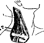
of the sides to interrupt the conductivity of the sensory nerves innervating the given area. Depending on the planned operation and the field of intervention, various forms and shapes of spraying are necessary (pyramidal, trough-shaped). The infiltration method and, especially, the method of spraying the operative field, are the most common methods of local anesthesia. They are simple in technique, do not require precise knowledge of the course of nerve trunks, are safe, and provide good anesthesia. The infiltration method of local anesthesia competes with another, increasingly developing but more difficult method of regional anesthesia. Regional anesthesia is based on the idea of temporarily interrupting the conductivity of a sensory nerve along its length and, thus, excluding the area to be operated on. To interrupt the conductivity of a nerve, thereby anesthetizing the area innervated by it, one can inject an anesthetic substance directly into the nerve—intraneural anesthesia, or into the surrounding nerve tissues—perineural anesthesia. The latter type of regional anesthesia is used more frequently. The nerve along its length is surrounded by a sheath—the perineurium, which prevents the anesthetic substance from acting more quickly, therefore, with a perineural injection, a stronger solution of novocaine is used—1-2% with adrenaline; the amount of solution is 10-20-40 cubic cm. Technically, regional anesthesia is more complex than infiltration. It requires precise knowledge of the topographical features of the area to be anesthetized and the course of the nerves. The anesthesia is performed in the depths of the tissues, where one must use reference points, such as: the sensation of bone, the depth, the direction of the needle, and the sensations of the patient (paresthesias from the needle touching the nerve). In areas of large vessels, when advancing the needle and injecting, one must be careful. It is recommended to locate the nerve only with the help of the needle, without a syringe. If a vessel is accidentally injured, blood will immediately appear from the needle, then the needle is withdrawn back. If the bleeding is severe, it is better not to perform anesthesia at this site.
A typical regional anesthesia is the interruption of the conductivity of the brachial nerve plexus (plexus brachialis; Kulenkampf, 1911); with this, complete insensitivity of the upper extremity is achieved. Anesthesia of the brachial plexus according to Kulenkampf is performed in the supraclavicular fossa. With a finger above the middle of the clavicle, the first rib is palpated in the depth; with more superficial pressure, the pulse of the supraclavicular artery is clearly felt—it is located medial to the brachial plexus (see Figure 2). The site of needle insertion is located 1/2-1 finger's breadth above the middle of the clavicle and lateral to the a. subclavia. After making a skin wheal at the injection site, a thin long needle is inserted into the depth past the supraclavicular artery; at a depth of 2-3 cm, the needle rests against the bone, the first rib. Then the needle is withdrawn back slightly and the nerve plexus is carefully sought with it. Entry into the nerve plexus is determined by characteristic paresthesias (tingling) in the forearm and fingers. When paresthesias appear, without changing the position of the needle, a syringe is attached and 10-20 cubic cm of 1/2-2% solution of novocaine with adrenaline, or 30-40 cubic cm of 1% solution, is injected. In addition to arterial injuries, with this anesthesia, one should avoid dangerous injury to the pleura, which is possible with deeper, careless insertion of the needle. With anesthesia of the brachial plexus, all major operations on the shoulder, elbow, forearm, and hand (amputations, resections, etc.) can be performed successfully. - Conductive anesthesia also typically occurs on the head for all branches of the trigeminal nerve and the nerves of the lower extremity (n. ischiadicus, n. femoralis). Figure 3 shows anesthesia of the sciatic nerve (n. ischiadicus). The intersection of lines ab and cd—the site of needle insertion; on the right—the location of the sciatic nerve. Performing these anesthesias requires precise anatomical knowledge and a technique developed through experience.
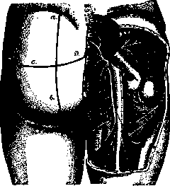
Figure 3. Conductive anesthesia of the sciatic nerve-
the intersection of lines ab and cd—site of needle insertion, on the right—location of the sciatic nerve. To the conductive anesthesia also belongs anesthesia of the splanchnic nerves (nn. splanchnici), which has recently been acquiring ever greater importance in operations in the abdominal cavity and, mainly, on the stomach. The greater and lesser splanchnic nerves (splanchnicus major et minor) are formed from the connections of rami communicantes of the 6-12 thoracic nerves. Both nerves, which contain sensory fibers for the stomach, liver, spleen, and the upper part of the small intestine, approach the aorta at the level of the exit of the a. coeliaca and form a paired semilunar node here (ganglion coeliacum). By means of short gray threads, both nodes are brought together, forming a ring-shaped double node—the solar plexus (ganglion solare). Near this solar plexus, anesthesia of the splanchnic nerves has been proposed. Kappis first (in 1918) proposed performing anesthesia of the n. splanchnici from behind, from the side of the back. At the lower edge of the XII rib, an injection is made with a needle 12-15 cm long, the tip of the needle penetrates at a certain angle to the lateral surface of the XII thoracic vertebra. Near this place in the retroperitoneal tissue lie the splanchnic nerves; here 30-40 cubic cm of 0.5% solution of novocaine is injected. Anesthesia of the n. splanchnici
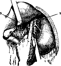
Figure 4. Anesthesia of the n. splanchnici according to Braun' during open abdominal cavity: 1 - aorta; 2 - v. cava.
According to Kappus, it is difficult in technique and not without dangers (possibility of damage to large vessels or the needle entering the spinal canal). The method of Brown with anesthesia of the celiac nerves from the abdominal side, after opening the latter, is simpler and safer. After infiltration anesthesia of the abdominal wall above the navel, it is opened along the midline. The wound is expanded with blunt hooks, the left lobe of the liver is pulled upward. With the right hand, the stomach is pulled downward so that the lesser omentum tenses. With the index finger of the left hand, the aorta is palpated. By pressure of the index finger, the lesser omentum is pressed against the spine (to the XII vertebra). Under pressure, the aorta moves to the left, the v. cava to the right. After this, under visual control, a special thin needle (see Figure 4) is inserted along the index finger, it rests against the bone, against the vertebra. After slightly withdrawing the needle, it is fixed and, without changing direction, 40 cubic cm of 0.5% novocaine solution is injected. If a vein is accidentally injured, blood appears from the needle, in which case anesthesia is better not performed. In cases of severe intra-abdominal adhesions, anesthesia according to Brown cannot be performed. At present, several thousand major operations have already been published under anesthesia of the celiac nerves from the abdominal side; operations of gastrointestinal anastomosis, resections of the stomach, cholecystectomy and others have been performed successfully. Instruments and solutions for local anesthesia. The success of local anesthesia depends greatly on proper technique and appropriate instruments. There is no need for complex devices. For anesthesia in all areas of the body, it is sufficient to have a syringe, a set of needles, and a vessel for the anesthetic solution. Ordinary 2-gram and 5-gram glass "Record" syringes are quite suitable for injections. One can use the special metal syringe of Brown. Needles should be of different lengths (from 2-3 to 15-20 cm) and different thicknesses (0.5 - 1.5 mm). Needles should be thin, with a short-cut end. Curved needles and cannulas are of little use, as curvature interferes with the correct sensation of the needle's position. The most common for all types of local anesthesia is a 0.5-1% solution of novocaine prepared in a 0.6-0.7% solution of NaCl. Ready-made Brown's tablets of novocaine with adrenaline (novocaine 0.125, synthetic adrenaline 0.00012) are available for sale. Eight such tablets, dissolved in 200 g of physiological solution, give a 0.5% solution of novocaine. The novocaine solution with adrenaline is unstable and quickly spoils when exposed to air, becoming colorless, pink, and brown; in this form, the solution cannot be used for anesthesia, so it is better to add adrenaline to the solution immediately before anesthesia. Preparation of the patient. Before operation under local anesthesia, the patient must be studied as thoroughly as before general anesthesia, attention must be paid to the heart, vascular system, and lungs. Although the substances used in local anesthesia act much more weakly on the listed organs, still to the body during operation increased demands are made; therefore, preparation of weak patients before the operation with preparations of strophanthus and digitalis is desirable. Laxatives that weaken the patient before the operation are even more unnecessary than under general anesthesia. Complete abstinence from drinking and eating before local anesthesia does not seem necessary. The "psychic preparation" of the patient for local anesthesia is important. In the past, too little attention was paid to this important moment. In the clinic, in the department, a corresponding atmosphere should be created, the patient should be confident that he will not feel pain. It is incorrect to tell the patient: "You will not feel anything," this is not justified during the operation, undermines the authority of the physician and discredits the method. The patient should be informed that he will feel and be aware that he is being operated on, but at the same time he will not feel pain. In clinics where many operations are performed under local anesthesia, such a favorable psychic atmosphere is easily created, patients themselves point out to each other the advantages of local anesthesia: the severe postoperative condition is eliminated, patients have a cheerful appearance, they can drink and eat immediately. Nervous patients are given bromide preparations for several days before the operation. 1/2 hour before the start of anesthesia, the patient is given morphine subcutaneously (Morph. mur. 0.01). After the completion of anesthesia, i.e., after the last injection of solution, one should wait at least 10 minutes to allow the anesthetic solution to be well absorbed and act on the nerve endings. During the operation, in some cases, it is desirable to distract the patient's attention from the course of the operation itself, which is often easily achieved by ordinary conversation ("psychic narcotizer"). Indications and contraindications. The main significance of local anesthesia is that compared to general inhalation anesthesia, it is a much safer method. With ordinary methods of infiltration and regional anesthesia, with proper technique and normal doses, local anesthesia is a completely safe method. With local anesthesia, a number of unpleasant sides inevitable with general anesthesia are eliminated, such as strong excitement, asphyxia, falling back of the tongue, decline of cardiac activity. With general anesthesia, the attention and care of the operator are constantly divided, with local anesthesia the patient himself is an active assistant to the operator. On the skull, on the face, in the oral cavity and on the neck-operations under local anesthesia present special conveniences, here the narcotizer with a mask always interferes. In some operations, especially those lasting for several hours, the advantages of local anesthesia are even more evident, as it has little effect on the general condition of the patient, which cannot be said about anesthesia lasting for hours. With local anesthesia, the distressing post-anesthesia state of the patient with vomiting and poor health, requiring special nursing personnel, is eliminated. In addition, local anesthesia has a number of special advantages, such as significant de-vascularization of the operative field, which in some operations on the skull, face, and in the mouth is of decisive importance. Post-anesthesia complications, according to many authors, are observed less frequently after local anesthesia. Pulmonary complications are observed with local anesthesia, but they are less frequent and run milder. According to the 1926 statistics, Shaak out of 1,063 operations under local anesthesia had 5.6% pulmonary complications, and under general anesthesia-8%. All the advantages of local anesthesia appear even more clearly when the surgeon works in modern conditions with limited personnel, in the field and in provincial settings. Therefore, we now notice a wider application of local anesthesia. On statistical material, one can trace the gradual increase in the use of local anesthesia. In the clinics of Wilms and Narath in Heidelberg and Brown in Zwickau before the world war, 50.5 - 54.2% of all operations were under local anesthesia. Shaak's clinic in Leningrad went from 33% of all operations under local anesthesia in 1918 to 88% in 1926. Therefore, the old rule that local anesthesia should be applied only when general anesthesia is contraindicated must be significantly expanded. Local anesthesia will be contraindicated, or rather, unfeasible, in cases where its performance encounters technical obstacles related either to the operative field or to the nature of the operation itself. In progressive purulent-inflammatory processes, if the needle has to pass through infected tissues, in sepsis and pyemia, where injections can lead to new metastases, local anesthesia is contraindicated. Local anesthesia is contraindicated in subjects with an especially vulnerable and unstable nervous system, where shock phenomena can be expected. According to recent data, childhood cannot be considered in all cases a contraindication to the use of local anesthesia. In a number of cases, many authors have successfully used local anesthesia in children, starting from infancy to 13 years (Shaak, Farr, Brown). Local anesthesia is indicated in children in cases with pulmonary complications and generally in very weak and exhausted children. Operations performed under local anesthesia. In all areas of the body, a large number of operations are now successfully performed under local anesthesia. On the head and face, with the help of regional anesthesia of the branches of the trigeminal nerve and with simple infiltration anesthesia, almost all operations are performed, starting from small and ending with large bone-plastic trepanations of the skull in neurosurgery. Operations on the jaws (resections, trepanations) are performed successfully under local anesthesia. In operations on the ear, throat, and nose, local anesthesia is widely used: trepanation of the mastoid process in mastoiditis, operations on the maxillary cavity and other operations in the oral cavity, all tooth extractions are performed under local anesthesia. On the larynx, tracheotomy, partial or complete removal of the larynx are also performed under local anesthesia. On the neck, the vast majority of all operations are performed under local anesthesia. In goiter, strumectomy under local anesthesia is the general rule, usually infiltration of the line of the collar incision and spraying of the goiter from several lateral points on the neck is sufficient. In large goiters, conduction anesthesia of the cervical plexus (plex. cervicalis) should be performed.
For all major operations on the upper extremity, the method of choice for local anesthesia is conduction anesthesia of the brachial plexus, as described above, but for many operations, the methods of spraying and infiltration are also sufficient. For operations on the fingers of the hand, the old method of Oberst is used: an anesthetic solution is injected at the base of the finger from two lateral points, thus interrupting the conductivity of the digital nerves. For operations on the chest, the sensitivity of the intercostal nerves is interrupted (intercostal anesthesia). If the injections are brought close to the spine, into the area of the transverse processes of the vertebrae, we interrupt the sensitivity of all branches of the spinal nerve (rami anterior, posterior and communicans); such anesthesia is called paravertebral, and it can be unilateral or bilateral. Depending on the size of the operative field, various segments of the thoracic and lumbar nerves are blocked. Insensitivity of the chest wall, abdominal wall, back, and lumbar region is achieved. The technique of paravertebral anesthesia is quite difficult; therefore, it has not become widely used. The operation of typical amputation of the entire breast gland with the pectoral muscle and removal of the glands of the axillary region in breast cancer can also be performed under local anesthesia. For this, a combination of intercostal and infiltration methods is necessary, with interruption of the sensitivity of the brachial plexus according to Kulenkampf in the supraclavicular fossa. For operations in the abdominal cavity, simple infiltration anesthesia of the abdominal wall is used for small operations, for example, gastrostomy. In a number of cases, gastroenterostomy and appendectomy are also successful under such anesthesia. In other cases, for example, in resections of the stomach, mesenteric anesthesia is required, in which the lesser omentum is infiltrated with a novocaine solution, or anesthesia of the splanchnic nerves. For appendectomy, in addition to anesthesia of the abdominal wall at the incision site, deep infiltration of the retrocecal area in the iliac fossa should also be performed. All operations for various types of hernias (inguinal, femoral, umbilical) are conveniently performed under infiltration anesthesia in combination with conduction anesthesia, as well as operations on the rectum, for example, in hemorrhoids and fistulas. Large operations on the rectum, such as amputation for cancer, are best performed under parasacral anesthesia. Operations on the lower extremities are performed under conduction anesthesia of the major nerve trunks: sciatic nerve, femoral nerve, obturator nerve, or under infiltration anesthesia with spraying. Amputations and resections on the lower extremities are also successfully performed under anesthesia of the so-called 'transverse incision,' as proposed by Sievers (1915) and Kemir (König); in this case (for example, in amputation of the thigh), a 0.5-1% solution of novocaine infiltrates all soft tissues to the bone at a certain level above the line of incision. It should also be mentioned that in the treatment of dislocations and fractures of bones, local anesthesia has come to play a prominent role. The reduction of a dislocated joint or the setting of broken bone fragments in the correct position is easily and painlessly accomplished under local anesthesia, in which case the simplest method is to inject the anesthetic solution between the displaced bone parts (30-60 cubic cm of a 0.5-1% solution of novocaine); complete anesthesia is achieved during all manipulations in dislocation and fracture necessary for reduction and application of bandages. Complications in local anesthesia. Regarding possible complications as a result of the use of various types of local anesthesia and their treatment, reference should be made to special manuals on local anesthesia. Here, only the postoperative course in patients operated on under local anesthesia and general complications that may occur with any method of local anesthesia can be mentioned. Small operations requiring a small amount of anesthetic substance are usually not accompanied by any consequences. Headaches, dizziness, nausea, and other phenomena sometimes observed should be attributed to individual peculiarities of the organism. Treatment is symptomatic: camphor, caffeine under the skin. For headaches - Aspirin 0.5, Pyramidon 0.2. With large amounts of injected anesthetic solution, complications may occur from the kidneys, as they mainly bear the work of excreting the anesthetic poison. Acute-nephritic phenomena (protein, cylinders in the urine, etc.) are sometimes observed, sometimes anuria, which with proper treatment (milk diet, warm baths) pass very quickly and leave no consequences. The course of the wound after operations under local anesthesia does not differ from that after general anesthesia. A difference in healing of the operative wound was observed only where there were defects in the preparation of anesthetic solutions in terms of their sterility. Hematomas and secondary bleeding are more often observed, as the strongly anesthetized wound masks bleeding during the operation. Careful ligation of small vessels prevents this type of complication. As for other postoperative complications, they may occur both with local anesthesia and with general anesthesia (for example, thromboses and emboli). Spinal anesthesia. The essence of spinal, or lumbar, anesthesia is that the anesthetic substance is injected in the lumbar region into the dural sac (cavum subarachnoidale) of the spinal cord. The substance introduced spreads in the cerebrospinal fluid, acts on the nerve roots and trunks, interrupting the conductivity of the cauda equina trunks and those roots of the spinal nerves with which the anesthetic liquid comes into contact. As a result of such injection of anesthetic solution into the subarachnoid space of the spinal cord, complete anesthesia of the lower half of the body is achieved. In its action, spinal anesthesia is actually a type of conduction anesthesia, but in its effect and extent of anesthesia, it occupies a place between general anesthesia and local anesthesia. The credit for the discovery of spinal anesthesia and its introduction into surgical practice belongs to the German surgeon Bier, who in 1899 published his first observations on spinal anesthesia with cocaine. Only later did it become known that Corning as early as 1885 had performed experiments with the introduction of cocaine between the spinous processes of the vertebrae in order to obtain anesthesia of the legs, but these experiments did not give practical results. Having proposed spinal anesthesia, Bier used the lumbar puncture suggested by Quincke in 1891. The new method of anesthesia was met with great interest in the medical world. In France, Tuffier was a fervent defender of the method, developing and improving the technique. Over time, the initial enthusiasm somewhat cooled, as the method nevertheless presents a number of dangers. Nevertheless, spinal anesthesia now occupies a firm place among the methods of anesthesia. The spinal cord occupies the space from the upper edge of the first cervical vertebra (atlas) to the second lumbar vertebra. Fluid for spinal anesthesia must be introduced into the subarachnoid space of the spinal cord. To enter this cavity, it is necessary to pierce the dura mater of the spinal cord with a needle, first passing between the spinous processes of the lumbar vertebrae. The instruments necessary for spinal anesthesia consist of a special Bier needle 1-1.5 mm thick, 12 cm long, with a short-cut end. The needle must be provided with a mandrel and a Record syringe of 10-15 cubic cm capacity must fit it well. To perform the anesthesia, the patient is seated across the table, legs hanging down, resting on elbows on thighs, bending the back and head as far forward as possible (see Figure 5). In this position, the spinous processes of the vertebrae stand out clearly. Lateral curvature of the spine should be avoided. It is possible to perform the anesthesia, especially in seriously ill patients, in the lying position, for which the patient is laid on the side, legs are bent at the knees and hips, and the body is sharply bent forward. An assistant fixes the patient in this position so that there are no accidental movements during the injection. The injection site is disinfected in the usual way - smearing with tincture of iodine and subsequent wiping with alcohol or ether to remove the iodine. Of anesthetic substances for spinal anesthesia, novocaine, stovaine, and tropococaine are currently used. Cocaine, with which Bier performed his first anesthesia, has only historical interest due to its strong toxic action. The most widespread is the use of novocaine. Usually a 2-5% solution is used. A five percent solution of novocaine is taken in a dose of 1.5 cubic cm; however, doses up to 3 cubic cm have been used for high and widespread anesthesia. Usually a dose of 0.06 novocaine (for example, 3 cubic cm of 2% solution) is used. Adrenaline should not be added to the novocaine solution in spinal anesthesia, as its sterilization is unreliable and its introduction into the subarachnoid space is undesirable. Tropococaine also has strong anesthetic action without any unpleasant consequences during and after anesthesia. The normal dose according to Bier and Donitz is 0.05, i.e., 1 cubic cm of 5% solution.
Maximum dose 0.06 of tropococaine. The site of needle insertion can be the space between the I and II, II and III, and III and IV lumbar vertebrae. Typically, the spaces between the II and III vertebrae or between the III and IV are used; here there is the least chance of damaging the spinal cord. To find the insertion site, a sterile towel is placed and stretched over the crests of both iliac bones. The point where the line of the spinous processes intersects with the towel will correspond to the IV lumbar vertebra, immediately above which, LOCAL
124 strictly along the midline, a needle with a stylet is inserted (see Figure 5). The direction of the needle point is somewhat upward. The first obstacle the needle encounters is the resistance of the lig. interspinalis. Often without much effort this resistance is overcome, and the needle enters further. The next obstacle the needle encounters is the tense dura mater; after puncturing it, cerebrospinal fluid begins to flow through the needle, indicating that the needle is in the subarachnoid space. The fluid flows either in a strong stream or in separate drops, depending on the amount of fluid, its pressure, and the position of the needle. After several drops of clear cerebrospinal fluid have flowed out, a syringe with an anesthetic solution is inserted into the cannula and 1 cubic cm of cerebrospinal fluid is drawn into the syringe;
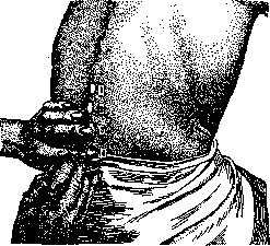
Figure 5. Spinal anesthesia; insertion of the needle between the 3rd and 4th lumbar vertebrae.
it is mixed with a warm solution of the anesthetic substance and slowly injected back. After injection, the needle is quickly withdrawn, and the opening is sealed with adhesive tape or collodion. The position of the patient after injection determines the degree of spread of A. and the speed of its onset. For operations in the pelvic area and on the extremities, a horizontal position of the patient with the upper part of the body slightly elevated and the head is quite sufficient. For "high A.", i.e., A. around and above the navel, the patient is placed in a high pelvic position. After injection, A. occurs in 6-10 minutes and lasts for about 1-2 hours. Failures occur in 9% of cases. During A., disturbances of the circulatory organs (weak pulse, pallor of the face, cold sweat, fainting) and respiratory organs (from mild shortness of breath to severe collapse and complete respiratory arrest) may be observed. Among the complications of spinal anesthesia observed in severe cases, mention should be made of vomiting after surgery, headaches, symptoms of irritation of the meninges, and rarely, prolonged paralysis of the lower extremities. Mortality from spinal anesthesia varies among different authors, averaging from one death per 500 cases to one death per several thousand cases. Due to the relatively high mortality and severe complications that sometimes occur after spinal A., it should be considered more dangerous than general anesthesia, not to mention the simple methods of local A.; therefore, the scope of its application in most clinics is not particularly large. It is more widely used in operative gynecology and in operations on the rectum. It is indicated in elderly and old people where local A. is necessary and general anesthesia is contraindicated. In these cases, spinal A. is an extremely valuable, sometimes indispensable method. Contraindications to spinal A. may be: 1) increased excitability of the nervous system, 2) presence of septic foci, 3) organic diseases of the central nervous system (brain tumors) and partly curvature of the spine. In children under 15 years of age, spinal A. should not be resorted to. In 1908, the Romanian surgeon Ionesc (Jonnesco) proposed general spinal A. (rachianesthesia general). He injects a solution of stovaine with strychnine into the spinal canal, and depending on the site of injection, obtains A. of all areas of the body, from head to toe (Stovain 0.005 to 0.06 + Strychn. sulfur. 0.0005 to 0.002). This method has not yet become widespread outside of Romania, and in any case, its application requires great caution. For completeness of presentation, it should be mentioned that there are some types of analgesia that partly belong to spinal A. and partly to local conduction, in which the areas of the pelvis, perineum and organs located in the pelvis (rectum, urethra, bladder, etc.) are anesthetized. These include: 1) epidural or sacral A. and 2) parasacral A. These methods have not yet found wide distribution. -Epidural, sacral, or caudal A. was developed by Lawen; with it, the anesthetic substance is introduced through the sacral hiatus into the sacral canal and the epidural space. This cavity is located between the wall of the bony vertebral canal and the dura mater; the injection is performed with the patient in the knee-elbow position. An injection is made into the epidural space, in which the spinal nerves pass, enveloped by the dura mater. Since the latter is dense and prevents direct action on the nerves, anesthetics of considerable concentration are required. Lawen injects 20 cubic cm of a 2% solution of novocaine with adrenaline. A. occurs slowly, after 15-20 minutes, anesthetizing the skin of the anus, perineum, scrotum, genital organs, and the lower part of the rectum.-Parasacral A. was introduced into practice by Brown. It is convenient and relatively simple in technique, completely safe, and sufficient for operations on the organs of the genital sphere and pelvis. The inner surface of the sacral bone is concave, on its anterior surface is located the sacral, pudendal, and coccygeal plexus. From here originate the ischiadic nerve, nerves of the gluteal region, nerves intended for the walls of the lower end of the trunk and for the pelvic organs. With parasacral A., the conductivity of these nerve plexuses is interrupted. When performing parasacral A., the patient is in the supine position with the thighs strongly bent, as in hemorrhoid operations. To the right and left of the coccygeal bone, at a distance of 1 1/2 - 2 cm, a deep injection of novocaine solution is made, guided by the sacral bone, the concave inner surface of which is filled with an anesthetic solution; about 80 cubic cm of a 0.5% solution of novocaine is used on each side. A. covers the anus, rectum, perineum, and urogenital organs, and is indicated for operations in these areas.
Related articles
Mentioned in
Cite this page
“Local Anesthesia.” Soviet Medical Encyclopedia. English translation of Bolshaya Meditsinskaya Entsiklopediya, 1st ed. (Moscow, 1928–1936), ed. N. A. Semashko. https://sovietmedicalencyclopedia.pages.dev/article/local-anesthesia/