Laminectomy
Historical document, translated for reference. It reflects medical knowledge of the 1920s–30s and is not medical advice.
Summary
Laminectomy is a surgical procedure to open the spinal canal, initially performed for spinal injuries and later expanded for various conditions including tumors, tuberculosis, and neurological disorders. The operation has become increasingly refined and widely applied, particularly after World War I due to spinal injuries.
Encyclopedia article (1928–1936)
LAMINECTOMY (from Latin lamina-plate and Greek ectomia-excision), an operation for opening the spinal canal; it serves as a preliminary step for any kind of operative procedures on the spinal cord, its membranes, or on the posterior surface of the vertebral bodies. The operation with complete removal was first performed by the Scottish surgeon Macewen (1886) for a fracture of the spine; in the following year (1887), the English surgeon Horsley by means of laminectomy successfully removed a tumor of the spinal cord. During the next 20 years, laminectomy remained a little-used operation, but in subsequent years and especially in the last 15 years, the operation has become very popular partly in connection with the world war, where it found the widest application due to the enormous number of injuries to the spine and spinal cord. Parallel with traumatic cases, the field of application of laminectomy expanded for other indications, and at present the number of such operations can be counted in the thousands. For its effectiveness, this operation is one of the most brilliant, quickly freeing patients from severe pain or restoring to those who seemed to be hopeless paralytics. For details on indications for this operation and its results-see Spine, Spinal Cord. The field of application of laminectomy is now already sufficiently extensive, and indications for the operation continue to gradually expand. The operation itself in its technique and safety has been so perfected that the question of its application as a diagnostic method ('trial laminectomy') is often raised in practice. The most frequent application of laminectomy is in injuries complicated by injury to the spinal cord and spine, especially by projectiles, or in fractures of vertebrae, where the operation aims to eliminate compression of the spinal cord by bone fragments, projectiles, blood clots, or displaced vertebral bodies; but here laminectomy is sometimes performed atypically, and often the matter is limited only to the exposure of broken arches and removal of displaced bone fragments or opening of the spinal canal for drainage in infected injuries. New growths developing from the bodies of vertebrae, from the tissue of the spinal canal, membranes, or the substance of the spinal cord itself constitute the second most frequent area of application for laminectomy. Tuberculous lesions of the spine causing compression of the spinal cord by cold abscesses or tuberculous granulomas can also be included here. The next group of cases for laminectomy is given by those forms of diseases in which it is necessary to sever the sensory roots of the spinal cord, such as persistent neuralgic pains in the extremities, certain types of spastic paralyses, tabetic crises, etc. The technique of laminectomy consists of two moments: 1) laminectomy proper and 2) special subsequent procedures determined by the main indication and peculiarities of the individual case. The operation is usually performed under general anesthesia, but in many cases it is also possible to use local infiltrative anesthesia with a 1/2% solution of novocain with adrenaline; however, moments associated with manipulation on the spinal nerve roots and on the spinal cord itself are painful, and additionally, lubrication with a novocain solution should be applied or one should switch to general anesthesia. The position of the patient during the operation is usually on the stomach (more convenient on the right), and an attempt is made to keep the spine curved backward as much as possible to increase the divergence of the spinous processes. The prone position gives some advantage only in operations on the cervical region. Some surgeons, using local anesthesia, operate on patients in a sitting position in order to avoid the accumulation of blood and spinal fluid in the depth of the wound, but these considerations are of no great importance, since the operation is usually not accompanied by any significant bleeding.
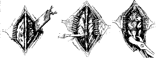
Figure 1.
Figure 2.
Figure 3. 481
LAMINECTOMY
482 not having great significance in details. The simplest method is as follows: above and below the spinous processes marked for removal, the interspinous ligaments are incised with a scalpel (Figure 2), and then the spinous processes are bitten off at their base with curved Liston forceps (Figure 3). Removal of the arches is simplest, fastest, and least traumatic when performed with Luers bone forceps (Figure 4). Special 'laminectomies' find less application. The most important thing is to make the first 'window' through the lowest located arch, for which it is necessary to make an incision with a scalpel along the lower edge of the arch, separate the soft tissues from the lower edge of the arch and its anterior surface with a knee-shaped elevator, and then the arch is bitten out through this slit. After the window is made, its further expansion along the entire length no longer encounters difficulties and can be performed very quickly. Backward, the posterior arches are bitten off up to the articular or transverse processes (Fig. 5) or even with part of the first. In the cervical part, provided accurate localization of the
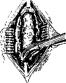
Figure 4.
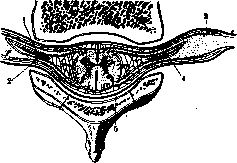
Figure 5. Schematic section of the spinal cord with a vertebra: 1 and 2-anterior and posterior root; 3-dura mater; 4-space between the roots; 5-epidural space.
process, due to the width of the spinal canal, it is sometimes possible to limit oneself to excising only half of the lamina (hemic laminectomy); this is sometimes possible in other parts of the vertebral canal under special conditions and indications, for example for removing foreign bodies, severing roots (Taylor); but as a rule the entire lamina is removed in order to allow for a free examination and investigation of the underlying part of the spinal cord. In the most favorable cases with precisely localized pathology, it may be sufficient to remove two laminae, but more often 3 to 5 and sometimes even more need to be excised. Bleeding from bone is usually negligible; only in rare cases of pathological vascular development can it present a significant obstacle to further surgery and in exceptional cases even forces the interruption of the operation and its completion in a second stage after an interval of several days. To stop bone bleeding, one resorts to chiseling the bone with a blunt chisel, compressing it with sequestration forceps, or pressing wax or Stens' mass into the bone. If bleeding does not stop completely by the time the wound is being sutured, a piece of muscle taken at that moment is applied to the bleeding sites. During operations in the region of the upper thoracic vertebrae, fatal outcomes have been observed on the operating table due to air embolism from air entering through the anastomoses of the veins of the spinal canal with the v. azygos. The epidural fat with the venous network contained within it is bluntly shifted away from the dura mater in both directions. If during the further course of the operation it becomes necessary to open the dura mater, this is done along the midline through a small initial incision in the membrane, which is then enlarged using a grooved probe or with blunt-tipped scissors (Fig. 6). Rapid outflow of cerebrospinal fluid can cause a drop in blood pressure and shock phenomena; to avoid this, one should initially make only a small hole in the membranes and tilt the table with the head end downward. At the upper end of the wound, the dura mater can be pressed with a small tampon, and these measures are usually sufficient to avoid the danger of rapid and profuse fluid outflow. Osteoplastic methods of L. consist in that they preserve either the entire spinous processes and laminae or only the spinous processes, while the laminae are removed in the usual manner. In the first method, the operation is performed by various surgeons with diverse modifications differing from each other in the form of the skin incision, the method of separating the laminae, but the basic principle consists in removing the posterior wall of the spinal canal with one single flap, which at the end of the operation is placed back and fixed with sutures. An intermediate position between these methods and the ordinary method of L. is occupied by the method of Cavicchia, proposed as early as 1898 and consisting of the following: an incision of the skin coverings as in ordinary L., then an incision through the aponeurosis and muscles down to the bone, detachment of the muscles on both sides of the spinous processes and exposure of the laminae; then with the help of broad bone forceps the spinous processes are severed at their base and the entire chain, connected with each other by ligaments (lig. interspinalia), is shifted to one side, after which the laminae of the vertebrae are removed in the usual manner. Alessandri modified this method in that he makes the incision through the muscles on one side of the spinous processes and, after detaching the muscles from the laminae, severs the spinous processes at their base and then separates the muscles from the laminae on the opposite side with a periosteal elevator, after which the entire chain of spinous processes is deflected to the side; the operation is then completed in the usual manner. At present, osteoplastic methods in all their modifications have lost their significance and are almost not used due to their complexity and painstaking execution without substantial benefits. The flap itself restricts the operator, narrows the operative field, and complicates the extension of the wound upward or downward if that should become necessary. Meanwhile, clinical experience has shown that complete removal of 3-5 and even 6-7 laminae does not substantially affect the strength of the vertebral column, does not cause sequential curvatures, and does not affect statics. Only in operations in the cervical region should one avoid excessive trauma to the muscles and their supplying nerve branches and too wide excision of the laminae. After L. has been performed, all further operative procedures depend on the indications and nature of the process and are described in the respective sections. Here it is necessary to note several more aspects of a general nature. Each time L. is performed, it is necessary to orient oneself with the utmost precision regarding the level of the lesion, i.e., the segment, based on neurological and other data; once the segment is determined, it must be precisely projected onto the skin, i.e., one must calculate which vertebra it corresponds to and mark the corresponding spinous process on the skin. Extreme care must be taken when counting the vertebrae; an error in this respect leads to L. being performed at an improper level and consequently the lesion is not exposed at the site where L. was done, and one has to extend the wound upward or downward, i.e., remove additional laminae; in some cases due to incorrectly localized pathology or an error in counting the vertebrae, the operation remains unfinished or is performed in vain. Nevertheless, despite all precautions, inaccuracies may occur either in the diagnosis of the nature of the disease, or in topographic diagnosis, or direct errors of other kinds, due to which no visible lesion is found at the site where L. was performed. Then it is necessary to perform additional examinations in situ: the spinal canal is probed both upward and downward before opening the dura mater with a flexible metal probe or Nelaton's catheter; to examine the lateral and anterior surfaces of the canal, the spinal cord, still covered by the dura mater, is shifted to one side or the other and slightly elevated with curved spatulas. If even then no lesion is found, the meninges are opened and if necessary here again the same manipulations are performed on the exposed spinal cord, i.e., the subdural space is probed and the lateral and anterior surfaces of the spinal cord are examined, elevating it on spatulas and rotating it on its axis after first grasping the ligamentum denticulum with forceps.
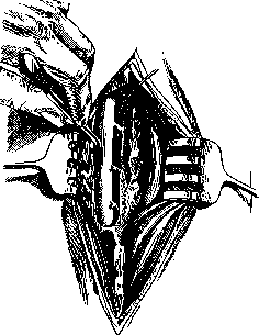
Fig. 7. Operation for laminectomy for severing the roots of the spinal cord.
of the brain, one or two posterior roots can be severed, and in the thoracic section, the anterior roots as well, which does not lead to significant subsequent paresis (Fig. 7). At the end of the operation, before proceeding to suture the dura mater, it is necessary to remove all blood clots, as their presence can subsequently cause irritation of the membranes and increased transudation of fluid or contribute to the development of adhesions. Some surgeons resort to careful washing of the subarachnoid space with physiol. solution through a thin, soft catheter. Suturing the dura mater is almost always necessary except in special cases and indications. The suture should be placed frequently, preferably continuous, with fine silk. With a loosely placed suture, subsequent leakage of cerebrospinal fluid may occur, which accumulates in the depths of the wound, then under the skin in the form of a fluctuating swelling, and finally may lead to an external fistula, sometimes with abundant and prolonged discharge; the danger of the fistula lies not so much in the loss of fluid as in the possibility of infection and the development of meningitis. Therefore, such subcutaneous accumulations should never be opened; in most cases they resolve on their own or can be emptied by aspiration with a syringe through a puncture made away from the scar. - If during the operation it was necessary to remove part of the dura mater, then when suturing, attention should be paid to not pulling the membrane too tightly, as this will cause either rupture of the sutures and separation of the edges or compression of the spinal cord due to accumulation of cerebrospinal fluid above. With large defects of the dura mater, one can resort to free plasty with fascia taken from right above the back muscles. The muscle wound is sutured tightly so that no gaps remain, with deep catgut sutures. For hermeticism and tight apposition of the muscle flaps, it is additionally recommended to place several removable wire sutures, tightened at the end over rollers. Draining the wound in aseptic cases is contraindicated and only harmful. After a blind skin suture, the wound is covered with a narrow strip of gauze, the edges of which are glued with collodion, mastisol, gutta-percha putty, or adhesive tape. After the operation, the patient is placed horizontally in bed on his back, but after coming out of anesthesia, he can, and it is even better, be turned onto his stomach, placing pillows under the pelvis, abdomen, and chest, or alternately kept on the right and left sides. In this position, it is easier to avoid subsequent leakage of fluid and the formation of bedsores; existing bedsores are more accessible for care, and changing the dressing is facilitated. - The normal method of L. is a one-stage operation. Only in exceptional cases is the operation performed in two stages: this is necessitated either by shock phenomena that have already developed in the first moment, or by profuse bleeding, or other unexpected complications during anesthesia; further - uncertainty about the aseptic state of the wound (in trauma cases). Finally, the two-stage method is sometimes used when removing spinal cord tumors.
Related articles
Mentioned in
Cite this page
“Laminectomy.” Soviet Medical Encyclopedia. English translation of Bolshaya Meditsinskaya Entsiklopediya, 1st ed. (Moscow, 1928–1936), ed. N. A. Semashko. https://sovietmedicalencyclopedia.pages.dev/article/laminectomy/