SPINELLI
Historical document, translated for reference. It reflects medical knowledge of the 1920s–30s and is not medical advice.
Summary
Spinelli (Pier Giuseppe Spinelli, 1862-1929) was an prominent Italian gynecologist and brilliant surgeon, one of the pioneers of operative gynecology. He was a former assistant to the renowned Morisani and left a significant literary legacy, particularly known for his works on tuberculosis of the female reproductive sphere, ectopic pregnancy, and uterine inversion.
Encyclopedia article (1928–1936)
SPINELLI (Pier Giuseppe Spinelli, 1862-1929), prominent Italian gynecologist, brilliant surgeon, one of the pioneers of operative gynecology, former assistant to the renowned Morisani. Spinelli received his medical education in Naples, where from 1900 until his death he headed the gynecological department at Ospedale di S.M. della Pace. He left a large literary legacy; among his works, the most famous are those on tuberculosis of the female reproductive sphere, ectopic pregnancy, and uterine inversion. Spinelli was one of the first to persistently point out the need for social struggle with syphilis. SPINAL CORD. Contents: I. Phylogenesis.....................279 II. Ontogenesis.....................284 III. Anatomy.....................284 IV. Physiology..............t......302 V. Pathology.....................310 VI. Surgery.....................321 Spinal cord (medulla spinalis), the posterior, or lower part of the central nervous system, located in the vertebral canal. I. Phylogenesis. The spinal cord progressively develops at various stages of the animal kingdom. At the most primitive stage of development in Amphioxus, it has the shape of an irregularly shaped tube (Fig. 1), in the center of which passes the central canal. The anterior and posterior roots, which do not arise at the same level of their peripheral branches, branch strongly. The initial cells of the anterior roots are not yet localized with precision, but lie not at the level of the exit of the roots. The posterior roots contain fibers of three types-somatic sensory (for the skin), visceral sensory (for mucous membranes) and motor fibers (for visceral musculature). The cells of the posterior roots are bipolar and lie partially intramedullary, partially on the extramedullary segment of the root; at the entrance to the roots are cells, the axons of which form the second sensory neuron. In cyclostomes, the cross-section of the spinal cord has an oval shape, as does the central canal; the anterior and posterior roots also alternate2-(Fig. 2). The gray matter surrounds the canal and corresponds to the mixed anterior and posterior roots (Fig. 3); the gray matter contains cells that give rise to the anterior roots. The posterior root also contains three types of fibers-somatic sensory, visceral sensory, visceral motor; part of the root fibers originate from intramedullary bipolar cells, and the rest from cells lying outside the brain matter. The central process of these cells goes to the dorso-lateral area of the spinal cord and sends collaterals into the gray matter. The location of visceral motor fiber cells is precisely unknown. The secondary systems of the spinal cord are better developed
than in Amphioxus: ventral arcuate fibers of secondary sensory paths, going in the anterior columns, short intersegmental fibers, short ascending paths to cells of the reticular substance of the medulla oblongata 6^
„
4 5 'Д S n/ Figure 4. Cross-section of the spinal cord of Plagiostomes: 1-posterior root fibers; 2-posterior column; 3-central fibers; 4 and 6-anterior roots; 5-anterior columns; 7-tractus Oct. spinalis lateral, cruciata; *-lateral column; 9-marginal plexus of dendrites; 10-ascending and descending fibers of posterior roots. and finally the system of fibers formed by Müllerian fibers, going from the frontal zone of the brain through the entire spinal cord. The fibers are devoid of myelin. The spinal cord of Plagiostomes is the prototype of the spinal cord of higher animals, its fibers are surrounded by myelin, and the gray matter has a structure similar to that of higher animals (Fig. 4), the anterior and posterior roots connect and give fibers to the viscera and to the vessels; there is no sympathetic border column yet, and there are also no cervical and lumbar enlargements. The posterior sensory part of the spinal cord differs from the anterior by the arrangement of the posterior horns, which connect with each other, while the anterior ones are clearly delimited. In the anterior roots, in addition to somatic motor fibers, there are visceral motor fibers; the posterior roots contain the same fibers as in cyclostomes; they originate from spinal ganglia, the central processes partly end in the posterior horns, and partly go in the spinal cord in an ascending direction. The cells of the anterior horns connect with fibers or collaterals by dendrites, forming a reflex arc. There is a decussation of secondary sensory neurons (tr. spino-bulbaris, spino-mesencephalicus); on the same side goes tr. spinocerebellaris. The spinal cord is under the influence of higher centers. In fish (Teleostei) the spinal cord is somewhat reduced (in cross-section and in length) compared to Plagiostomes. They have a true sympathetic border column. The cells of the anterior horns are located closer to the anterior surface
(Fig. 5), their dendrites, as in previous animals, form a plexus on the lateral surface of the spinal cord. In the anterior roots, in addition to somatic motor fibers, there are motor fibers to the viscera. In the posterior roots, the same fibers as in previous ones; they originate not only from spinal ganglia, but also from cells of the spinal cord. The central processes of cells go in the lateral columns, which is why the posterior columns
anterior roots; 4-lateral column.
are very small. Ascending endogenous fibers conducting sensitivity, homo- and hetero-lateral (tract, spino-bulbaris, spino-mesencephalicus, spino-cerebellaris). - In amphibians (Amphibien) the spinal cord is very reduced
Figure 8. Cross-section of the spinal cord of Reptiles (schematic representation of cells and fibers): 1-fasc. dors.; 2-posterior horn; 3-posterior root; 4-lateral horn; 5-marginal plexus of dendrites; 6-commissura ant.; 7-sensory-motor collaterals.
compared to the spinal cord of Plagiostomes (Fig. 6); the number of spinal nerves in tailless amphibians is 10-11 (while in a shark about 100), in tailed amphibians there are more nerves, since the tail is also innervated. For the first time, cervical and lumbar enlargements appear. In the anterior horns, two groups of cells can be distinguished: one, internal, extends throughout the spinal cord, the other, external,-only at the levels of the enlargements. The cells give rise to the anterior roots; the dendrites of cells, forming plexuses, cover the surface of the spinal cord. The posterior roots originate from cells of spinal ganglia (in the embryo from intramedullary cells); their central process forms two bundles: one is located in the marginal zone of the posterior horns, the other-in the posterior columns; the reflex arc is carried out by collaterals from the fibers of the posterior columns, ending at the cells of the anterior horns. From the secondary paths, crossed sensory paths are known (tract, spino-bulbaris, spino-meseneephalicus), in addition to uncrossed spino-cerebellaris. The short paths are the same as in fish. From the descending fibers, the roots of nn. V-VI-VIII-X enter the spinal cord; the root of n. V extends to the lumbar segment.
The spinal cord of reptiles (Reptilien) can be considered as a preliminary stage in the structure of the spinal cord of mammals (Fig. 7). It differs from the spinal cord of a frog (amphibian) in that it fills the entire vertebral canal, which is associated with the existence of metamerous musculature of the tail. In this class, three types of spinal cord can be distinguished depending on the development of musculature: 1) in a lizard, the musculature of the back and limbs is well developed, the spinal cord has enlargements; 2) in snakes, only the back musculature is developed, there are no enlargements in the spinal cord; 3) in a turtle, the back musculature is not developed at all, only that of the neck, limbs and tail, the spinal cord in the thoracic part is very thin. In the anterior horns of the cervical region, in addition to motor cells, there are also sympathetic cells. Figure 9. General view of the spinal cord of an ostrich: 1 and 7-sulcus mfdianus post; 2-posterior roots; 3-posterior column; 4-lumbo-sacral sinus (rhomboid sinus); 6-sulcus lateralis; 6-sulc. lateralis pest.; 8-14-anterior roots. The marginal plexus covering the periphery of the spinal cord is very reduced, in its place appear marginal cells. The posterior roots originate from spinal ganglia, the number of fibers in them is increased; the ascending central process goes in the posterior columns in a lizard and in the lateral-in snakes and turtles. For the first time, nuclei of the posterior columns appear (Fig. 8), which send fibers to the medial
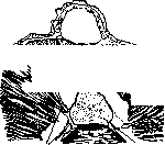
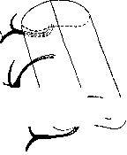
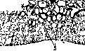

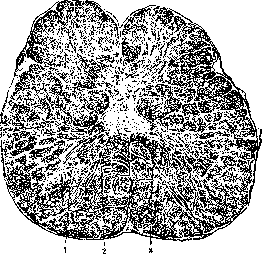
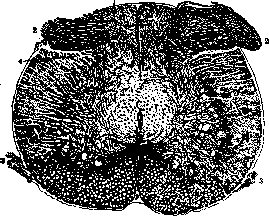
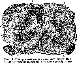
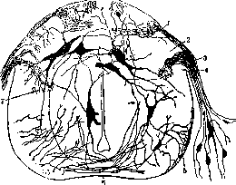

Figure 10. Cross-section of the spinal cord of birds: 1-posterior horn; 2-cells of the anterior horn. The complication of the central sensory apparatus occurs in parallel with the development of the peripheral sensory apparatus, which depends on the way of life. In all other respects, the same structure as in amphibians. The spinal cord of birds (Aves) differs from the spinal cord of reptiles by the greater length of the cervical region, the presence of a lumbosacral sinus between the posterior columns (Fig. 9). The enlargements are very well developed; in flying birds, the lumbosacral enlargement is better developed; the anterior roots and horns are better developed than the posterior ones; in the anterior horns, the internal and external groups of cells are well developed (Fig. 10). The marginal plexus is weakly expressed, in its place the number of marginal nuclei is increased. The posterior roots originate in the spinal ganglia and their fibers in the spinal cord go in the posterior columns and in the marginal zone; due to the small size of the posterior roots, the posterior columns are rather poorly developed. The nuclei of the posterior columns are small. Endogenous fibers are more numerous than in reptiles, especially short intrasegmental fibers. Tractus spino-bulbaris and spino-mesencephalicus are very well developed, the size of tr. spino-cerebellaris is also increased; descending bundles tractus cerebro-spinalis, tr. vestibulo-spinalis, rubro-spinalis, tr. tecto-spinalis appear, they pass in the anterior-lateral column. The spinal cord of birds is greatly influenced by higher centers. In mammals, in general, the structure of the spinal cord corresponds to the structure of the human spinal cord, there are only minor changes in details depending on the presence of a tail, on the way of life, on the absence of lower limbs. For example, in a whale due to the absence of lower limbs there is no lumbar enlargement; it is very well developed in a kangaroo. The nucleus and bundle of Goll are less developed in animals deprived of lower limbs. The size and length of some bundles also change. The pyramidal bundles gradually increase: in dogs they make up 10% of all the white matter of the spinal cord, in monkeys 20% and in humans 30%. II. Ontogenesis. The rudiment of the central nervous system is the neural or medullary plate - a long and wide strip of ectodermal cells, consisting of two halves, divided by a neural groove (Fig. 11); the edges of the plate rise, come towards each other and connect, due to which first a neural groove is formed, and then a medullary, or neural, plate
j medullary plate; e - medullary, or neural groove. 4-provertebrae; b-chor-
da dorsalis; b-neural groove; _?-neural groove; 2-ganglionic plate-
covering the surface
of the body; at the place of pe-
retraction emerges a
groove; 2-entodermal plate; 3-ectodermal plate; 4 - double mesodermal
plate. j-cardiac pit; ganglionic plate, giving rise to
ganglia com-
plexes, which divide into ganglia spinalia and ganglia sympathica. The medullary plate, groove and tube lie on the chorda dorsalis. The closure of the neural groove begins at the level of the midbrain, and last of all occurs at the level of the spinal cord. In the early period of development, the medullary tube is in communication with the epidermal tube and with the gastral-canalis neurentericus.
Cross-section 288 On the fourth week of development, the spinal cord in the embryo is formed by two thick lateral walls (Fig. 13), connected above and below by poorly developed commissures. The longitudinal lateral groove divides the lateral walls into two plates: the anterior-fundamental (lame fondamentale de
Figure 12. Cross-section of the medullary plate at a later stage of development: 1-medullary tube; 2-intervertebral node; 3-pharynx; 4-somato-peritoneal cavity; 5-sepi-pericardial part; 6-primordia of heart cells; 7-ectodermal pocket of the lower surface of the head. His), which gives rise to all motor nerves, and the posterior (lame alaire de His), which receives sensory fibers. The ventral commissure connects the fundamental plates, and the dorsal commissure connects the posterior ones. The cavity of the spinal cord has the shape of a slit, elongated in the anterior-posterior direction. In the second month of development, the lateral walls bend, due to which the shape of the central canal changes (Fig. 14); in it one can note 4 grooves-anterior and posterior, very narrow, and lateral grooves, wider. The anterior and posterior parts of the wall are connected by an intermediate plate. The anterior plate is bounded by a cylindrical groove, from which the anterior and lateral horns, the anterior
Figure 13. Cross-section
of the colum n of the spinal cord of a 4-week human embryo: 1- part of the lateral horns, posterior plate; 2-anterior plate; 3-ventral commissure; 4-motor roots; 5-meningeal border partition; 3 forms the posterior horns and the root zone of white matter (root zone); 9-place of dorsal commissure; and the root zone (cordon ovulaire de His). The intermediate plate, located between the cylindrical groove and the marginal, corresponds to the wide part of the canal; it gives rise to the neck of the posterior horn, Clark's column, processus reticularis and the posterior part of the lateral column; in this area later appears the pyramidal crossed bundle and the bundles of Flexig. With the increase of gray matter of the spinal cord, sending fibers to the columns, the anterior-lateral columns also increase, which in development outpace the posterior columns, developing only by the end of the 2nd month; at the same time changes occur in the central canal, which narrows and occupies the central part of the spinal cord and connects with the periphery along the posterior surface of septi postici (Fig. 15). The development of gray and white sub-
stances does not occur simultaneously throughout the spinal cord, but from top to bottom: the cervical region in development is ahead of the lower parts of the spinal cord. During the 2nd and 3rd months of intrauterine life, the spinal cord occupies the entire spine, and starting from the 4th, the spine develops faster, due to which the segments of the spinal cord do not retain relations with the corresponding vertebrae; on the 6th month, the end of the spinal cord reaches the sacrum, and at birth to the III lumbar vertebra. The roots and membranes grow, like the spine, due to which the lower roots depart from the spinal cord not at a right angle, as in the cervical region, but with a downward and outward inclination, and this inclination is the greater, the lower the roots. All fibers of the spinal cord are covered with myelin not simultaneously, but in a strict sequence. On the 5th month of intrauterine life, the fibers of the main bundle are covered with myelin. Starting from the 6th month,
Figure 14.
Figure 15.
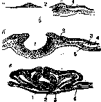
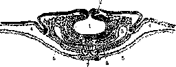
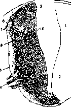
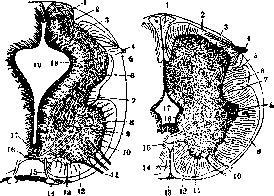
Figure 14. Cross-section of the spinal cord of a human embryo 41/2 weeks, 12.5 mm in length (thoracic region); 1 - outer gray lamina; 2 - inner gray lamina; 3 - root zone; 4 - posterior, sensory roots; 5 - marginal groove; 6 - intermediate lamina; 7 - cylindrical groove; 8 - lateral horns; 9 - arcuate fibers; 10 - anterolateral column; 11 - anterior roots; 12 - anterior column; 13 - a. spinalis ant.; 14 - groove artery; 15 - anterior commissure; 16 - base of the central canal; 17 - ependyma; 18 - central canal. Figure 15. Cross-section of the upper thoracic region of the spinal cord of a 3-month human embryo: 1 - Goll's tract; 2 - Burdach's tract; 3 - posterior horn; 4 - posterior, sensory root; 5 - marginal groove; 6 - lateral column; 7 - cylindrical groove; 8 - lateral horn; 9 - anterolateral column; 10 - anterior, motor root; 11 - anterior horn; 12 - anterior column; 13 - a. spinalis ant.; 14 - anterior median groove; 15 - anterior commissure; 16 - ependyma; 17 - central canal. Myelin appears in the fibers of the flexor tract, while in the Govers tract the fibers become myelinated later and in the newborn are not yet fully covered with myelin. The fibers of Goll's and Burdach's tracts do not become myelinated simultaneously; in Goll's tract it appears later. The fibers of the pyramidal tract are covered with myelin last (even in the newborn it is still poor in myelin), the development of which ends in the 2nd year of life.
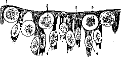
Histogenesis. At the moment of development, the medullary groove or tube consists of cylindrical epithelium, the neuroepithelial cells of which are arranged in a single layer. The cells are located between two partitions (membr. limitans externa and membr. limitans interna), which limit the cavity of the central canal. The cells consist of ectoplasm and endoplasm containing a nucleus, which lies closer to the outer partition; near the inner partition 'among the epithelial cells there are very large round cells, lying at some distance from each other; they have very large nuclei, some in a state of rest, others in a state of karyokinesis; these are cellulae germinativae (Keimzellen de His); these cells besides the neural plate are also in the ganglionic plate. The epithelial cells multiply rapidly and due to this they are compressed and stretched, the nuclei begin to be placed at different levels, thereby giving a multilayered appearance to the lateral wall (fig. 16), but in reality the layer remains one, and each cell retains connection with both partitions; in this layer three parts, or zones, can be distinguished: the middle - nuclear zone, and two lateral, devoid of nuclei: internal - columnar zone, in which the germinal cells are located; it forms the supporting framework for the gray matter; outer - marginal zone, forming supporting tissue for the white matter.
The number of germinal cells in a very small embryo (4 weeks) is very large, in places they form a continuous row, later their number decreases. Their size varies from 10 to 14 μ. Epithelial and germinal cells give rise to the elements of the nervous system: epithelial cells turn into spongioblasts, forming the ependyma, and from germinal cells develop neuroblasts - nerve cells and neuroglia. Neuroblasts have a pear-shaped form, and on the thin end the axial cylinder develops by the displacement of protoplasm from the cell. Neuroblasts partially migrate into the deeper layers of the wall.
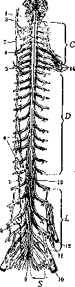
Figure 16. Internal spongioblastic and germinal cells: 1 - membrana limitans externa; 2 - germinal cell; 3 - columnar zone; 4 - spongioblasts.
III. Anatomy. The spinal cord above without sharp boundaries passes into the medulla oblongata, below it ends in a cone (conus medullaris), from which a thread-like continuation (filum terminale) extends (fig. 17). In filum terminale there are 2 parts - internum and externum. Fil. terminale internum, 16 cm in length, is located inside the sac of the dura mater between the lumbar and sacral roots, extends to the II sacral vertebra. Fil. terminale externum, 8 cm in length, is surrounded by the dense dura mater (vagina terminalis), which forms filum durae matris spinalis and ends in an expansion on the II coccygeal vertebra. The upper end of the spinal cord is considered the place of exit of the first cervical roots and the beginning of the decussation of the pyramidal fibers. Along its entire length, nerves originate from the spinal cord, beginning with two roots - the anterior motor (radix anterior medullae spinalis) and the posterior sensory (radix posterior); both roots of the spinal cord, connecting, form the spinal nerves, which exit through the intervertebral foramina.
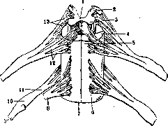
The spinal cord has a symmetrical cylindrical shape, flattened in the dorsoventral direction, especially on the anterior surface. Along its length there are two bends - cervical and thoracic, merging into each other at the level of the VII cervical vertebra. The spinal cord is divided into departments or parts (pars): cervical (pars cervicalis medull. spinalis), thoracic (pars thoracalis), lumbar (p. lumbalis), sacral (p. sacralis) and coccygeal (p. coccygea). Each department in turn is divided into segments, of which there are 31-33. A segment is a portion of the spinal cord (fig. 18) giving rise to two pairs of roots; thus the number of segments corresponds to the number of roots: in the cervical department there are 8 segments, in the thoracic - 12, in the lumbar - 5, in the sacral - 5 and in the coccygeal - 1-3 segments. The first cervical nerve leaves the vertebral canal between the occipital bone and the I cervical vertebra, the second between the I and II cervical vertebrae, etc. Since the spinal cord is shorter than the spine (it ends at the level of the II lumbar vertebra), the roots must pass a more or less long distance in the vertebral canal (fig. 19) to reach their exit opening; this distance is particularly significant for the lumbar, sacral and coccygeal roots; together with filum terminale these roots form the so-called 'horse's tail' (cauda equina).
The spinal cord does not maintain the same thickness, width and shape throughout its entire length; in those places where large nerves for the limbs depart from the spinal cord, there are spindle-shaped thickenings; the upper, cervical Figure 17. Spinal cord, above connected to the medulla oblongata: 1 - p. accessories spinalis; 2 - border column; 3,4,5 - upper, middle and lower cervical sympathetic nodes; 6 - thoracic sympathetic nodes; 7 - lumbar sympathetic nodes; 8 - rami communicantes; 9 - filum terminale; 10 - n. ischiadicus; 11 - n. obturatorius; 12 - n. cruralis; 13 - conus terminalis and beginning of filum terminale; 14 - plexus brachialis. C, D, L, S - cervical, thoracic, lumbar and sacral roots. thickening (intumescentia cervicalis) is formed by the four lower cervical segments and the first thoracic; the lower, lumbar thickening (intumescentia lumbalis) includes lumbar segments from II to V and sacral I and II. On the spinal cord there are several longitudinal grooves (fig. 17,18 and 20); the anterior longitudinal groove (fissura mediana ant.) penetrates to a depth of 4 mm, lies on the anterior surface, extends upward to the foramen caecum
spinal cord
of men and 41-42 cm in women. The transverse diameter in the thoracic region is 10 mm, the sagittal is 8 mm; in the widest part of the cervical thickening 13-14 mm, in the lumbar thickening 12 mm. The weight of the spinal cord is 34-38 g; specific gravity 1,034, volume 39 cm3. Topography. In relation to the skeleton, the upper end of the spinal cord corresponds to the upper edge of the 1st cervical vertebra, and the conus terminalis to the upper edge of the 2nd lumbar vertebra. The cervical thickening begins at the level of the 3rd cervical vertebra and ends at the 2nd thoracic. The lumbar thickening begins in the region of the 10th thoracic vertebra, the 12th thoracic corresponds to the widest part of the thickening. Structure of the spinal cord. On cross-sections of the spinal cord, it can be seen that it consists of two symmetrical halves, connected in the middle by commissures. In the spinal cord, two substances are distinguished - gray and white (substantia grisea and substantia alba) (fig. 20); the gray matter lies in the center and is surrounded by white matter. The gray matter
12 11
Figure 20. Cross-section of the spinal cord: 1- fissura mediana post.; 2-septum posticum; 3-posterior columns; 4-commissura alba post.; 5-apex; 6-cervix of the posterior horn; 7-lateral horn; 8-lateral column; 9-commissura grisea; 10-commissura alba ant.; 11-anterior column; 12-fissura mediana ant.; 13-anterior horn; 14-sulcus lateralis ant.; 15-formatio reticularis; 16-column of Clark; 17-substantia gelatinosa Rolandi; 18-sulcus lateralis post.; 19-central canal. forms paired projections, horns (cornua), syn. gray columns or columns (funiculi cinerei, s. columnae griseae). The large projection of gray matter, turned toward the anterior surface, forms the anterior horns (cornu anterius, s. columna anterior, s. funiculus cinereus anterior), its posterior part passes into the posterior horn (cornu posterior, s. columna posterior, s. funiculus cinereus posterior); the posterior horn first narrows into the neck (cervix columnae posterioris), and further back widens into the head (caput columnae posterioris) and finally narrows again into the apex (apex cornu posterioris), which corresponds to sulcus lateralis posterior; at the apex is substantia gelatinosa columnae posterioris Rolandi, s. substantia gelatinosa Rolandi, to which posteriorly is attached a narrow layer of zona spongiosa, or the belt layer of the posterior column. In addition to the anterior and posterior horns, at some levels of the spinal cord (thoracic region), the lateral horns (columnae laterales) are also well expressed, which depart from the base of the anterior horn; from the lateral horn to the posterior horn through the lateral columns go gray matter crossbars, forming the forma or processus reticularis. The gray matter of both halves of the spinal cord is connected by a bridge, a isthmus of gray matter (commissura grisea), in the center of which is the central or spinal canal (canalis centralis medullae spinalis), lined with ependyma. The shape of the central canal is very varied - slit-like, round, oval, triangular; sometimes the canal is filled with ependymal cells; it is surrounded by gelatinous substance (substantia gelatinosa centralis). Closer to the medulla oblongata, the central canal turns into a sagittal fissure. In the conus medullaris, the canal approaches the posterior central sulcus and at the end of the cone expands into the ventriculus terminalis Krause. In front of commissura grisea is the anterior commissure, or commissure (commissura alba anterior), and behind it the posterior commissure (commissura alba posterior), formed by a small number of fibers. The white matter of the spinal cord consists of longitudinal ascending and descending myelin fibers, separated by partitions of neuroglia; in the transverse direction go, t
bundles of roots, fibers in commissures and fibers or their collaterals, ending in the gray matter. The white \ matter of the spinal cord is divided _ into columns or funi-";.g __ culi: the anterior column lies between fissura mediana anterior and the anterior roots; the lateral column - between the anterior and posterior horns and the posterior column - between the posterior horn and sulcus medianus posterior and its continuation in depth to commissura alba posterior-septum posticum. Figure 21. groups of cells in the gray matter of the spinal cord of the thoracic region (Г),,). 1- the first group of cells of the anterior horn; 2- the second group of cells of the anterior horn; 3- the third group of cells of the anterior horn; 4- the fourth group of cells of the anterior horn; 5- the fifth group of cells of the anterior horn; 6- the sixth group of cells of the anterior horn; 7- the seventh group of cells of the anterior horn; 8- cells of the posterior horn; 9- nucleus sympathicus lateralis sup.,- 4- white matter; in the thoracic region the size of the anterior horns decreases, the figure of the gray column resembles the letter "H"; in the lumbar-sacral region not only the anterior but also the posterior horns increase and their shape has great similarity with a butterfly (see separate table, pp. 295-296). The spinal cord is formed by nerve cells and fibers, surrounded by a supporting framework consisting of two tissues - connective tissue processes of pia mater and neuroglia (see). Neuroglia does not have the same appearance everywhere; three of its types can be distinguished: ependyma (see), neuroglia of gray matter and neuroglia of white matter. In the gray matter - a large number of fibers and cells of neuroglia; in the white matter they have a more regular arrangement between longitudinally running nerve fibers; on the outer surface of the white matter, neuroglia forms a thin dense layer - the cortical layer, the subpial layer of Waldeyer, covered on the outside by pia mater. The nerve cells of the spinal cord are scattered throughout the gray matter, in places forming accumulations or groups or nuclei of the spinal cord. The basis for the classification of nerve cells can be either their topographical location or the course and termination of their axon. Based on the topographical location of nerve cells, the following are distinguished (fig. 21): 1) Groups of the anterior horn - anterior and posterior medial group, anterior and posterior lateral; they extend throughout the entire spinal cord continuously to
Figure 22. Groups of cells in the gray matter of the cervical region of the spinal cord (CVI); 1-anterior-medial group; 2-middle group; 3-anterior-lateral group; 4-central group; 5-center of m. deltoidei; 6 and 7-outer and inner parts of the posterior group; 8-cells of the posterior horn; 9-commissura grisea. Ill sacral segment, at the level of the thickening other groups are added (fig. 22 and 23); the cells of these groups belong to the largest nerve cells, they reach a size of 100 μ; the group of cells at the base of the anterior horn, near the anterior commissure - tractus cellularum medio-ventralis (Jacobson's).
Figure 23. Groups of cells of the lumbar region of the spinal cord (L): 1-anterior-medial group of cells; 2-central group of cells (for m. semitendinosus); 3-anterior-lateral group (for m. glutaeus sup.; 4-group of cells for m. gastrocnemius; 5-group for m. tibialis anticus; 6-group for extensors of fingers; 7-group for flexors; 8-group for m. plantaris; 9-central canal; 10-central group of cells (for m. biceps femoris). 2) Group of the lateral horn (s. intermedius lateral. tract. Clarke's, s. nucleus sympathicus lateralis superior Jacobson's) consists of small elongated cells, which can be found in the processus reticularis; this nucleus is located between the VIII cervical segment and the II lumbar (fig. 21); another vegetative nucleus (nucleus sympathicus lateralis inferior) lies between the anterior and posterior horn from the II sacral segment to the coccygeal and the third (nucleus sympathicus medialis inferior)-on the inner wall of the posterior horn from the IV lumbar segment to the coccygeal. Between the posterior and lateral horn there are scattered cells (tractus cellularis intercommissuralis Jacobson's). 3) Clarke's cells (nucleus dorsalis, s. nucl. columnae Clarki, or column of Clarke) - a group of cells at the base of the posterior horn inward (fig. 21 and 24); very well expressed in the thoracic segments and the two upper lumbar. In other regions of the spinal cord - in the cervical, lumbar-sacral - in the corresponding places of the gray matter there are only individual cells - Stilling's cells, or nucleus dorsalis Stillingi; they are considered as a continuation of Clarke's column; the cells of this column contain pigment. 4) In the posterior horn, similarly large pigmented cells are partly scattered on the periphery
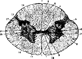

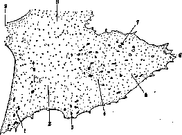
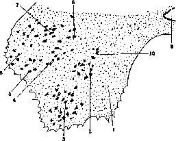
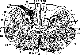
Figure 24. Cross-section of the spinal cord; schematic representation of the main cell groups and main bundles: 1-internal-anterior group of the anterior horn; 2-anterior roots; 3-external-anterior group of cells; 4-central group; 5-external-posterior group; 6-group of cells of Clark's column; 7-group of cells 'caput' of the posterior horn; 8-substantia gelatinosa; 9-stratum zonale; 10-main bundle of the posterior column; 11-oval field of Flechsig; 12-bundle of Goll; 13-comma of Schultze; 14-bundle of Burdach; 15-posterior root; 16-zona Lissaueri; 17-pyramidal crossed bundle; 18-bundle of Flechsig; 19-bundle of Monakow; 20-main bundle of the lateral column; 21-bundle of Gowers; 22-fasc. testo-spinalis along with fasc. vestibulo-spinalis and fasc. spino-reticularis post.; 23-fasc. Helwegi; 24-fasc. spino-reticularis ant.; 25-main bundle of the anterior column; 26-fasc. commissuralis; 27-direct pyramidal bundle; 28-fasc. sulco-marginalis. (cellulae marginales Waldeyeri, nucleus pericornualis Jacobsoni), and partly form the central group in the posterior horn (nucleus magnocellularis centralis cornus posterioris Jacobsoni). In substantia gelatinosa there are small nerve cells-Girke-Virchow bodies, and in zona spongiosa-individual marginal or border cells. According to their location and axon termination, nerve cells are divided into (fig. 25 and 26): 1) Root cells (cellulae radiculares) in the anterior-lateral parts of the anterior horns, multipolar in shape, the largest in size, motor in function; their axon cylinder is directed into the anterior root. 2) Commissural cells (cellulae commissurales), located in the internal parts of the anterior horn, in close proximity to the central canal; they are also in the posterior horn; their axon crosses through the commissure to the anterior column of the opposite side; they are thus commissural-columnar cells (van Gehuchten). Cells are described whose axon branches in the anterior horn of the opposite side-commissural cells with short axons, commissural cells of the 'type'
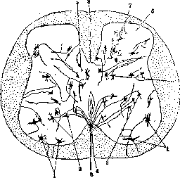
Figure 25. Cells of the spinal cord and the course of their axon cylinders: 1-root cells; 2-commissural cells; 3-commissura ant.; 4-fissura mediana ant.; 5-posterior column cells; 6-substantia gelatinosa Rolandi; 7-cells with short processes; 8-fissura mediana post. , Golgi. 3) Columnar cells (cellulae funiculares), constituting the largest number of cells; they are found everywhere, their neurites are directed to the anterior, lateral and posterior columns; most cells are in the lateral column, especially many in the external parts of the gray
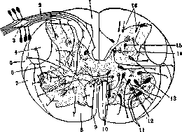
Figure 26. Diagram of the structure of the spinal cord: 1-posterior column; 2-posterior roots; 3-intervertebral ganglion; 4-crossed pyramidal tract; 6-bundle of Flechsig; 6-main bundle of the lateral column; 7-bundle of Gowers; 8-anterior column; 9-direct pyramidal tract; 10-commissural cells; 11-anterior root; 12-columnar cells (of the anterior-lateral bundle); 13-root cells; 14-cells of Clark's column; 15-cells of type II Golgi; 16-cells of the posterior horn- substantia gelatinosa Rolandi.
substance; very few cells in the posterior column; they lie in the posterior horns and in substantia gelatinosa. The neurites of these cells form centripetal pathways; there are short and long pathways. The neurite of a cell is directed to the corresponding column and there, bending, goes upward or divides in a T-shape, with one branch going in the ascending direction, the other in the descending, or one goes to the lateral column and the other to the anterior. 4) Cells with short axons (syn. cells of Golgi type II, s. cellulae axiramificatae); they are most often found in the posterior horn and in substantia gelatinosa; their neurite divides near its origin; sometimes along the anterior commissure the neurite goes to the gray matter of the opposite side, where it ends with branches (s. cellulae commissurales axiramificatae); these cells are interneurons, their purpose is to transmit irritation from the posterior roots to other cells of the gray matter. 5) A special group is formed by the cells of the posterior roots (cellulae radiculares post.); they lie outside the spinal cord in the spinal ganglia or spinal ganglia. Some authors assume that there is a small part in the anterior horns, and their neurite penetrates into the posterior root. The cells of the spinal ganglia are pseudounipolar; their process divides in a T-shape; the central end of the process-neurite-forms the posterior roots, while the peripheral process-a long dendrite-ends in peripheral organs (fig. 27). The root cells of the anterior horn are the motor centers of the spinal cord, as indicated by their connection with the anterior roots. Two groups of cells in the anterior horns-internal and external-are found throughout the spinal cord, while at the level of the enlargements other groups are added to them (fig. 21-23). The first group is related to the muscles of the trunk: the anterior-internal innervates the muscles of the spine, it extends to the upper sacral segments; the anterior-external is related to other muscles of the neck and trunk. The groups of cells that appear only in the enlargements must be related to the muscles of the limbs; there are several such groups in each enlargement, but they do not form regular columns-some groups disappear, being replaced by others. In the upper cervical segments, in addition to the usual two groups, a central and a posterior-external are also distinguished, which serve as the beginning of the spinal root of the n. accessorius, being its nuclei-nucleus n. accessorius pars spinalis. The question of what the groups of cells correspond to-whether to individual muscles, peripheral nerves, or segments of the limbs-has not yet been finally resolved; in recent years, there has been a tendency towards the functional
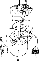
Figure 27. Course of the pyramidal crossed tract and the sensory tract of the posterior bundles: 1-cerebral cortex; 2-Betz cell; 3-pyramidal tract; 4-its decussation; 5-its collaterals; 6-pyramidal tract in the spinal cord and its termination in the cells of the anterior horns; 7 and 8-anterior root; 9-muscles; 10-spinal cord; 11-nerve endings in the skin; 12-intervertebral ganglion1; 13-posterior root, which enters the posterior column (bundle of Goll), sends collaterals (14 and 15) to the gray matter of the spinal cord; 16-nucleus funiculi gracilis; 17-cerebral cortex.
theories—each cellular group innervates muscles connected by one function. Groups controlling movements of more peripheral parts of the body lie outside and behind groups controlling more central muscles. Neuritis of root cells in the anterior horn of the spinal cord forms the anterior roots (radix motoria), while neuritis of cells of the spinal ganglia forms the posterior roots (radix sensi-tiva). The anterior roots consist of centrifugal fibers, while the posterior ones consist mainly of centripetal fibers (the presence of a small number of centrifugal fibers is noted in them). Outside the spinal ganglia, in the intervertebral foramen, the anterior and posterior roots connect to form a mixed spinal nerve (n. spinalis), which, upon exiting the spinal column, divides into ramus posterior (posterior branch), ramus anterior (anterior), ramus com-municans (to the sympathetic border column), and ramus meningeus. The anterior branches, being more significant, connect with each other except in the thoracic region to form plexuses—cervical plexus (see), brachial plexus (see), lumbosacral and coccygeal (see Lumbosacral plexus). The posterior branches innervate the skin and muscles of the occiput and back. From the plexuses arise peripheral nerves, which thus contain fibers from various segments of the spinal cord. White matter of the spinal cord. The white matter of the spinal cord consists of separate bundles, of separate systems. These bundles were identified by special research methods—Flexig's embryological method and the method of secondary degeneration. The first method is based on the asynchronous development of myelin sheaths in the fibers of different bundles; this development occurs strictly sequentially for different bundles and always in the same order. The method of secondary degeneration is even more valuable; thanks to it, many bundles were identified, the direction in which a given bundle passes was studied, and even its origin was determined. All fibers of the spinal cord can be divided into endogenous, or intrinsic fibers of the spinal cord, and exogenous, or foreign ones. Endogenous fibers include those originating in the spinal cord; they can be long and short: long ones—fibrae spino-cerebrales and spino-cerebel-lares, short ones—intersegmental fibers (fibrae intersegmentares), connecting different segments of the spinal cord. Short endogenous fibers lie in the inner parts of the white matter of the spinal cord, while long ones lie in the outer parts. Exogenous fibers are divided into descending (fibrae cerebro-spinales) and ascending—fibers of the posterior roots (fibrae ganglio-spi-nales and ganglio-bulbares). Fibers of the posterior roots enter the spinal cord in the area of Lissauer's marginal belt (syn. marginal zone of Lissauer, s. zona Lissauer'a), located between the end of the posterior horn and the periphery, and in the adjacent part of the posterior column—root zone, then divide into ascending and descending branches. The ascending fibers, varying in length, also differ in direction and termination (fig. 24 and 28): 1) the longest of them from the root zone deviate somewhat inward and occupy the posterior columns, forming two bundles—Goll's bundle and Burdach's bundle; in the lower parts of the spinal cord, only Goll's bundle (funi-culus Golli) exists, which carries fibers from the lower
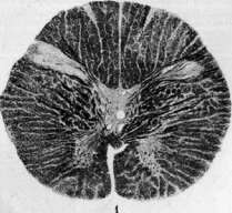
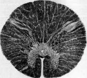
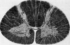
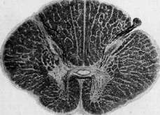
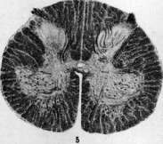
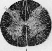
Fig. 1-6, Cross-sections of the spinal cord at various levels. Fig. 1. Transition from the first cervical segment to the medulla oblongata. Fig. 3. First cervical segment. Fig. 3. Seventh cervical segment. Fig. i. \ thoracic segment. Fig. b. Third lumbar segment. Fig. c. First sacral segment. 2»7 limbs and lower parts of the trunk, in the upper parts of it Burdach's bundle (funiculus Burdachi) is added, which carries fibers from the upper parts of the trunk and from the upper limbs. In the upper cervical regions, the boundary between them is very clear—septum paramedianum; there they are renamed: funiculus Burdachi into funiculus cuneatus (cuneate bundle), funiculus Golli into funiculus gra-ciiis (thin, delicate bundle) and end in
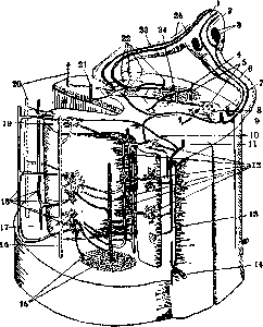
Fig. 28. Main formations of the spinal cord on cross-section and longitudinal section: 1-nervus spinalis; 2-ganglion spinale and cell in it; 3 and 4-fasc. spino-cerebellaris dorsalis; 5-secondary reflex pathway; 6-posterior root; 7-collaterals to posterior horns; 8-bundles of the posterior column; 9-descending pathways of the posterior column; 10-reflex pathway; 11-ascending fibers of the posterior column; 12-secondary reflex pathways; 13-descending fibers of the posterior column; 7 s!-fibers of the posterior root; 15-fasc. cerebro-spinalis laterals; 16-fasc. rubro-spinalis; 17-fasc. vestibulo-spinalis; J 8-fasc. spino-cerebellaris ventralis et fasc. spino-tectalis; 19 - substantia grisea; 20 - fasc. spino-reticnlaris ventralis; SI-fasc. cerebro-spinalis ant.; 22-fasc. reticulo-spinalis; 2Я and 85- anterior root; 2i-fasc. cerebro-spinalis late-ralis.
in the medulla oblongata in the nuclei of the posterior columns or funiculi—in nucl. funiculi cuneati and in nucl. funiculi gracilis. All the time fibers from Burdach's bundle pass into Goll's bundle, and new root fibers take their place. 2) Shorter fibers from the root zone go to the cells of the anterior horns, these are reflex fibers; together with the cell and anterior roots they form a reflex arc, in which they are the efferent part (fig. 28). Besides special fibers, collaterals also end near the cells of the anterior horns, these are reflex collaterals of Kölliker. Not only do collaterals end near the cells of the anterior horns, but also near the cells of Clarke and other cells of the posterior horn on the same and opposite sides. Besides the direct reflex arc—there is also an indirect reflex arc, which is carried out by the inclusion of a reflex cell or rather a reflex neuron between the motor and sensory roots. 3) Part of the fibers ends at the cells of Clarke's column and in the cells of the posterior horns. Descending fibers of the posterior roots are shorter, in the lower parts of the spinal cord they are located closer to the midline, and in the upper parts—closer to the gray matter. In the posterior column, besides posterior root fibers, there are also endogenous fibers; the shorter ones occupy the more anterior part of the posterior column zona ventralis, syn. anterior girdle; long endogenous fibers go in the posterior part of Goll's bundle, at the same time they occupy different positions at different levels: in the cervical and thoracic region the bundle of fibers has the shape of a comma—'Schultze's comma', occupies the space between Goll's and Burdach's bundles. At the same level in the posterior part of the column there are scattered fibers, which downward gather at the septum posticum-tractus or fasc. septo-marginalis Ho-che. In the lumbar region they have the shape of a semi-oval—Flexig's oval field, s. tractus cervico-lumbalis dorsalis (Flexig), located at the posterior septum; in the sacral region on the periphery there is Gombault et Philippe's sacral triangle; the last three areas pass into each other and possibly belong to the same endogenous system, while 'Schultze's comma' is a mixed formation consisting of descending posterior root fibers and endogenous ones. In the anterior-lateral column, fibers go in ascending and descending directions. Of the bundles going in the ascending direction, the following bundles should be noted: 1. Fasciculus spino-cerebellaris dorsalis, s. fasc. lateralis, syn. direct cerebellar bundle or Flexig's bundle (see); it consists of neurites of Clarke's column cells of the same side, goes to the cerebellum. 2. Fascicul. spino-cerebellaris ventralis, s. fasc. anterolateralis superficialis, syn. crossed cerebellar bundle, Gowers' bundle (see); originates from cells of the posterior horn of the opposite side. Part of the fibers of Gowers' bundle ends already in the cervical region of the spinal cord, in the nuclei of the medulla oblongata (fibrae spino-bulbares dorsalae), in the quadrigeminal tubercles (fibrae spino-tectales) and finally the large bundle goes to the thalamus (fibrae spino-thalamicae, s. fasc. spino-thalamicus lateralis). It is located inward from Gowers' own bundle, which goes to the cerebellum through its superior peduncles. 3. Part of the fibers of fasc. spino-thala-mici after the decussation does not go into the lateral column, but into the anterior, forming fasc. spino-thalamicus anterior. 4. At the boundary between the anterior and lateral columns, between the anterior roots lies fasc. or tractus spino-olivaris, going to the base of the medulla oblongata; it is separated from the periphery by Goll's bundle (see below). 5. The ascending bundle of the anterior column [fasc. sulco-marginalis ascendens (Marie)] occupies the edge of the anterior sulcus and the nearest part of the anterior column; this bundle is crossed, originates from cells of the opposite side, part of the fibers goes to the medulla oblongata, forming fasc. spino-bulbaris ventralis, part—to the higher parts of the brainstem. Of the bundles going in the descending direction, the following should be noted: 1. Pyramidal bundles, s. tractus cerebro-spinalis, which after decussation in the medulla oblongata divide into two bundles: one goes into the lateral column of the opposite side—tractus cerebro-spinalis lat., syn. lateral or crossed pyramidal
bundle; it lies outward from the posterior horn, inward from the bundle of Flexig, and ends in the cells of the anterior horn on its side (fig. 27); another pyramidal bundle goes in the anterior column of the same side along the fis. mediana anterior, part of its fibers ends in the anterior horn of the opposite side, passing through the commissura alba anterior, while another part ends in the anterior horn of its side. The anterior pyramidal bundle is significantly smaller than the crossed one; both bundles gradually decrease downward, the anterior one ends in the lower thoracic region, while the crossed one reaches the lower end of the spinal cord. 2. Fasc. rubro-spinalis, synonym bundle of Monakoff, lies in front of the lateral pyramidal bundle and inward from the bundle of Govers, 3 takes its origin from the red nuclei, after which it undergoes a decussation (Forel), and ends in the cells of the anterior horns. Figure 29. Spinal cord and its membranes: 1-dura mater and its continuation to the roots; 2
1 arachnoidea; 3-pia mater; 4
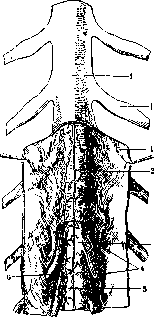
les are located near and between the pyramidal bundles and the bundle of Monakoff, originate from the substantia reticularis of the opposite side and end in the cells of the anterior horns. 4. Fibrae reticulo-spinales ventrales go in the lateral parts of the anterior column, in its main bundle, take origin from the substantia reticularis of the same side. 5. Fasciculus or tractus vestibulo-spinalis, s. fasc. Lowenthal's, synonym anterior marginal bundle, lies along the periphery of the anterior column at the exit of the anterior roots, takes origin from the cells of Deiters' nucleus and ends in the cells of the anterior horns; this bundle is distinguished as tractus vestibulo-spinalis anterior, while part of the fibers or tractus vestibulo-spinalis lateralis passes in the lateral column. 6. Bundle of Helweg (syn triangular bundle, s. tractus praeolivaris) is located along the periphery on the border of the anterior and lateral columns, between the anterior roots, descends only to the cervical enlargement, constitutes a continuation of the central bundle of the tectum. 7. Fascic. longitudinalis posterior, s. dorsalis, synonym posterior longitudinal bundle, occupies the inner part of the anterior column near the pyramidal bundle, and in its absence lies along the anterior groove, consists of fibers originating from various cellular groups of the brainstem, ends in the cells of the anterior horns. 8. Tractus tecto-spina-lis (s. fascic- praedorsalis, s. tractus marginalis anterior, synonym pretectal bundle) takes origin from the quadrigeminal bodies, forms the fountain-like decussation of Meynert and ends in the anterior horns of the spinal cord. 9. Fasc. praepyramidalis Thomas begins in the outer parts of the substantia reticularis of the medulla oblongata, goes parallel to the outer edge of the anterior horn and ends in the anterior horns of the cervical region. The descending fibers in the anterior column together have the shape of a crescent-faisceau en croissant Marie et Guillain. In the lateral columns, in addition to those described, pass vegetative paths, but not in separate bundles, but as individual fibers, mixed with other systems. In the inner parts of the anterolateral column, directly near the gray matter, lie short endogenous fibers, forming the main bundles (fasciculus proprius) for each column-anterior main and lateral main; in these bundles pass intersegmental fibers connecting different segments of the spinal cord. The membranes of the spinal cord (meninges spinales). The spinal cord is covered by three membranes-the soft (pia mater), the arachnoid (arachnoidea) and the hard (dura mater). Pia mater and arachnoidea together form the leptomeninx (fig. 29). In the region of the foramen occipitale magnum, the spinal part of the membranes passes into the cranial. Pia mater or the vascular membrane is tightly applied to the surface of the spinal cord and penetrates into the grooves; it consists of 2 layers separated by a very narrow space, blood vessels lie between both layers and penetrate into the adventitial sheath from the inner layer into the brain substance; the initial parts of the sheath form funnels of the soft membrane, connecting with the systems of grooves of the soft cerebral membrane, which are its lymph spaces. The nerves of the soft membrane originate from the sympathetic nervous system, there are also spinal nerves; they form the plexus nervosus piaematris in the outer layer of the membrane. Arachnoidea is applied with one surface to the dura mater, being separated from it by a slit-like subdural space (spatium subdurale, s. cavum subdurale medullae spinalis); with its inner surface, arachnoidea fuses with pia mater with the help of numerous crossbars and partitions; between both membranes is a wide subarachnoid space, filled with cerebrospinal fluid, thus arachnoidea covers the spinal cord in the form of a wide loosely fitting bag. Dura mater forms a long and wide bag (dural bag) of cylindrical shape, which is wider than the spinal cord; above it is attached to the edge of the foramen occipitalis, and below at the level of the III lumbar vertebra it narrows and descends as the filum terminale of the spinal cord to the coccygeal bone, forming the filum durae matris spinalis (fig. 30). Dura mater is connected to the walls of the spinal cord with the help of connecting cords in the form of ligamenta anteriora durae matris, lig. dorso-lateralia durae matris, ligamenta intervertebralia cervicalia, and the hard membrane on the roots. Inside it is connected by subdural threads with the outer surface of the arachnoidea. From pia mater to dura mater go two symmetrical rows of flat teeth (19-23 teeth)-the toothed plate or ligamentum denticulatum, s. ligamentum serratum medullae spinalis; this ligament serves for the more direct position of the spinal cord (fig. 31). The first tooth is attached under the I root, and the last between the XII thoracic and I lumbar roots; below to the end goes the lateral stripe. By these ligaments the subarachnoid space is divided into anterior and posterior parts; the anterior subarachnoid space, crossing the anterior roots, represents a continuous free space; in the posterior subarachnoid space pass the posterior roots; with the help of the septum subarachnoidale posterius it is divided into right and left halves. Blood supply - of the spinal cord. The arteries of the spinal cord belong to a. spinalis anterior and two a. spinales posteriores. A. spinalis ant. takes origin from a. vertebralis at its exit from the canal in the transverse processes; both arteries go downward and to the middle and merge into a single artery located in the fis. mediana anterior (fig. 32), which descends to the filum terminale. Aa. spinales posteriores depart at the same level from a. vertebralis, do not merge into one artery, but go along the sides of the posterior column at the entrance of the posterior roots. A. spinalis anterior along its entire path sends branches through the anterior groove into the gray matter of the brain, into its anterior horns, to the group of cells of Clarke; from a. spinalis poster. go branches into the posterior horns of the spinal cord. In addition to these two arteries, the spinal cord is also nourished by
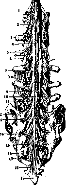
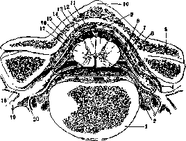
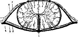
Figure 32. Diagram of arterial circulation on a cross-section of the spinal cord: 1-art. spinalis ant.; 2-anterior root of the spinal cord; 3-anterior branch of the root arteries; i-root artery; 5-intervertebral node; 6-posterior branch of the root artery; 7-posterior root of the spinal cord; 8-art. spinalis post. ramuli medii-spinal branches from the aa. vertebralis, intercostales, lumbales, ilio-lumbales, sacrales, which penetrate along the roots to the spinal cord and, anastomosing with each other and with the aa. spinales, form an arterial network on the surface of the spinal cord, mainly nourishing the white matter. From the vascular network in the spinal cord, radial branches penetrate, which give rise to a network of capillaries densely covering the white and especially the gray matter. From the capillary networks, venous blood collects into two large internal veins-vv. centrales. The external veins are vena spinalis anterior and posterior. e. Konova. IV. Physiology. The spinal cord represents a department of the central nervous system that has to the greatest extent preserved, both in morphological and physiological aspects, the features of the primitive central organ of the nervous system-the segmental apparatus, which is the original organ of the evolution of the central nervous system. The basic fact of this evolution is the superstructure over the segmental apparatus of the mantle department of the brain. One of the anatomophysiological features of the suprasegmental apparatus is that it has no direct connection with the periphery, but is always connected with the latter through the segmental apparatus. Accordingly, in the spinal cord two main functions can be distinguished: 1) reflex activity or the function of the spinal cord's own apparatus and 2) conductive function, i.e., the role of the spinal cord as an intermediate stage between the higher brain and the periphery. The relationship between the brain and the spinal cord can be generally defined as the inhibition by the brain of the mechanisms of the spinal cord's own apparatus and the subordination of the latter to cortical innervations. The basic essence of the function of the spinal cord's own apparatus is determined by its participation in the reflex act. The gray matter of the spinal cord is part of the segmental reflex arc, being the area of synaptic switching of the impulse from the receptor peripheral apparatus, i.e., sensory nerves entering the spinal cord through the posterior roots, to the effector peripheral apparatus represented by the motor cell of the anterior horn of the spinal cord and its process, i.e., the motor fiber of the peripheral nerve. The propagation of the impulse from the periphery along the receptor fiber through the gray matter of the spinal cord to the motor cell and along its process to the muscle determines the role of the spinal cord as a mechanism carrying out the reflex function. The reflex act represents a segmental function of the spinal cord in the sense that the basic mechanism for carrying out this function is the spinal cord's own apparatus and that this function is not directly dependent on the conductive apparatus of the spinal cord. Segmentation should not however be understood in this case in the sense that the reflex is carried out by any single segment. In view of the existence of anatomophysiological connections between individual segments by means of the so-called internsegmental fibers, irritation of a given segment spreads to a greater or lesser number of neighboring segments: reflex reactions are represented not only as "isomeric", i.e., corresponding directly to the irritated segment, but also as "allomeric", i.e., corresponding to several segments. The physiological significance of this plurisegmentality or allomericity of spinal cord reactions lies in the fact that both in reflex and in voluntary motor acts, the contraction of not individual muscle fibers and not individual muscles, but of entire groups of muscles is required; the anatomophysiological basis for such combined participation of a group of muscles consists of internsegmental associative connections. The cells of the anterior horns of the spinal cord have significance for the trophicity of the contractile substance of the muscle fibers innervated by them. Isolation of the muscle from these cells, as occurs when the latter die or when conductivity in the peripheral motor nerve is impaired, leads not only to the loss of motor function but also to a special disturbance in the vital activity of the muscle, consisting in the gradual decrease of the contractile substance. This process of so-called "muscular atrophy" can reach the degree of complete disappearance of muscle substance with its replacement by connective tissue. If the anterior horns represent the segmental motor centers of the spinal cord, then the posterior horns have the sensory segmental function: with the exception of the fibers of conscious proprioceptive receptors ("muscle-joint sensitivity"), which enter directly into the white matter, forming the bundles of the posterior columns (Burdach's bundles), all other sensory fibers of peripheral nerves enter the posterior horns of the gray matter. Here, in the posterior horns, the synapse between the first and second sensory neurons occurs; from the cells of the posterior horns begin fibers which, proceeding to the lateral and anterior columns of the white matter of the spinal cord, form the system of ascending conductors of the spinal cord, conductors carrying impulses from the segmental centers of the spinal cord to various departments of the suprasegmental area (spinothalamic, spinocerebellar, spinotectal, spinobulbar bundles). But besides the aforementioned role of the posterior horns as the area of switching impulses from the first (peripheral) neuron to the second (spinosuprasegmental), the posterior horns must also be attributed their own conductive function. Among the conductors passing in the gray matter of the posterior horns are the internsegmental fibers mentioned above, establishing connections between different segments of the spinal cord. In addition, part of the conductors of pain and temperature sensitivity, without undergoing decussation and not entering the spinothalamic bundle (see below), is directed upward through the gray matter of the spinal cord, namely through the area of the posterior horns. The existence of such conduction of sensitivity in the area of gray matter (Grisealleitung, Forster) apparently explains the cases of failure of chordotomy, i.e., cases of preservation of pain despite the transection of the spinothalamic bundle. The relative amount of sensitivity conductors passing in the gray matter of the homonymous half of the spinal cord is apparently subject to individual fluctuations, which may explain why the results of chordotomy are not in all cases identical.
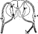
- Sympathetic cells of the lateral horns with their processes emerging through the anterior roots constitute the efferent element of the spinal sympathetic centers. Haskell considers these elements analogous to internuncial neurons establishing connection between the posterior and anterior horns (Fig. 33). Afferent fibers of the sympathetic system enter the spinal cord through the posterior roots and posterior horns. Thus the gray matter of the spinal cord, along with somatic reflexes, also carries out reflexes of the sympathetic nervous system. Summarizing the data concerning the physiology of the gray matter of the spinal cord, one can characterize the function of this department of the spinal cord as an active process of formation, combination, and distribution of impulses. In contrast to this, the white matter represents a system of fibers carrying out the passive function of conductors, serving for communication between the segmental centers of the spinal cord and suprasegmental departments of the central nervous system. The entire system of conductors of the spinal cord can be subdivided into two main groups, corresponding to the direction in which the propagation of impulses occurs: 1) ascending conductors, serving for conducting impulses from the segments of the spinal cord to suprasegmental departments, 2) descending conductors, serving for conducting impulses from suprasegmental departments to segmental centers of the spinal cord.
The system of ascending conductors performs the function of conducting receptor impulses from segmental centers of the spinal cord to suprasegmental divisions. With regard to the correspondence of individual conductors to certain types of sensitivity, the following data can be considered established. The main conductor of pain sensitivity is the spino-thalamic tract. The aforementioned tract is a common conductor for all forms of pain sensitivity—deep and superficial, protopathic and epicritic. This commonality of spinal cord conductivity for all types of pain sensitivity is the reason that in lesions of the spino-thalamic tract, there is an even loss of all the aforementioned types of sensitivity, and the dissociation between deep and superficial or protopathic and epicritic sensitivity, often observed in lesions of peripheral nerves and the brain, is not observed in spinal cord lesions. In contrast to the previous view of the spino-thalamic tract as the only conductor of pain impulses in the spinal cord, at present the conviction is becoming increasingly firm that pain impulses can also spread along other pathways. In addition to the aforementioned possibility of the spread of pain impulses by fibers passing in the area of the posterior horns of gray matter, the possibility (Forster) of their conduction in the posterior columns and the posterior-lateral parts of the lateral columns is admitted. The degree of development of such accessory pain conductivity in the spinal cord is subject to individual variations; however, their existence is beyond doubt and is important for explaining certain, at first glance paradoxical phenomena, when e.g. in lesions of the spinal cord, not affecting the spino-thalamic tract, there are phenomena of loss of pain sensitivity, or conversely, when there is a definite impairment of the conductivity of the spino-thalamic tract, pain sensitivity is not completely lost. If we take into account that according to recent data, the point of entry of pain impulses into the spinal cord is not only the posterior roots but also the anterior ones (Forster), then, without denying the importance of the spino-thalamic tract as the main and predominant conductor of pain impulses, we must keep in mind the possibility of the passage of pain impulses, at least in some individuals, in other areas of the spinal cord. Forster summarizes the modern data on the conduction of pain sensitivity as follows: "pain impulses penetrate the central nervous system not through a narrowly limited trunk of fibers, but through a multitude of winding paths, and if we block the main path, there always remains some hidden path, by which pain finds access to the brain." In the area of the spino-thalamic tract, fibers corresponding to temperature sensations immediately adjoin the pain fibers; however, pain and temperature conductors remain isolated from each other, therefore in spinal cord lesions it is possible to have loss of only pain or only temperature sensitivity; similarly, there are isolated conductors for heat and cold stimuli, which explains the possibility of dissociation of disorders of heat and cold sensitivity in lesions of the conducting apparatus of the spinal cord. Similar to pain sensitivity, temperature sensitivity does not have separate fibers for protopathic and epicritic stimuli; therefore in spinal cord lesions, protopathic and epicritic temperature sensitivity are lost to the same degree.--With regard to the conduction in the spinal cord of impulses of tactile sensitivity, there is a difference depending on the protopathic or epicritic character of the tactile impulses. Impulses arising from touch to the skin surface and from pressure on the skin, like the conductors of pain and temperature sensitivity, undergo decussation in the spinal cord, but upon entering the white matter of the opposite side, they separate from the pain and temperature conductors, not being located in the lateral columns as the latter are, but in the anterior ones. This bundle, conducting protopathic tactile elements (pressure and touch), is designated by the term "anterior" or "ventral" spino-thalamic tract, in contrast to the lateral spino-thalamic tract, described above as the conductor of pain and temperature sensitivity (tractus spino-thalamicus lateralis). It should be mentioned that the fibers of the lateral spino-thalamic tract, i.e. the conductors of pain and temperature sensitivity, undergo decussation very soon after their entry into the spinal cord, whereas the fibers corresponding to the anterior spino-thalamic tract, for several segments, run on the side of their entry into the spinal cord before undergoing decussation. This explains the fact that in some lesions of the spinal cord, the level of loss of tactile sensitivity does not coincide with the level of loss of pain and temperature sensitivity. Epicritic, or "discriminative" forms of tactile sensitivity (light touch, precise recognition of Weber's compass points, localization of touch) are conducted in the spinal cord in the posterior columns. In addition to conducting certain types of tactile sensitivity, the function of the posterior columns is to conduct impulses of proprioceptive sensitivity, so-called muscle-joint feeling or sense of position. Finally, in the posterior columns, impulses of vibratory sensitivity are conducted. In the most lateral parts of the lateral columns, impulses of reflex proprioceptors are conducted, which form the basis of the automatic regulation of movements and have the cerebellum as their central apparatus. According to their direction, these conductors are designated by the term "spino-cerebellar tracts". Topographically: a ventral (Gowers) and a dorsal (Flexig) spino-cerebellar tract are distinguished (fig. 34). There is no difference in function between these tracts. The descending system of conductors of the spinal cord is a collection of bundles, the fibers of which conduct impulses from the suprasegmental apparatus to the spinal cord. The final stage of the spinal cord, through which impulses from the suprasegmental apparatus spread to the muscles, are the cells of the anterior horns (see Human Motorics, Motor Centers). Functionally, the descending conductors can be characterized as a system of fibers that perform the motor function. The conductors of cortical "volitional" impulses are the so-called pyramidal or cortico-spinal tracts (tr. pyramidalis, cortico-spinalis), located in the posterior parts of the lateral columns (fig. 34). Directly with their terminal branches or with the help of short intercalary neurons, the fibers of the pyramidal tract come into anatomophysiological contact with the cells of the anterior horns. The functional effect on the latter from the pyramidal tracts consists of a combination of stimulating influences with inhibitory ones. This at first glance paradoxical fact of the simultaneous performance of two opposite functions by the same apparatus is a common feature of the relationship
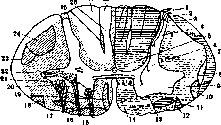
Figure 34. Diagram of a cross-section of the spinal cord in the middle part of the cervical region: 1-dorsal root; 2-dorsolateral tract; 3-dorsal spino-cerebellar tract; 4-posterior horns; 5-dorsal proprius fasciculus; 6-lateral proprius fasciculus; 7-ventral spino-cerebellar tract; 8-lateral spino-thalamic tract; 9-anterior horns; 10-spino-tectal tract; 11-ventral spino-thalamic tract; 12-spino-olivary tract; 13-ventral proprius fasciculus; 14-sulcomarginal fasciculus; 15-anterior roots; 16-vestibulo-spinal tract; 17-tecto-spinal tract; 18-olivo-spinal tract; 19-anterior cortico-spinal tract; 20-ventrolateral nucleus; 21-ventromedial nucleus; 22-dorsolateral nucleus; 23-rubro-spinal tract; 24-lateral cortico-spinal tract; 25-interfascicular fasciculus; 26-septomarginal fasciculus; 27-Burdach's fasciculus [the inner part of Burdach's fasciculus (see the right half of the figure) corresponds to segments I-IV thoracic, and the outer part corresponds to cervical segments]; 28-Goll's fasciculus [the innermost part of Goll's fasciculus (see the right half of the figure), separated by the first groove, indicates the shape of this fasciculus in the sacral region; the second groove delimits the part of Goll's fasciculus corresponding to the lumbar region, and the outermost part of Goll's fasciculus corresponds to segments V-XII thoracic]]
between the segmental mechanisms of S. m. and suprasegmental centers. As indicated above, the cerebral cortex in the process of phylogenetic evolution is built upon the segmental apparatus, and the latter does not lose its inherent automatic function, but merely comes under cortical innervation. However, to implement cortical innervation, a certain degree of inhibition of the reflex mechanisms of the segmental apparatus is necessary. Thus, the cerebral cortex, while sending stimulating impulses to S. m. in the sense of activating the activity of motor spinal cord cells, at the same time exerts an inhibitory effect on the reflex function of its own apparatus. In other words, the cerebral cortex through the pyramidal tracts moderates the reflex activity of S. m. The role of the pyramidal tract in the function of tone is connected with this. Muscle tone, i.e., a certain degree of tension of muscle tissue, is determined by a number of different factors. The own apparatus of S. m., i.e., the reflex mechanism, is a factor increasing tone: constant impulses arising on the periphery, spreading along reflex arcs to the muscles, maintain a certain degree of contraction in the muscles; thus, the reflex function is a tone-generating factor. On the contrary, the pyramidal tracts, inhibiting reflex activity, moderate muscle tone. This explains the increase in tone observed with lesions of the pyramidal tract. The descending conductor of cerebellar impulses to S. m. is the so-called Monakovsky tract (tr. rubro-spinalis), located in the lateral columns in front of the pyramidal tract (fig. 34); the intermediate stage between the cerebellum and S. m. are the red nuclei. But the same red nuclei are also an intermediate stage between S. m. and corpus striatum. Thus, the rubrospinal tract contains two different systems of descending fibers: cerebellospinal and striospinal. Physiologically, cerebellospinal impulses are the main factor of automatic coordination of movements, synergy of muscle contractions, and regulation of tone in the sense of proper distribution of tension in the muscles participating in the execution of a given motor act. The function of striospinal impulses consists in regulating the tone of the musculature and carrying out amorphous diffuse contractions of the musculature, against the background of which stimulating and inhibiting cortical innervations create fine movements of a limited number of segments. On the basis of experimental data, the red nucleus and the rubrospinal tract should be attributed the function of inhibiting postural tone (see Postural reflexes). The data presented above on the role of various spinal cord apparatuses in muscle tone function can be summarized as follows. The influence of S. m. on muscle tone is carried out through the cells of the anterior horns of S. m. These cells are subjected to a stimulating effect on tone from the receptor part of the reflex arcs. Constantly arriving peripheral irritations, spreading from the receptor part of the reflex arc to the motor part, form the basis of the so-called "reflex tone" of the musculature. Thus, the reflex act is a factor stimulating tone. From the suprasegmental apparatuses, the cerebellum exerts a stimulating effect on tone via tr. rubro-spinalis, and the Deiters nucleus of the vestibular nerve via tr. deitero-spinalis (s. vestibulo-spinalis; fig. 34), located in S. m. in the anterior columns. Factors inhibiting tone are: the cerebral cortex via tr. cortico-spinalis and corpus striatum (pallidum) via striospinal fibers that are part of tr. rubro-spinalis. Thus, in connection with lesions of S. m., the following changes in tone can be observed: disturbances in the conductivity of the reflex arc lead to weakening or loss of tone; disturbances in the conductivity of the pyramidal tract and loss of pallidal influence lead to an increase in tone. In addition to suprasegmental conductors in S. m., there are intersegmental fibers connecting various spinal cord segments; these conductors are also designated by the term "spinospinal." The presence of these connections constitutes the anatomical-physiological basis for the combined action of a group of segments. Most of the intersegmental conductors are located in the immediate vicinity of the gray matter, forming the so-called "main bundle," or fasciculus proprius (fig. 34), but some of them pass as part of the white matter; these include: the Schulze bundle (fig. 34) and fasciculus septo-marginalis (fig. 34). Part of the intersegmental fibers is located in the anterior columns of tr. sulco-marginalis; functionally, it is a continuation of the posterior longitudinal bundle of the brain stem (function of combined rotation of the eyes, head, and trunk). As for the localization of functions in the spinal cord, the relationship between the segments of the spinal cord and the distribution of skin sensitivity see Metamerism. The correspondence between the segments of S. m. and certain muscle groups can be summarized as follows: I-IV cervical segments correspond to the cervical musculature; V-VIII cervical and I-II thoracic segments correspond to the muscles of the upper extremities; from III thoracic to I lumbar - to the muscles of the trunk; II-V lumbar and I-II sacral segments innervate the lower extremities, III-V sacral segments - the muscles of the perineum, anus, and urogenital organs. As indicated above, each muscle is innervated not by one, but by 2-3 segments; however, there is a predominant dependence of individual muscles on certain segments. This connection between individual muscles and segments of S. m. is presented in table 1. Table 1. Segments of S. m. Muscles Segments of S. m. Muscles CIV Diaphragm Lr Heo-psoas CV Deltoideus Lin Quadriceps CVI Biceps Liv Adductors of thigh CVN Triceps Lv TiMalis ant. CVNI Flexors of hand fingers SI Gastrocnemius DII Hypothenar Sn Small muscles of foot Dm-xn Intercostal muscles Sin-v Muscles of perineum On reflex centers of S. m. see Reflexes. The localization of centers of the sympathetic nervous system corresponding to internal organs is given in table 2. Table 2. Internal organs Segments of S. m. Internal organs Segments of S. m. Heart . . . Aorta . . . Stomach : Intestine . Rectum . . . DI-DIII DVI-IX UI-XI SII-III Liver . . . Kidney . . . Bladder and sexual organs . . . DVN-X DXN and LI SII-IV The indications given in table 2 need to be supplemented with the following explanations. The centers of urinary and sexual organs located in the II, III, and IV sacral segments, also designated by the terms centrum "vesico-spinale" and centrum "genito-spinale," are not sympathetic but parasympathetic centers. The sympathetic innervation of the urinary and sexual organs is carried out by fibers originating in the sympathetic centers of the I and II lumbar segments. These fibers pass through gangl. mesentericum inferius and then continue in the form of n. hypogastricus to the bladder and internal sexual organs. Irritation of n. hypogastrici causes contraction of the sphincter and relaxation of the detrusor; irritation of parasympathetic fibers (n. pelvicus), on the contrary, leads to contraction of the detrusor and relaxation of the sphincter. Special mention is deserved by the cilio-spinale center - the spinal cord center for pupil dilation. This center and the corresponding fibers form part of the cervical sympathetic nerve. The cilio-spinal center is localized in S. m. at the level of I-II thoracic segments; fibers starting from here exit S. m. as part of the anterior roots of the VIII cervical, I and II thoracic nerves and then, as part of the cervical sympathetic nerve, reach the superior cervical sympathetic ganglion, from where, by wrapping the internal carotid artery in the form of a periarterial sympathetic plexus, they enter the composition of the superior branch of the trigeminal nerve, with the fibers of which they enter the eyeball, ending in the radial musculature of the pupil. Irritation of these fibers causes pupil dilation; cilio-spinal fibers are antagonists to parasympathetic fibers innervating the sphincter of the pupil and entering the composition of the oculomotor nerve. V. Pathology. Clinical manifestations of lesions of S. m. can be subdivided, according to the two main apparatuses of S. m., into two groups: segmental and conductor disorders. In segmental lesions, functional disorders are observed only from the side of the directly affected segments. In conductor lesions, due to disruption at a certain level of communication between the periphery and the brain, there is a diffuse loss of functions corresponding in their innervation to the parts of S. m. located below the level of the lesion. Since the motor central conductor and the conductors of muscle feeling in S. m. do not undergo decussation, with their lesions, disorders are observed on the side, homonymous to the lesion; in contrast to this, with conductor lesions of the spinothalamic tract, loss of pain and temperature sensitivity is observed on the side opposite to the localization of the lesion, since these conductors undergo decussation in S. m.
The main data for topical diagnosis of lesions of the spinal cord are as follows: I. Segmental disorders (lesion of gray matter and roots). 1) Symptoms of posterior root lesions: pain corresponding to the area of innervation of the given posterior root; disturbance of all types of sensitivity, loss or weakening of reflexes. 2) Posterior horns: loss of pain and temperature sensitivity in the given segment with preservation of tactile and muscular sensation; weakening or loss of reflexes. 3) Anterior horns and anterior roots: motor paralysis, muscle atrophy, loss of reflex, reaction of degeneration in the area of the affected segment. 4) Area of gray matter anterior to the central canal: bilateral loss of pain and temperature sensitivity at the level of the affected segment. II. Conductor disorders. 1) Posterior columns: disturbance of muscular sensation and tactile sensitivity below the level of the lesion. 2) Lateral columns: central (spastic) paralysis on the side of the lesion, loss of pain and temperature sensitivity on the opposite side. The clinical picture of spinal cord lesions is determined, in addition to the spread of this lesion to various parts of the cross-section of the spinal cord, also by the height of localization of this lesion. In this regard, the following main types of symptom complexes can be distinguished. Complete diffuse lesion of the spinal cord leads to so-called paralytic syndromes, which differ as follows depending on the height of localization of the pathological process: 1) Conus medullaris syndrome: anesthesia in the perineal area and around the anus; disturbance of urination; defecation (paralysis of sphincters); loss of erection. 2) Lumbar-sacral enlargement syndrome: paralysis of the lower extremities of the atrophic type; disturbance of urination in the form of urinary retention or intermittent incontinence; anesthesia of the lower extremities. 3) Thoracic segment syndrome of the spinal cord: spastic paralysis of the lower extremities and anesthesia below the level of the lesion; disturbance of urination. 4) Cervical enlargement syndrome: atrophic paralysis of the upper extremities; spastic paralysis of the lower extremities; anesthesia below the level of the lesion; disturbance of urination. 5) Syndrome of the upper cervical segment of the spinal cord: spastic paralysis of the upper and lower extremities; anesthesia below the level of the lesion; paralysis of the diaphragm; disturbance of urination. 6) Syndrome of half-lesion of the spinal cord (Brown-Séquard paralysis): central paralysis and disturbance of muscular sensation on the side of the lesion; loss of pain and temperature sensitivity on the opposite side. In the initial periods of acute diseases of the spinal cord (trauma, inflammation), a picture is sometimes observed that at first glance contradicts the general rule about the spastic nature of paralysis of the lower extremities in high lesions of the spinal cord; in the acute period of spinal cord lesions, despite the high localization of the process (thoracic, cervical segments), there is hypotonia and loss of all reflexes in the lower extremities. This fact is explained by the state of shock developing in the segmental centers located below the site of injury. This state of shock usually lasts from several days to 3-4 weeks, after which the automatic activity of the reflex centers that were in a state of shock is restored, there is a gradual increase in muscle tone and an increase in reflexes; at the same time, the symptom of automaticity of the spinal cord appears in the form of so-called "protective reflexes": in response to skin irritations applied to areas below the level of the brain
SPINAL
cord
lesions, there occurs flexion of the lower limb in the hip and knee joints and dorsal flexion of the foot and toes. The main principle of classification of spinal cord diseases is their division into systemic and diffuse. By systemic are meant such pathological forms in which there is selective damage to a certain specific system, i.e., a complex of anatomical elements that perform a specific function; for example, there exist diseases in which pathological-anatomical changes affect only the central or peripheral motor neurons (lateral sclerosis, spinal progressive muscular atrophy). In other cases, the anatomical changes affect only the sensory neurons (for example, tabes dorsalis, which is based on degeneration of the posterior columns). Finally, in some of the systemic diseases there is a combination of damage to two or more systems, for example, funicular myelitis, in which there is a combination of degeneration of the pyramidal tracts and conductors of muscle-joint sense in the posterior columns. In diffuse diseases, the matter concerns lesions that spread diffusely throughout the substance of the spinal cord, regardless of the functional properties of the affected elements. The main signs of systemic and diffuse diseases of the spinal cord can be characterized by the following features. Systemic diseases are based on degenerative processes; their etiology consists of chronic intoxications and chronic infections. Clinically, systemic diseases develop slowly, gradually, and have a chronic progressive course. For the diagnosis of a systemic disease, the limitation of clinical phenomena to certain functional systems is of importance. The prognosis in systemic diseases, due to their progressive nature, is unfavorable; treatment can have only palliative significance. Diffuse diseases of the spinal cord are characterized by an acute onset; their etiology consists of acute infections, injuries, hemorrhages, tumors. In diffuse diseases, the maximum of symptoms usually occurs at the beginning of the disease; subsequently, death or recovery is observed, but there is no chronic progression of the disease. In this sense, the prognosis in diffuse diseases can be considered more favorable. In the clinical picture of diffuse diseases, there is a mixture of symptoms from the segmental and conduction apparatus and a combination of disorders from various functional systems. It goes without saying that this division of spinal cord diseases into systemic and diffuse is artificial and schematic, since there are forms of diseases that do not quite correspond to any of these groups. For example, brain tumors, representing a diffuse disease, have a chronic progressive course. Syringomyelia, also representing a diffuse disease, has a progressive course and gradual development. On the other hand, some of the systemic diseases do not always limit their manifestations to a specific functional system. In view of this, some authors use the term 'pseudosystemic' diseases instead of the term 'systemic'. Many diseases of the spinal cord, both diffuse and systemic, do not limit themselves to the spinal cord, but also extend to the brain. Of the diffuse diseases of the spinal cord, the most common are injuries, inflammations, vascular disorders, and tumors. Traumatic injuries of the spinal cord, according to their mechanism of origin, can be divided into two main groups: 1) Direct injuries, in which there is penetration of foreign piercing objects into the spinal cavity with direct violation of the integrity of the spinal cord. 2) Indirect injuries, occurring without violation of the integrity of the covering tissues and without penetration of foreign bodies into the spinal canal; the essence of these injuries consists in severe concussions causing contusion of the spinal cord (contusio medullae spinalis), its ruptures, hemorrhages into the substance of the spinal cord or its membranes due to rupture of vessels. The causes of such indirect injuries are severe bruises to the back, falls from a height, etc. It should be especially noted that bullet, bayonet, etc., wounds occurring in the vicinity of the spine can cause injuries to the spinal cord, mainly hemorrhages, by simple concussion without direct penetration of a foreign body into the spinal canal. It goes without saying that injuries to the spinal cord can also occur under the influence of the introduction of bone fragments in fractures of the vertebrae, compression in dislocations and displacements of the vertebrae, etc. Hemorrhage into the spinal cord, so-called hematomyelia (haematomyelia), usually affects the gray matter, which is explained by its greater looseness and more abundant blood supply. Therefore, one of the main symptoms of hematomyelia is segmental dissociated sensory disorder; to this is added pressure from the spilled blood on the white matter; in cases where this pressure is significant, it can lead to complete disruption of conductivity at a certain level. Subsequently, under the influence of absorption of the hemorrhage, the initial picture of complete 'transection' of the spinal cord may be replaced by a picture of combination of segmental disorders with partial disruption of conductivity. Symptoms characteristic of hematomyelia are the absence of blood in the cerebrospinal fluid and the absence of radiating 'radicular' pains. Thus, the presence of a complex of symptoms of disruption of spinal cord conductivity (paraplegia, urinary retention, etc.) after traumatic injury, in the absence of blood in the cerebrospinal fluid and absence of radicular pains, speaks in favor of hematomyelia. Hemorrhage into the membranes of the spinal cord (haematorrhachis) can also lead to a picture of complete disruption of conductivity of the spinal cord, but unlike hematomyelia, in meningeal hemorrhage there are always present radicular pains, signs of irritation of the membranes (Kernig's symptom), painful tension of the neck muscles, and the cerebrospinal fluid is found to be colored with blood. However, since hemorrhage into the membranes can be combined with hematomyelia and the latter may be hidden behind the signs of meningeal hemorrhage, the diagnosis of 'pure' meningeal hemorrhage in most cases appears not entirely reliable. This same combination of symptoms of paraplegia with irritation of the roots and membranes is observed in cases of direct injuries to the spinal cord, i.e., in cases of penetration of foreign bodies into the spinal canal. Such injuries, in addition to violation of the integrity of the spinal cord, are almost always accompanied by hemorrhages and irritation of the roots and membranes. Only simple pressure of a foreign body on the spinal cord without violation of the integrity of the brain membranes may not be accompanied by the phenomena mentioned above, but such injuries are a rarity. The considerations presented show that in many cases the exact determination of the nature of traumatic injury to the spinal cord presents significant difficulties. Thus, with clear signs of irritation of the roots and membranes and the presence of blood in the cerebrospinal fluid, one can with confidence recognize meningeal hemorrhage, but one cannot with full categoricality decide the question of whether, in addition to meningeal hemorrhage, there is hematomyelia, whether there is penetration of bone fragments, a foreign body, etc., into the spinal canal. In all these cases, the same picture will be observed: paraplegia with signs of irritation of the roots and membranes. Data from radiography are of substantial help for clinical diagnosis in some of these cases. The clinical picture of traumatic injuries to the spinal cord can be presented in general terms as follows. At the moment of injury, unconsciousness is often observed, which is explained by concussion of the brain occurring under the influence of the injury. In cases where consciousness is preserved, the victim usually experiences a peculiar sensation 'as if the body were cut in half'. The victim instantly falls or, if he was in a lying position, finds himself unable to get up. In the presence of injury to the roots, patients experience severe pains. It should be noted that localized pains in the spine, no matter how intense, have no significance for the diagnosis of spinal cord injury; on the contrary, radicular pains radiating along the course of certain nerves should always arouse suspicion of intraspinal irritation. Sometimes the symptoms of spinal cord injury do not develop immediately after the injury, but after several hours; in these cases, the matter usually concerns hematomyelia; the delayed development of symptoms is explained in such cases by the fact that at the moment of traumatic injury only the vessel wall is torn and only after some time, under the influence of accidental increase in blood pressure, complete disruption of the vessel wall occurs with hemorrhage into the substance of the spinal cord. The prognosis in traumatic gunshot wounds of the spinal cord, of course, depends on the degree of injury and its localization. In general, injuries manifesting as a picture of complete disruption of spinal cord conductivity (paraplegia, disorders of urination) give a fairly high percentage of mortality. Particularly unfavorable course is observed in injuries of the cervical region and the region of the conus.
A significant percentage of patients with injuries to the spinal cord die from complications in the form of bedsores, cystitis, and sepsis. In wartime, wounds and injuries to the spinal cord constitute about 5% of all wounds to the nervous system. Wounded persons with spinal cord injuries present considerable difficulties in front-line conditions because, on the one hand, they require rapid evacuation, and on the other hand, their evacuation in the first period is dangerous due to the possibility of increasing the existing injury, especially hemorrhages, because of shaking, sharp changes in body position, etc. The treatment of spinal cord injuries in the initial stages consists in maintaining complete rest and measures to prevent complications in the form of bedsores and cystitis: water or air mattress, maintenance of cleanliness; when a bedsore begins (epithelial detachment, redness)-rubbing with camphor alcohol, dusting with powder. For a developed bedsore: ointments: Bals. Peruv. 1.0, Lanolini 30.0; or Dermatol 3.0, Camph. tr. 2.0, Vaselini 30.0. Blue light, heated air. After the initial shock has passed-treatment of paralysis with massage and passive gymnastics. For atrophic paralyses-electrization, baths. In later periods, especially in cases with meningeal hemorrhages, spa treatment in the form of local mud and sulfur baths. Regarding surgical treatment, the following must be kept in mind: 1) Structural injuries to the spinal cord, such as: direct destruction by foreign bodies, hemorrhages, contusions, etc., cannot be eliminated surgically. 2) Hematomyelia is not subject to surgical treatment, because the removal of spilled blood or clot cannot restore the functions of nerve elements destroyed by hemorrhage. The restoration of elements that are not destroyed but only involved in the process due to edema, pressure, concussion, etc., occurs even without surgical treatment. 3) Meningeal hemorrhages usually resolve quite quickly and completely, and therefore the phenomena they cause, such as compression of the roots and spinal cord, do not require surgical treatment. If adhesions, limited accumulations of fluid, etc., form, they can be the subject of surgical treatment 2-3 months after the injury, not in the acute period. 4) In the initial periods (the first 2-3 weeks) after injury, laminectomy is associated with the danger of new hemorrhage. Therefore, early surgical intervention should in any case be considered undesirable. 5) A direct indication for surgical intervention is cases of unquestionable pressure on the spinal cord from bullets, fragments of vertebrae, etc. However, even in these cases, the operation can only eliminate symptoms that depend on compression of the spinal cord; the consequences of ruptures and destructions of the spinal cord and hemorrhages into its substance cannot be eliminated by surgery; if there is an immediate threat to life (pressure of a foreign body on the cervical section of the spinal cord), then early surgery, although dangerous, is still the only method that offers a chance of saving life. Inflammatory diseases of the spinal cord are usually secondary, i.e., they arise as complications of various infectious diseases (see Myelitis); they represent acute diseases. Chronic inflammatory processes of the spinal cord are observed in syphilis of the spinal cord (see Nervous diseases).- Abscesses of the spinal cord, especially compared with abscesses of the brain, represent a very rare disease. The main etiological factor of spinal cord abscess is trauma; in some cases, the development of spinal cord abscess was associated with bronchiectasis, tuberculous spondylitis, purulent meningitis, etc. Clinically, the disease is expressed by a rapidly developing picture of transverse myelitis with high temperature. The diagnosis presents difficulties in distinguishing it from myelitis, extradural abscess, edema of the spinal cord in spondylitis, etc. Treatment is surgical-see also below. Vascular lesions are observed in the spinal cord much less frequently than in the brain. Hemorrhage into the spinal cord, if we do not count the traumatic hematomyelias mentioned above, is such a rare phenomenon that it has almost no practical significance. Nevertheless, the existence of such 'spontaneous' hematomyelias cannot be doubted. In most cases, they occurred under the influence of a factor, in its nature close to trauma, namely during excessive physical exertion; a predisposing factor are blood anomalies (purpura, malignant anemia, hemophilia, alcoholism). Even rarer than spontaneous hematomyelia are embolisms of the spinal cord. The latter can include caisson disease. The most common vascular disease of the spinal cord is thrombosis, which usually develops on the basis of syphilitic lesions of the spinal cord vessels. There is no doubt that a significant part of the changes underlying the so-called syphilis of the spinal cord is nothing other than thrombotic softening of the spinal cord (myelomalacia). Vascular lesions of the spinal cord on the basis of arteriosclerosis cannot of course be denied, but their practical significance appears immeasurably smaller compared to similar diseases of the brain. In any case, arteriosclerotic changes in the blood supply to the spinal cord must be attributed a significant role in the pathogenesis of paretic phenomena characteristic of old age. Tumors of the spinal cord represent a rather rare disease: according to Schlesinger's statistics, out of 35,000 autopsies, spinal cord tumors were found in 104 cases, including in this number tumors of the vertebrae, secondarily compressing the spinal cord. Regarding the anatomical nature of spinal cord tumors, they represent considerable diversity. Intramedullary, i.e., originating from the substance of the spinal cord, tumors usually belong to the group of gliomas; tuberculomas are also observed intramedullary. The majority of extramedullary tumors grow from the meninges or roots. Regarding the terminology of these tumors, there is no unity. Tumors originating from the meninges were designated by the terms: 'sarcoma', 'meningioma', 'meningoblastoma', etc. According to the latest classification by Elsberg, these tumors are designated by the term 'meningeal fibroblastoma'. The same diversity of terminology exists also for tumors originating from the roots: 'fibroma', 'Schwannoma', 'neuroma', 'fibroglioma', etc. Elsberg designates these tumors by the term 'perineural fibroblastoma'. The tumor groups mentioned above, developing inside the dura mater, can be isolated into a special group called intradural tumors. Compression of the spinal cord can also occur from tumors developing outside the dura mater; these are so-called extradural tumors. Extradural tumors can originate from the dura mater itself, from vertebrae, etc. Metastatic tumors of the spinal cord usually have an extradural location; their number includes carcinoma, sarcoma, hypernephroma. Lipomas growing from the epidural fatty tissue are also located extradurally; however, cases of intradural location of lipomas are known. Cysts of the spinal cord represent accumulations of fluid in encapsulated cavities of the pia mater (meningitis serosa circumscripta spinalis). Tumors of the spinal cord are usually solitary and limited in their spread. As multiple tumors, fibromas are observed, more rarely sarcomas. The latter sometimes grow diffusely in the form of 'diffuse sarcomatosis of the pia mater' and surround the substance of the spinal cord in the form of a sheath. Cases of such sarcomatosis of the meninges, occupying the entire length of the spinal cord, have been described. The clinical picture of spinal cord tumors is characterized by a slowly increasing symptom complex of transverse damage to the spinal cord at a certain level. The overwhelming majority of extramedullary tumors begin with root pains due to irritation of the posterior roots. These pains, radiating to the area of innervation of the root corresponding to the level of the tumor's location, can long precede the appearance of spinal cord symptoms, which are detected when the tumor begins to compress the spinal cord; since such compression is often unilateral, the initial form of compression of the spinal cord by a tumor is often expressed by a more or less typical picture of Brown-Séquard paralysis, after which the symptom complex of complete diffuse transverse section of the spinal cord at a certain level develops (paraplegia, loss of sensation, disturbance of pelvic organs). In addition to root pains in extramedullary tumors, there is often tenderness on tapping the spine at the level corresponding to the tumor's location. Often, with such tapping, the pains radiate to the area of innervation of the root corresponding to this level. Intramedullary tumors as a rule are not accompanied by pains. Starting mostly from the gray matter, they initially give a picture resembling syringomyelia: dissociated disturbance of sensation, atrophic paralysis; later, a gradual diffuse lesion of the spinal cord develops. It should be noted that compression of the pain-conducting fibers usually does not cause pain. Only in rare cases have pains been observed that had to be attributed to pressure on the spinothalamic tract at the level of the growing tumor.
In even rarer cases, during the stage of Brown-Séquard paralysis, a combination of convulsions on the side of the tumor (pressure on the pyramidal tract) with pains on the opposite side below the level of the tumor (pressure on the spinothalamic tract) was observed. This syndrome, described under the name 'spasmodynia cruciata', represents a great rarity. The diagnosis of a spinal cord tumor is based on the gradual development of compression of the spinal cord. In regard to differential diagnosis, one should keep in mind syphilis, disseminated sclerosis, syringomyelia, and tuberculous spondylitis. Certain auxiliary diagnostic methods are of great importance for establishing a spinal cord tumor. Particularly valuable indications in this regard are provided by the examination of the cerebrospinal fluid. Characteristic changes in the cerebrospinal fluid in spinal cord tumors are: a sharp increase in protein without lymphocytosis and a yellowish color of the fluid, which retains its transparency (Nonne's compression syndrome); often the fluid soon after its extraction takes on a gelatinous consistency; characteristic of spinal cord tumors is also the manner of fluid outflow during lumbar puncture: at first the fluid flows out in a continuous stream, after which the flow of fluid completely ceases. The Queckenstedt test (see Queckenstedt's symptom) gives a negative result in spinal cord tumors (pressure on the jugular veins does not increase the outflow of fluid). The localization of the tumor is determined on the basis of general data on topical diagnosis of the spinal cord. The introduction into the spinal canal of solutions that give shadowing on the X-ray can serve as an essential aid for the precise localization of the tumor. The most commonly used substance is lipiodol (see). It is introduced in the amount of 1 cm3 through a suboccipital puncture. Descending, the solution stops at the site of impaired patency in the spinal canal and can be detected by X-ray examination. In addition to this 'descending lipiodol' procedure, an 'ascending lipiodol' is sometimes used, i.e., a solution of lipiodol with a specific gravity less than that of the cerebrospinal fluid is introduced by lumbar puncture; when rising upward, the introduced solution stops at the lower end of the tumor if there is impaired conductivity. The treatment of spinal cord tumors is exclusively surgical (see below). Systemic diseases of the spinal cord, in regard to their nature and basic features, have been characterized above. Spinal tabes (see Tabes dorsalis) is considered the typical systemic disease of the sensory conductors, in which there is degeneration of the posterior roots and posterior columns. Systemic diseases of the motor apparatus of the spinal cord can affect both the peripheral neuron and the central one. Chronic progressive degeneration of the anterior horns of the spinal cord, so-called Aran-Duchenne muscular atrophy (see Atrophy of muscles in diseases of the nervous system, Aran-Duchenne muscular atrophy), can serve as an example of a systemic disease of the peripheral motor neuron. Isolated lesion of the central motor neuron is clinically expressed by the picture of so-called 'lateral sclerosis': a chronically developing spastic paralysis without sensory disturbances, without disturbances of pelvic organs, without change in abdominal reflexes. There also exist forms of diseases of the spinal cord that represent a combination of progressive degeneration of peripheral and central motor neurons—amyotrophic lateral sclerosis. Some systemic diseases of the spinal cord simultaneously affect known motor and sensory conductors. Thus, in Friedreich's disease or 'hereditary ataxia,' there is chronic degeneration of the posterior columns, pyramidal and cerebellar pathways (tr. spino-cerebellaris). Clinically, the disease is expressed by a combination of cerebellar, tabetic and pyramidal symptoms. The disease has hereditary-familial distribution. Combined degenerations of the motor and sensory systems also include funicular myelitis (synonyms: myelosis, combined sclerosis of the lateral and posterior columns, ataxia spastica progressiva, anemic paralysis, etc.). This disease attracts special attention due to its marked increase in frequency in all countries in the post-war period—a fact which many put in parallel with the increase in cases of pernicious anemia at the same time. The connection between funicular myelitis and pernicious anemia is not subject to doubt at present; however, there are disagreements regarding the nature of this connection: some consider funicular myelitis a consequence of the blood disease. Such a view is contradicted by the circumstance that funicular myelitis can for a long time precede the phenomena of anemia; moreover, the phenomena of funicular myelitis often increase despite improvement in the course of anemia; finally, it should be borne in mind that in many cases of pernicious anemia, phenomena from the spinal cord are not observed. A very frequent symptom accompanying funicular myelitis is also achylia, often accompanied by changes in the mucous membrane of the tongue, characterized by smoothing of the papillae and the associated 'lacquered' appearance of it (so-called 'Hunter's tongue'). Clinically, the disease is expressed by a combination of ataxia with spastic paraparesis. The following triad of symptoms is considered cardinal: paresthesias, disturbance of deep sensation (early loss of vibratory sensation), and muscular weakness. Depending on the degree of lesion of the lateral and posterior columns, the clinical picture is dominated by phenomena of spastic paralysis or tabetic symptoms. The disease has a slow, progressive course. The diagnosis in typical cases, with clearly expressed anemia, presents no difficulties. In the early stages, especially in cases where the disease develops in the pre-anemic period, confusion with disseminated sclerosis, spinal tabes, or neuritis is possible. The prognosis in most cases is unfavorable; remissions are sometimes observed in the course of the disease; the possibility of complete recovery in rare cases is not subject to doubt. The treatment consists in liver therapy, i.e., treatment of anemia. In recent years, the systematic consumption of brains in food has been recommended. As a preventive measure, hydrochloric acid is proposed in the presence of achylia. The symptom complex of combined sclerosis is observed besides anemia in pellagra, scurvy, latirism, and cancerous cachexia. Diseases of the membranes of the spinal cord usually accompany changes in the spinal cord or are only the initial stage in the development of their combined lesion. Therefore, as an isolated disease, they have no practical significance. However, as was indicated in the example of hemorrhages of the spinal cord, recognizing the presence of membrane lesions (see above) has substantial practical significance. Establishing phenomena of membrane irritation also has important significance in phenomena of compression of the spinal cord, e.g., as one of the early signs of gradual spread of the process from the vertebrae to the surface of the spinal cord (tuberculous spondylitis, tumors). Signs of involvement of the membranes in the process are: Kernig's symptom, pain on sharp forward bending of the head; in limited lesions of the spinal cord membranes, pain from both these procedures radiates to the area corresponding to the level of the membrane lesion. Tapping on the vertebrae corresponding to this level also causes pain. The presence of these symptoms, especially if it is accompanied by root pains, indicates, for example, in tuberculous spondylitis, the spread of the process from the vertebra into the spinal canal. In recent times, the value of the specific symptom of membrane lesion was attributed to so-called spinal spots—taches spinales. The essence of this symptom is the easy appearance of long-lasting strips of cutaneous hyperemia with slight irritation of the skin. This symptom, which represents a consequence of disturbance of the vasomotor innervation, is in essence identical to dermographism. It cannot be assigned substantial diagnostic value for membrane disease, since this symptom can also be observed independently of membrane lesions. Developmental defects of the spinal cord in the mildest degrees may not manifest with definite clinical symptoms, but may only serve as a soil for the development of various spinal cord diseases. Among such defects should be included incomplete fusion of the posterior commissure of the spinal cord with cavities remaining in its substance. This developmental defect is considered a pathogenetic factor of syringomyelia (see). In some cases, developmental defects of the spinal cord are combined with external deviations that do not go beyond the limits of physiological variations, but represent an inadequacy of the organ and constitute a constitutional predisposition to the development of various pathological phenomena (status dysraphicus, myelodysplasia). In more severe developmental defects, there are present this or that disturbances of function. These include, for example, delays in development of the spinal cord, accompanied by non-fusion of the vertebrae (spina bifida).
The extreme degree of this developmental defect is so-called rachischisis—a delay in the development of the spinal cord at the stage before the formation of the neural tube; in this case, the spinal cord at a certain level is completely absent, and instead of nervous tissue, there is an amorphous mass of epithelial tissue. In this case, the developmental defect of the spinal cord is accompanied by a developmental defect of all other tissues: skin, vertebral bone substance, muscles, etc. Rachischisis is usually accompanied by other severe developmental defects and has no particular practical significance, as subjects with this defect are found to be nonviable. In other cases, where the vertebrae fail to close, the substance of the spinal cord is developed but not perfectly, and due to the lack of support from the vertebrae and the accumulation of cerebrospinal fluid in the corresponding area, a cystic protrusion (spina bifida cystica) forms; this phenomenon is most often observed in the lumbo-sacral region (see Spina bifida). There are no independent, isolated functional diseases of the spinal cord. The term 'spinal neurasthenia' (myelasthenia) should be considered inconsistent with modern views on the nature of functional diseases; in the past, this term was applied to forms of neuroses in which phenomena of rapid fatigue in the lower extremities, pain in the back, etc., predominated—phenomena generally characteristic of subjects with an asthenic constitution, suffering from overwork, etc. Similarly, the term 'spinalgia' or 'spinal neuralgia' should be considered inappropriate. The conditions denoted by these terms represent various kinds of pain and paresthesias in the back region. The basis of these disorders is not 'hyperemia of the spinal cord,' as was previously assumed, but neuralgia of the posterior branches of the spinal nerves and myalgia (myositis) of the back muscles, which are associated with metabolic disorders, overwork, injuries, etc.; in some of these cases, it is a matter of hysterical suggestibility and autosuggestion (rachialgia hysterica). To the category of functional disorders, incorrectly previously attributed to lesions of the spinal cord, should also be included the so-called 'spinal reflex paralysis,' the occurrence of which was attributed to various peripheral irritations, for example, catheterization of the bladder ('paraplegia urinaria'). The paralyzes described under this name are not 'reflex' disorders, but either organic diseases (myelitis) or hysterical paralysis. The disorder of gait previously attributed to 'irritation of the spinal cord,' designated by the term pseudoparalysis agitans or dystaxia agitans, represents a variety of hysterical abasia (spastic abasia). This is a hysterical disorder of gait, often observed after hysterical paraplegia, characterized by the appearance of severe tension in all muscles of the lower extremities when attempting to walk; the patient makes considerable efforts to lift the feet off the ground and balances with the arms; at the same time, tremor is observed in the lower extremities (abasia trepidans). This disorder responds well to psychotherapy.
M. Astiatsaturov. VI. Surgery. The beginning of spinal cord surgery was laid in 1886 by the Scottish surgeon Macewen, who proposed laminectomy (see), which provided access to the spinal cord. As early as 1887, Horsley first removed a spinal cord tumor, and from this moment, spinal cord surgery began to develop rapidly and in the last 20 years has become extremely common and widely available. With the improvement of both surgical technique and the technique of the operation itself and the application of strictest asepsis, laminectomy has become almost a safe operation, especially if it is performed under local anesthesia, and at present the question is being raised about trial laminectomy in diagnostically unclear cases. Laminectomy is a preliminary step in operations on the contents of the vertebral canal or a therapeutic decompression operation for various types of injuries and diseases that cause compression of the spinal cord, such as fractures of the spine, tuberculous spondylitis, and inoperable tumors of the meninges and spinal cord. Spinal cord surgery is divided: 1) into surgical treatment of injuries and diseases of the meninges and the spinal cord itself, 2) into surgical treatment of diseases of the peripheral nervous system. Surgical treatment of injuries and wounds of the spinal cord before the imperialist war was strictly conservative, and surgery was undertaken only after 1-2 months. The experience of American and French surgeons during the imperialist war showed that early intervention has great advantages, preventing those complications that often lead to the death of the wounded or leave them disabled for life. Particularly convincing are the experiments of Sozon-Yarovich on dogs, which showed that primary wound treatment within the first 6-8 hours after injury prevents the spread of infection and all complications associated with compression of the spinal cord. At present, the same measures should be taken for spinal cord injuries as are applied for injuries to other parts of the body. Usually, those wounded in the spinal cord are in severe shock, which often makes it difficult to determine the extent of the spinal cord injury, therefore, all efforts should first be directed at eliminating the shock by using stimulants, heat, blood transfusions, etc. (see Shock). After eliminating the shock and determining the extent of the spinal cord injury, within the first 6-8 hours after injury, primary wound treatment should be performed with excision of the wound canal, crushed tissues, removal of projectile fragments, damaged bones, and careful stopping of bleeding. If the dura mater is intact, it should be avoided to open the subarachnoid space to prevent the introduction of infection. Only indication of brain damage and bleeding gives the right to make an incision in the dura mater to stop bleeding and determine the extent of the spinal cord injury. After this, the dura mater is sutured. In case of injury to the dura mater, the wound opening is enlarged and the spinal cord is examined. If a rupture of the spinal cord is established, it is recommended to perform the connection of the separated ends of the spinal cord and suture them through the pia mater with catgut sutures, since observations on wounded who underwent suturing show partial restoration of spinal cord function and improvement of trophicity (Stewart, Hart, Grekov). When there is damage to the roots of the spinal cord, it is recommended to suture them or, if this is not possible, implant them into healthy roots. After this, bleeding is carefully stopped, all clots are removed from the subarachnoid space, and the dura mater is sutured. In case of a large defect of the dura mater, a flap can be formed by splitting it according to Bruning (Brunning). The muscles are sutured in several layers with catgut sutures, and the skin is closed tightly. It is recommended to avoid drains and tampons. Subsequently, complete immobilization of the spine and avoidance of transport are carried out. When a wounded person in the spinal cord arrives more than 8 hours after injury, after refreshing the wound, removing fragments and foreign bodies, open treatment is carried out. When a wounded person in the spinal cord was not given revision and primary wound treatment immediately after injury and the latter has healed, but symptoms of injury or compression of the spinal cord remain, there are two time frames for surgical intervention: early and late. Most consider the early time frame most convenient—from 6 to 8 days, others prefer a later time frame—4-6 weeks, when shock phenomena are already passing. The operation consists in removing foreign bodies, fragments and spinal cord
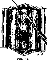
bone, freeing the membranes and S. m. from pressure, removal of scars and adhesions, restoration of circulation of cerebrospinal fluid with hermetic closure of the dura mater and soft tissues. Mortality from injury to the S. m. without surgical intervention is from 65% to 80% (Pansky). Early operations give 60% mortality, and it should be taken into account that the most severe cases are operated on, which without operation usually die or give severe complications requiring operation in the late period, which will give up to 30% mortality. Despite the high percentage of mortality in early operations, the advantage still lies with them, since more patients survive with them than with conservative treatment, and in addition, the development of persistent paralysis is prevented. In cases of damage to the S. m. with persistent paralysis existing for several years, decompressive laminectomy with freeing of the S. m. is also indicated, which sometimes gives good results (Polenov). Surgical treatment of inflammatory diseases of the S. m. in the acute stage--see Meningitis; in the chronic stage it amounts to treatment of meningitis serosa chr. circumscripta, which often occurs with symptoms of an extramedullary tumor, and consists in incising and excising adhesions, and restoring free circulation of cerebrospinal fluid and treatment of meningitis chronica fibrosa, which amounts to excision of scar bands on the dura mater sometimes over several vertebrae. The dura mater sometimes reaches up to 7 cm in thickness and constricts the spinal cord and roots so that it has to be split and the outer part excised, leaving only a thin layer of it. The results are sometimes remarkable. In the disease syringomyelia (see), the operation of Pussepp has recently been successfully applied. Treatment of abscesses of the spinal cord consists of laminectomy and opening of the abscess, care being taken to prevent the pus from leaking into the subarachnoid space of the S. m. The most developed chapter in the surgery of the S. m. is the surgical treatment of tumors. Depending on the location, size of the tumor and the disorders it causes, the operation can be of varying severity. Therefore, the earliest possible surgical intervention is necessary, and the indication for the operation should not only be the establishment of the diagnosis of a tumor, but even suspicion of a tumor. In the latter case, the operation has an exploratory character. The only contraindications may be the serious condition of the patient,
Figure 36.
advanced age and the presence of extensive paralysis with elevated temperature and bedsores. In most cases, the operation can be performed under local anesthesia. If local anesthesia is insufficient, ether anesthesia can be added. For intramedullary tumors, after laminectomy performed under local anesthesia, if necessary, ether anesthesia is added. The patient's position is usually on the side with a roll under the lumbar region with the foot end of the table elevated to prevent the outflow of cerebrospinal fluid. In the case of extradural location of the tumor, the operation consists in removing the tumor after laminectomy without opening the dura mater, if the tumor does not grow through it. In case of growth through the dura mater, the latter is excised and if it is impossible to suture the defect, it is left uncovered or closed with a flap formed
Figure 37. Subdural tumor: 1-dura mater; 2 and 3-parietal and visceral layers of the arachnoid membrane; 4-subdural space; 5-lig. denticulatum; 6-pia mater; 7-subarachnoidal space. tumor grows into the roots, transverse processes or body of the vertebra, it is excised with the roots and curetted from the bone with subsequent painting with Ac. carb. liq. and alcohol. After removal of the tumor and cessation of bleeding, the soft tissues are sutured tightly. In case of intradural location of the tumor, it may be extra- or intramedullary. Often, even upon exposure of the dura mater, it is easy to determine the presence of a tumor by the bulging, density and special bluish color of the dura mater at the site of the tumor. If this is not observed, the presence or absence of pulsation of the brain may serve as a sign of recognition. Absence of pulsation indicates the location of the tumor above the exposed dura mater, to which measures should be directed; in the presence of pulsation, the tumor is located below or it is absent. If with a tumor there is free circulation of cerebrospinal fluid, this sign may also be absent. Then upon opening the dura
Figure 38.
spinal cord, trying as much as possible not to open the arachnoid membrane, probing up and down with a special narrow curved spatula, which upon encountering
Figure 39.
an obstacle in its path. When the tumor is found, it is necessary to make the upper and lower ends of it accessible for removal. With extramedullary location and if the tumor is still behind, its removal usually presents no difficulty (fig. 35-37). If the tumor is adherent or has grown through the dura mater or root, it is removed together with them. To remove the tumor from the anterior surface of the S. m., it is usually necessary to cross 1 or 2 roots and, by turning the S. m. for the denticulate ligament, the tumor can be easily isolated (fig. 38). In the cervical region, care must be taken when crossing roots to avoid damage to the phrenic nerve. Operations are significantly more difficult with intramedullary location of the tumor, when it is necessary to incise and separate the tumor from the brain (fig. 39-41). Isolation of the tumor is facilitated by suturing the tumor with threads, by which the tumor is pulled and carefully extruded from the brain. After removal of the tumor, bleeding is carefully stopped, blood clots are removed and the dura mater is sutured (fig. 42). The muscles are sutured in several layers with catgut sutures and the skin is closed tightly. In non-removable, infiltrating tumors, the latter are left and the dura mater is not sutured to create decompression. Mortality from removal of tumors of the S. m., according to Adson, is 7%, according to Elsberg, out of 120 cases 11%, Heuer, out of 46 cases 19.5%, Petit-Dutail-lis, out of 20 cases 10% after operation and 30% from 2 months to 3 years after operation. "Mortality depending on the location of the tumor: according to Elsberg, out of 106 extramedullary tumors-9.5%, out of 14 intramedullary-21%; according to Petit-Dutail-lis, out of 4 extradural-0 deaths and 2 cases from 1 to 3 years after operation, out of 12 extramedullary-1 case and 2 from 3 to 4 months after operation, out of 4 intramedullary-1 case and 2 from 2 months to 2 years after operation. As can be seen from the results presented, the unfavorable outcome depends
Figure 40.
to a large extent on the location of the tumor extradurally, extra- or intramedullary. The long-term results are exactly the same. Some patients die from recurrences, others from the consequences of compression--paralysis, cystitis, pyelonephritis, bedsores. The prognosis depends on the time, degree and character of the paralysis. If the paralysis exists for more than 6 months, the prognosis is unfavorable, although in rare cases, paralysis existing for years undergoes regression after operation; the prognosis is better for spastic and worse for flaccid paralyzes. The long-term results are clearly presented by Elsberg: out of 95 removed tumors with 8 fatal outcomes, a good result was obtained in 72%, unsatisfactory-in 10.6%, recurrences-in 3% and unknown-5%. In addition to the above surgical interventions, a number of operations have been proposed for spastic paralyzes. This includes resection of the posterior roots of the S. m.- radicotomy (see) for shooting pains in tabetics, for neuralgic pains in cases of non-removable tumors, their metastases and consequences of injuries. Section of the posterior roots Dyi-ix is performed, according to
Figure 41.
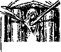
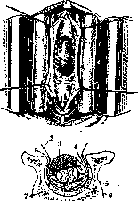
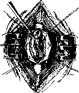
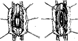
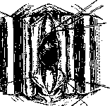
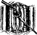
For theoretical crises, according to Förster, intradurally or, according to Guleke, extradurally. In view of the recurrence of pain in half of the cases, it is recommended to perform a more extensive resection from D10 to L1 and even with the severing of the anterior roots, where, according to Lehmann, pain-conducting fibers also pass. For spastic paralyzes of the upper extremities, the severing of the roots C4-5, C6-8 and D1 is performed, according to Förster; for the lower extremities-L2-3, L4 and S5. The results of the operation are not always good, therefore it is used only in severe cases not responsive to any other treatment. According to Eleckiy, out of 97 cases, death occurred in 13 and success only in 49 cases. Resection of the posterior roots for peripheral pains is now almost abandoned as not achieving success. A similar operation has been proposed for severe forms of spastic torticollis. With an incision in the upper part of the neck from behind, the spinous processes and arches of the I-III cervical vertebrae and the lower part of the occipital bone are exposed and excised. The dura mater is incised, the medulla oblongata with the upper part of the spinal cord is exposed, and one- or bilateral severing of the I-III spinal roots and one- or bilateral severing of the spinal part of the accessory nerves is performed. The best result is obtained from the severing of the 1-2 first cervical roots with bilateral severing of the accessory nerve (Olivecrona, Polenov).
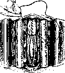
For the treatment of paralysis of the lower extremities, urinary bladder, anastomoses between the roots of the cauda equina have been proposed (Burdenko, Girgov, Shevkunenko), where the paralyzed root is sutured into a healthy one from the opposite side or into a root of the same side but lying above the injury. As a result of such operations, improvement in the function of the paralyzed organ is noted. For malignant tumors, recurrences of tumors causing unbearable pain,
tabetic crises, causalgias, pains in amputation stumps,
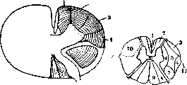
Figure 43.
Figure 44.
Figure 43. Location of conductors of various types of sensitivity: 1-pressure; 2-touch; 3-pain; 4-temperature sensitivity. Figure 44. Scheme of anterolateral cordotomy: 1- direct pyramidal tract; 2-anterior column; 3-Gowers' tract; 4-commissural fibers of gray matter; 5-lateral column; 6-tract of Flechsig; 7-lateral pyramidal tract; 8-tract of Burdach; 9-tract of Goll; 10-scheme of anterolateral cordotomy; 11-denticulate ligament. When irradiating pains do not respond to any conservative and operative treatment on peripheral nerves and posterior roots, severing of the spinothalamic anterolateral tract (fig. 43 and 44) in the spinal cord is proposed - cordotomy (Frazier, Robineau). The operation consists in the resection of 2-3 arches at the level of D8-9 or D10-11. The dura mater is carefully opened without damaging the arachnoid to avoid the outflow of cerebrospinal fluid for better orientation in the roots. The lateral surface of the spinal cord is exposed, the denticulate ligament is severed and the spinal cord is further turned with a clamp so that its anterolateral part is turned backward. Often a root is an obstacle to this, which has to be severed. Then it is easy to see in front of the denticulate ligament a light, glistening, poorly vascularized strip 2 mm wide between it and the anterior root. An incision is made on this space with a special Frazer knife to a depth of 2.5-3 mm. The operation is performed on the side opposite to the pain. In bilateral pains, the severing is done from both sides 1-2 cm below one another to avoid thinning of the cord. Immediately after the severing, pain sensitivity ceases to the corresponding level on the side opposite to the severing. If the incision is made to sufficient depth, the result is permanent. The mortality from the operation for malignant neoplasms reaches up to 25%, but is lower at present. Polenov has 2 deaths out of 40 cordotomies. The result in most cases is good, although in some cases a recurrence of pain occurs, but less intense than before. For pains from malignant neoplasms of internal organs and gastrointestinal crises, where it is necessary to bilateral cessation of pain sensitivity, Armour proposed to sever the anterior commissure, where all pain-conducting fibers terminate at the level of D11-12 and L1. The operation is extremely delicate, as it is difficult to make an incision strictly along the median line in front of the spinal cord and avoid injury to the anterior spinal artery. The operation was performed by Armour with a fatal outcome from pneumonia.
Related articles
Cite this page
“SPINELLI.” Soviet Medical Encyclopedia. English translation of Bolshaya Meditsinskaya Entsiklopediya, 1st ed. (Moscow, 1928–1936), ed. N. A. Semashko. https://sovietmedicalencyclopedia.pages.dev/article/spinelli/