Lumbosacral Plexus
Historical document, translated for reference. It reflects medical knowledge of the 1920s–30s and is not medical advice.
Summary
The lumbosacral plexus is a peripheral nervous system structure that gives rise to motor and sensory nerves of the pelvic girdle, perineum, pelvic organs, and lower limb. It is formed by the union of anterior branches of lumbar and sacral nerves and divides into secondary plexuses.
Encyclopedia article (1928–1936)
LUMBOSACRAL PLEXUS, plexus lumbosacralis, peripheral part of the nervous system, giving rise to motor and sensory nerves of the pelvic girdle, perineum, pelvic viscera, sexual organs, and finally the nerves of the lower limb. It is formed by the union of the anterior branches of nn. lumbales et sacrales and divides into four secondary plexuses: lumbar (plexus lumbalis), sacral (plexus sacralis), pudendal (plexus pudendus) and coccygeal (plexus coccygeus). The lumbar plexus is formed by the connection among themselves of the anterior branches of the first three lumbar roots, to which is joined part of the fourth lumbar, and sometimes also the twelfth thoracic (fig. 1). This connection occurs as follows: the anterior branch of L1, receiving an anastomosis from D12, divides into 3 branches, two of which form the peripheral nerves-nn. ilio-hypogastricus and ilio-inguinalis (fig. 2); the third connects with the anterior branch of the L2 root and also breaks down into 3 branches-n. femoro-cutaneus, n. genito-cruralis and an anastomosis for the L3 root, which, after giving off a branch for n. obturatorius, forms n. femoralis, s. cruralis. The anterior branch of the L3 root also divides into 3 branches-one enters into the composition of n. cruralis, another into n. obturatorius, and the third connects with the L4 root, forming truncus lumbo-sacralis, which enters into the composition of plexus sacralis. The size of the branches of the plexus increases downward: L1 has a diameter of about 2.5 li*, and L4 and L5 about 6 li*. -Position. The lumbar plexus is located in the thickness of m. psoas, between its two layers, in front of the proc. costo-transversus of the lumbar vertebrae; together with the plexus, the a. et v. lumbales are located there. The lumbar plexus forms anastomoses with n. intercostalis XII, with plexus sacralis and with n. sympathicus. On its path it gives short and long collaterals and terminal branches: short branches for m. quadratus lumborum (from L1) and for mm. psoas major et minor; of the long branches: 1) n. ilio-hypogastricus and 2) n. ilio-inguinalis, resembling nn. intercostales; they go in the thickness of the abdominal wall, give motor fibers to its muscles and sensory branches to the skin of the corresponding parts of the abdominal wall, to the upper-outer part of the buttock, to canalis inguinalis and to the upper parts of the inner surface of the thigh; 3) n. femoro-genitalis, almost exclusively sensory, innervates the skin on the front-inner surface of the thigh, on scrotum and the skin of the labia; 4) n. cutaneus femoris lat., sensory, innervates the upper-outer parts of the buttock and the outer surface of the thigh. The terminal nerves of the lumbar
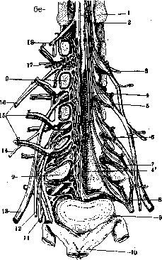
Figure 1. Lumbar plexus: 1-spinal cord; 2-dura mater; 3- L1, 4 and 4'-cauda equina; 5 - L2; 6-L3; 7 - L4; 8 - L5; 9 - spine; 10- v. cava inf.; 11- n. lumbo-sacralis; 12-n. obturatorius; 13-n: cruralis; 14-n. cutaneus femoris lat.; 15- a. et v. lumbalis; 16-n. ilio-hypogastricus; 17-plexus venosus in the spinal cord; 18-D12-
plexus: 1) n. femoralis and 2) n. obturatorius (see Human Nerves).,-Physiology. The lumbar plexus controls the movements of the pelvic girdle, hip and knee joints-flexes, extends the thigh; abducts, adducts, rotates outward, extends the leg, also participating in its flexion; complements the function of nn. intercostales-bends the spine to its side, flexes the trunk, acts
on the abdominal press. The sacral plexus is formed from the connection of truncus lumbo-sacralis with the anterior branches of S1, S2 and part of S3 (fig. 3). The composition of tr. lumbo-sacralis includes the anterior branches of L4 and L5; it descends into the pelvis and near foramen ischiadicum magnum connects with the remaining roots. The sacral plexus has the shape of a triangle, from the apex of which emerges the largest nerve of the body-n. ischiadicus. The length of the roots entering the plexus varies, downward it decreases, as does the thickness of the trunks. The sacral plexus
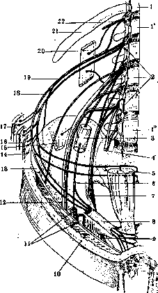
Figure 2. Lumbar plexus: 1-D12; Г - L2; 1" - L3; 2-truncus et ganglion sympathicus lumbalis; 3-m. psoas; 4- n. lumbo-sacralis; 5 - m. rectus abdominis; 6-n. genito-cruralis; 7-n. obturatorius; 8-canalis inguinalis; 9- for. obturatorium; 10-aponeurosis; 11-v. et a. iliaca ext.; 12- n. cruralis; 13-spina ossis ilii ant.; 14-m. transversus; 15-m. iliacus; 16-m. obliquus abdominis int.; 17-m.obliquus abdominis ext.; 18-n. ilio-hypogastricus; 19-n. ilio-inguinalis; 20-m. quadratus lumborum; 21-XII rib; 22-n. intercostalis. (After Testut) is located in the inner-posterior part of the pelvis on m. piriformis; truncus lumbo-sacralis extends beyond it upward, and below it-an anastomosis between S2 and S3 roots. Between the muscle and the plexus are a. et v. ischiadicae, a. et v. pudendae. The anterior surface of the plexus is covered with a fibrous plate, forming part of the aponeurosis of the pelvis and extending from the corresponding intervertebral foramina to foramen ischiadicum; inside from it is the parietal leaf of the peritoneum; both these leaves separate the plexus from a. et v. hypogastricae, from the sympathetic trunk, from the rectum and from the loops of the small intestine; in women-also from the uterus, ovaries and tubes. The apex of the plexus at the level of foram. ischiadici-without sharp boundaries passes into n. ischiadicus. Anastomoses: through truncus lumbo-sacralis the sacral plexus connects with the lumbar plexus, through S3-with the pudendal; through rami communicantes-with neighboring parts of the sympathetic trunk. From the sacral plexus fibers go for the pelvic
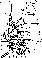
Figure 3. Formation of the sacral plexus: L4, L5, S1, S2, S3, S4, S5, C- lumbar, sacral and coccygeal roots of the spinal cord: /- n. sacralis; 2-nn. hypogastrici; 3-rami communicantes; 4- gngl. sympathicum sacralis; 5-anastomosis; 6-gngl. sympathicum "Walter; 7-gngl. hypogastricum; 8-a. et n. pudendus; 9-nerve for m. obturator internus; 10-nerve for m. levator ani; 11-plexus pudendus: 12-n. ischiadicus; 13-n. glutaeus superior.
girdle and to the lower limb (fig. 4). The first include nerves for mm. piriformis, obturator internus, gemelli superior et inferior, quadratus femoris; n. glutaeus superior to mm. glutaei medius et minimus, tensor fasciae latae; n. glutaeus inferior to m. glutaeus maximus. The nerves for the lower limb include 1) n. cutaneus femoris posterior, exclusively sensory, supplying the skin of the gluteal region, perineum, scrotum and posterior surface of the thigh, 2) n. ischiadicus (see Ischiadicus nervus). -Physiology of the sacral plexus. With its collateral branches it serves the pelvic girdle-abducts and rotates the thigh outward, extends it in the hip joint; when standing straightens the trunk or bends it to the corresponding side; controls the sensitivity of the posterior surface of the thigh and upper parts of the leg, partially the gluteal region, perineum and scrotum. The terminal branch controls the movements and sensitivity of the lower limb (see Ischiadicus nervus). The pudendal plexus (plexus pudendus) originates from the connection of the lower half of the anterior branch of the S3 root with S4; it lies under the lower edge of m. piriformis on the anterior surface of m. coccygeus. Through S3 it connects with plexus sacralis, S4 connects it with plexus coccygeus, and rami communicantes-with n. sympathicus. From the pudendal plexus depart 1) parietal branches-for the walls of the lower part of the trunk and 2) visceral branches-for the pelvic organs. From the branches should be noted: 1) rami musculares to m. levator ani and m. coccygeus; 2) nn. haemorrhoidales medii, branching on the rectum and in m. levator ani; 3) nn. vesicales inferiores, going to the bottom of the bladder and to the circular muscles of urethra vesicae; 4) nn. vaginales-to the vagina; 5) n. pudendus, giving n. perforans ligamentum sacro-tuberosum, which after piercing the ligament goes to the area of tuber ischiadicum and in the skin over m. glutaeus maximus, dividing into three terminal branches: a) n. haemorrhoidalis inferior, branching in the skin of the anus and in sphincter ani externus; b) n. perinaei, giving: a) rami musculares to m. transversus perinaei superficialis, m. sphincter ani externus, m. bulbo- and ischio-cavernosus; b) nn. scrotales (labiales) posteriores, s. nn. perinaei mediales, to the skin of the perineum and scrotum (labia majora);гv) n. perinaei lateralis to m. ischio-cavernosus and the lateral area of the perineum; g) n. dorsalis penis (clitoridis). The coccygeal plexus (plexus coccygeus) is formed from the anterior branches of S5 and Co. Represents a very small formation of very thin nerves; located
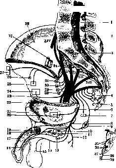
Figure 4. Sacral plexus: L1, L2, L3, L4, L5, S1, S2, S3, S4, S5. Lumbar, sacral, and coccygeal roots of the spinal cord; 1-cauda equina; 2-5th lumbar vertebra; 3-1st sacral vertebra; 4-nerves for internal organs; 5-coccyx; 6-n. pudendus; 7-m. levator ani; 8-tuber ischii; 9-m. sphincter ani; 10-m. ischio-cavernosus; 11-m. transversus; 12-n. perinaei; 13-m. bulbo-cavernosus; 14-n. perinealis superficialis; 15-n. scrotalis; 16-n. urethralis; 17-n. dorsalis penis; 18-urethra; 19-corpus cavernosum; 20-m. obturator int.; 21 and 23-os pubis; 22-n. ischiadicus; 24-m. glutaeus magnus; 25-m. glutaeus medius; 26-m. glutaeus minimus; 27-fascia lata; 28-m. quadratus lumborum; 29-m. gemellus inf.; 30-m. gemellus sup.; 31-m. pyramidalis; 32-a., et v. iliaca ext.; 33-iliac bone; 34-truncus lumbo-sacralis; 35-plexus hypogastricus; 36-ramus cutaneus; 37-ramus internus; 38-ramus externus; 39-m. ischio-coccygeus.
In front of m. coccygeus and lig. sacro-spinosum. In the upper parts, through S4 it is connected with plexus pudendus, and rami communicantes connect it with the lower sacral ganglia and with gngl. coccygeum. From the plexus arises n. anococcygeus, ending with branches in the skin between anus and coccyx. Rami musculares go to mm. coccygeus and levator ani. 21
Pathology of the lumbosacral plexus. The lumbosacral plexus, due to its great extent, can be affected by various processes and in various constituent parts. Diseases of the lumbosacral plexus (plexitis lumbo-sacralis, s. neuritis plexus lumbo-sacralis) are most often of traumatic origin - direct injury to the plexus by some piercing object; compression by bone fragments in fractures of the spine, pelvic bones, tumors developing in surrounding structures, or by aneurysms of nearby arteries; the uterus in an abnormal position or in pregnant women can press on one or another part of the plexus; the plexus can be involved in the process in diseases of the pelvic organs (ovaries, tubes, appendix, etc.). Various infections, intoxications, and auto-intoxications can be the cause of inflammation of the plexus. Various professions more likely cause diseases not of the plexus, but of its branches (see Ischiadicus nervus, Ischias, Femoralis nervus). Usually, the disease of the plexus is unilateral, but it is also observed on both sides in some infectious diseases, in lesions of the spine. (Path.-anat. changes--see Brachial plexus and Neuritides.) Clinically, plexitis lumbo-sacralis is detected in 2 forms: in paralytic form and in neuralgic form. The paralytic form in case of affection of the entire plexus is characterized by paralysis of all muscles innervated by the branches of the plexus, i.e., all muscles of the pelvic girdle and lower limb; the paralysis is flaccid, accompanied by atrophy and flabbiness of the paralyzed muscles. Tendon reflexes are absent, in the initial stage their increase can sometimes be observed. Sensibility is disturbed on the lower limb and on the skin of the pelvic girdle; usually all types of sensibility are affected; the degree of anesthesia depends on the intensity of the process. Spontaneous pains as well as pains on pressure on the plexus are also observed. For plexitis lumbo-sacralis, trophic disturbances are characteristic - abnormal growth of nails, swelling of the skin, increased sweating, stiffness of joints. Pelvic organs are disturbed if the process involves the pelvic plexus. In partial affection of the plexus, the clinical picture changes depending on the localization of the process in different parts of the plexus. In affection of the upper roots, lumbar plexus, whose motor function is limited, only pareses of the corresponding muscles are observed, disturbance of sensibility on the buttock and on the thigh (on the anterior and partly on the lateral surface). The lower roots of the lumbar plexus, on the contrary, have a very important motor function, as they innervate the anterior and internal muscles of the thigh, partly the posterior muscles and muscles of the leg (tibialis anticus), due to which flexion and adduction of the thigh, extension of the leg are disturbed; standing and walking are difficult; disturbance of sensibility is present on the anterior surface of the thigh, knee, on the internal surface of the leg and foot. Patellar reflex is absent. Affection of individual branches of the lumbar plexus is accompanied by more isolated and less intense symptoms. Disease of nn. ilio-hypogastrici and ilio-inguinalis causes disturbance of sensibility in the lower parts of the abdominal wall and some weakness of its muscles, in the innervation of which these nerves participate together with nn. intercostales. N. femoro-cutaneus is an exclusively sensory nerve, its affection causes anesthesia on the lateral surface of the thigh, and its irritation gives neuralgia known under the name meralgia paraesthetica (see). Disease of n. genito-cruralis is also expressed exclusively by disturbance of sensibility - painful hyperesthesia (hyperaesthesia dolorosa) in the area of the upper part of the thigh and scrotum. N. obturatorius is a mixed nerve; in its affection incomplete paralysis of the adductor muscles of the thigh is observed, as mm. adductores receive innervation also from other nerves (nn. femoralis et ischiadicus); the paralysis is accompanied by atrophy of the muscles. Clinically, the disease of n. obturatorii is expressed in that when walking the patient carries the leg outward; the affected leg cannot be placed over the other; disturbance of sensibility occupies a small triangle on the internal surface of the thigh. Affection of n. obturatorii is observed very rarely, as its trunk is very short and protected by the bones and muscles of the pelvic girdle. Disease of the sacral plexus is clinically manifested mainly by symptoms of affection of its terminal branch-n. ischiadici (see Ischiadicus nervus, Ischias), to which are added symptoms of collaterals innervating the muscles of the pelvis and perineum. The clearest picture is given by affection: 1) of n. glutaei inf., innervating m. glutaeus maximus, extending the thigh; paralysis of this muscle is revealed when running, jumping, ascending stairs or an inclined plane, but when walking on a level surface the paralysis is not especially noticeable; 2) of n. glutaei sup., in charge of abduction and rotation of the thigh. Paralysis of the muscles is accompanied by decrease in their tone, which causes on each step when walking dislocation of the thigh (dehanchement). Affection of n. cutanei femoris post. (exclusively sensory) is characterized by anesthesia in the area of the buttock, on the posterior surface of the thigh and upper part of the leg; its irritation causes neuralgia in the areas it innervates, differing from neuralgia of n. ischiadici by its topography, superficial pains, absence of Lassegue's symptom and painful points along the course of n. ischiadici and cutaneous hyperesthesia in the corresponding areas. - Affection of the pelvic and coccygeal plexuses is accompanied by disturbance of sphincters (incontinence of urine and feces), paralysis of the urinary bladder and muscles of the perineum (m. levator ani, mm. ischio- et bulbo-cavernosus), anesthesia of the rectum, bladder, skin, on the internal half of the buttock, area of the perineum and ani, posterior surface of the genital organs. Irritation of these plexuses causes neuralgia spermatica, neuralgia in the area of the sacrum (coccygodynia) and is observed in women in diseases or abnormal position of the genital organs. In the neuralgic form of inflammation of the lumbosacral plexus, the main symptom is painful sensations along the course of the nerve trunks emerging from the plexus; in general the clinical picture resembles the picture of the disease in inflammation of n. ischiadici (see Ischias).-The development and course of the disease depend on the etiological factor; in some cases the process can stop and all functions can be restored; in others the process stops but functions are not restored; finally there are also cases in which the process does not subside, progresses further, involving new parts of the plexus. The neuralgic form in one series of cases develops acutely, in another subacutely. In acute development the inflammation can later subside, lasting for a certain time, or takes a prolonged course, as in subacute forms, and lasts for a long time.---The differential diagnosis of plexitis lumbo-sacralis does not present great difficulties when the patient has all the symptoms of this disease and when accurate information about the course of the pathological process is obtained. In the absence of usual symptoms differentiation has to be made with poliomyelitis anterior, myopathies (see), with diseases of peripheral nerves (n. femoralis, n. ischiadicus), roots (see Radiculitides).-The prognosis is unfavorable for forms with progressive course, much better in cases where the disease does not have a progressive character. The prognosis also depends on the etiological factor: paralyses of the plexuses depending on injuries give a bad prognosis in the sense of recovery.--Treatment and prevention of diseases of the lumbosacral plexus are the same as in diseases of individual nerves (see Neuritides, Neuralgia, Polyneuritides, Brachial plexus, individual nerves).
Related articles
Mentioned in
Cite this page
“Lumbosacral Plexus.” Soviet Medical Encyclopedia. English translation of Bolshaya Meditsinskaya Entsiklopediya, 1st ed. (Moscow, 1928–1936), ed. N. A. Semashko. https://sovietmedicalencyclopedia.pages.dev/article/lumbosacral-plexus/