Human Nerves ([For anatomy, physiology and pathology of)
Historical document, translated for reference. It reflects medical knowledge of the 1920s–30s and is not medical advice.
Summary
This article provides a comprehensive overview of human nerves, covering their anatomy, physiology, and pathology. It details the systematic classification of peripheral nerves, their origins, pathways, branches, functions, and variations.
Encyclopedia article (1928–1936)
Human Nerves. [For anatomy, physiology and pathology of nerves, see article Nerves in Volume XX; there (pp. 667-782) are illustrations of Human Nerves]. Below is a table of nerves, systematically presenting the most important aspects of the anatomy and physiology of each peripheral nerve (its origin, relation to segments, course, branches, connections with other nerves, place of termination, general topography, function, variations). * When examining a patient, familiarity with all this data is necessary for establishing a topological diagnosis of a disease of a given nerve and especially for surgical intervention on it: * * Bilateral symmetry observed in spinal nerves, presence of a spinal ganglion at the beginning of each nerve - allows us to consider the nerve as a segmental formation, preserving rather clear traces of metamerism, which has almost disappeared in the central nervous system, where it can be traced only through comparative anatomical and embryological research. The nerve segment consists of a central organ, from which roots emerge - anterior (motor) and posterior (sensory); the latter has a thickening on its path - the intervertebral ganglion, outward from which both roots connect, forming a mixed spinal nerve - nervus spinalis, which divides into 4 branches (see Nerves). Each nerve segment innervates the corresponding segment of the body. This innervation is symmetrical on both sides, but on both the anterior and posterior surfaces, there is a transition of branches to the other side for a short distance (into the skin and muscles). The nerve segment in the indicated form is preserved only in the thoracic region, while in other regions (in the cervical, lumbo- * In Volume XX (pp. 667-782) are illustrations of most nerves listed in this table. All Latin terms in the illustrations are included in the Subject Index to this volume. Synonyms and Russian terms are also included in the Subject Index. ** Tables of plexuses and ganglia will be placed in the articles Nerve Plexuses and Nerve Ganglia. References printed in italics refer to these articles. References printed in regular font refer to the nerve table (for example: "See p. facialis", one should look for "Facialis"). sacral) there is a displacement of nerves over considerable distances (formation of plexuses - see Nerve Plexuses), and only through careful research can the connection of any peripheral nerve with the corresponding segments of the spinal cord be established. The second important point noted in the table is the anastomoses between nerves. Indeed, the term "anastomoses" in relation to nerves can be used only very conditionally (types of anastomoses - see Nerves, Vol. XX). The presence of these anastomoses explains how a nerve, motor in origin, becomes mixed, as well as how vegetative fibers enter the cerebrospinal nerves. At their beginning, nerves lie deeply and close to the skeleton, then, with few exceptions, become more superficial. Fibers in the nerve are arranged in a certain order: short fibers, for the most part, occupy a peripheral position, in the center are located the terminal long fibers; therefore, in a mixed nerve, sensory fibers occupy the central position in most nerves, while motor fibers are located more peripherally (see also Nerve Fibers). Such a deep position of sensory fibers in the nerve explains the preservation of sensitivity to a greater or lesser degree with contusion or compression of the corresponding nerve, and disturbance of sensitivity with transection of the nerve. g- As for the anatomy of the nerve, here it should be noted a very important fact - the presence of variations and anomalies that have great clinical significance. Anomalies in the formation of nerves are expressed in the fact that the nerve originates not from the same roots as usual. As for anastomoses, the latter may either be absent or, conversely, be present in greater numbers or in other places along the nerve. Variations in relations to surrounding structures, particularly to blood vessels, must be taken into account in operations on nerves, when it is easy to injure blood vessels located in unusual places. Anomalies in distribution are more often observed for sensory nerves, less frequently for motor nerves. Nerves Latin name Russian name Synonyms Origin. To which segment it corresponds With which nerves it anastomoses What branches arise from it 1: Abducens (abductor). Syn.: oculo-muscularis externus, par sextum nn. encephali Medulla oblongata - rhomboid fossa, colliculus facialis. Exits along the posterior edge of the pons between it and the pyramid Sympathetic fibers from sinus cavernosus and sensory fibers from n. ophthalmicus Peripheral Accessorius: r. internus and r. externus (accessory nerve, Willisii, XI cranial N.). Syn.: Willisi, recurrens, respiratorius externus, par undecimum The nucleus of the accessory nerve lies: a) in the medulla oblongata: in the mass of the floor of the IV ventricle, laterally from nucleus n. hypoglossi and the sensory nucleus of nn. vagi and glosso-pharyngei, behind their motor nucleus - nucleus ventralis, s. ambiguus, being a continuation of the latter backward; the nucleus extends forward to the posterior third of olivae inferioris; this nucleus gives r. internus; b) in the spinal cord: in the anterior horns of the spinal cord dorso-laterally from the medial cellular group in the form of a continuous cellular mass up to the level of the exit of V (VI or even VII) cervical nerves; this nucleus gives r. inferior, s. externus From the posterior roots of the three upper cervical nerves, receives motor roots from III(IV) cervical nerve and sensory fibers from the vagus and III and IV cervical 1) R. internus (accessorius vagi), 2) r. externus (accessorius spinalis) Acusticus: radix vestibularis et radix cochlearis (Russian: vestibular root, or vestibular nerve, or static nerve, or nerve of equilibrium, and cochlear root, or nerve of the cochlea or proper auditory nerve) Radix vestibularis begins in the nuclei of the vestibule from their ganglion cells; peripheral processes of these cells form five nerves and end in the following five places: I. Ampulla membranacea superior of the superior semicircular canal - its neuroepithelium (crista ampullaris) - this is nervus ampullaris superior. 2. Ampulla membranacea lateralis of the lateral semicircular canal - its neuroepithelium (crista ampullaris) - this is n. ampullaris lateralis. 3. Ampulla membranacea posterior of the posterior semicircular canal - its neuroepithelium (crista ampullaris) - this is n. ampullaris posterior. 4. Utriculus membranaceus - its neuroepithelium (macula acustica utriculi) - this is nervus utricularis. 5. Sacculus membranaceus - its neuroepithelium (macula acustica sacculi) - this is n. saccularis. Central fibers of these ganglion cells in the form of radix, s. n. vestibularis go as part of the auditory nerve to the medulla oblongata and end in three nuclei 1) R, communicans (Arnold!) externus and 2) internus, both with nervus facialis I. Radix, s. n. vestibularis: 1) n. ampullaris superior, 2) n. ampullaris lateralis, 3) n. ampullaris posterior, 4) n. utricularis, 5) n. saccularis. II. Radix, s. n. cocnlearis of humans. What do they innervate and where does the nerve end M. rectus lateralis oculi Topography of the nerve and its branches Character of the nerve and its function. Variations 1) Go into the path of n. vagi, 2) m. sterno-cleido-mastoideus and m. trapezius Having emerged from the medulla oblongata along the posterior edge of the pons, the nerve is directed along its ventral surface forward and laterally, pierces the dura mater, lies in the sinus cavernosus medially from the trigeminal nerve and laterally from the oculomotor nerve and laterally from the internal carotid artery, passes through the superior orbital fissure into the orbit, where it lies under the oculomotor nerve, and then between both origins of m. rectus lateralis oculi and penetrates this muscle from its medial side Having emerged from the medulla oblongata in two or three bundles from the sulcus lateralis posterior dorsally from the olive and caudally from the roots of the vagus nerve (ramus internus) and in several bundles from the spinal cord between the posterior and anterior roots of the upper cervical nerves (ramus externus) and merging into one trunk, the nerve in one common sheath of the dura mater together with the vagus nerve passes through the jugular foramen and divides outside the cranial cavity again into both its branches, which diverge: the external branch passes into nervus vagus and follows together with it in rami pharyngei, laryngei and cardiaci n. vagi; the external branch, having received sensory fibers from the vagus nerve and its nucleus, is directed outward backward and downward, lies laterally from the vagus nerve and caudally from the internal jugular vein, reaches the medial surface of m. sterno-cleidomastoidei, pierces the muscle, or, giving it some of its branches, passes with its other branches through regio lateralis colli to musculus levator scapulae, reaches musculus trapezius, lies on its medial surface and enters its mass, at the border of the middle and lower thirds Motor, but in its course sympathetic fibers from plexus caroticus internus and sensory fibers from the oculomotor nerve also pass. Abducts the eye outward.
N. may divide into two, may give a branch to ganglion ciliare, may be absent. Purely motor, but sensory fibers from the vagus nerve and from cervical nerves also pass through it. R. internus, going along the path of the vagus nerve, participates in the innervation of the pharynx, larynx, and heart. R. externus innervates m. sterno-cleido-mastoideus and musculus trapezius. N. may end in m. sterno-cleido-mastoideus or ramus externus, may divide into two separate branches for each muscle separately, may anastomose with ramus descendens nervi hypoglossi, with n. phrenicus, with ganglion cervicale superius. Having emerged from the medulla oblongata caudally from the pons and laterally from the facial nerve, the nerve is covered together with the facial nerve by a common process of the dura mater, enters the pons and meatus acusticus internus, connects here with the facial nerve by means of fila anastomotica and ends with both its roots in the above-mentioned places. In the depth of meatus acusticus internus it divides by means of crista transversa into two floors; in the upper, which in turn is also divided into two fossae, passes n. facialis in the anterior fossa-area nervi facialis, and in the posterior nervus vestibularis-area nervi vestibularis superior; in the lower - radix cochlearis: in the anterior fossa-area cochleae, which has the form of a spiral series of openings-tractus spiralis foraminosus and part of the fibers of n. vestibularis, in the posterior fossa-area vestibularis inferior.
Sensory. Radix vestibularis serves in the statics of the organism as a connection between the peripheral static apparatus (vestibule and semicircular canals) on the one hand and the brain on the other. Radix cochlearis- auditory nerve № Latin and Russian name- Synonyms Beginning. To which segment does it correspond With which nerves does it anastomose What branches arise from it of the rhomboid fossa: nucleus medialis (Schwalbe), lateralis (Deitersi) et superior (Bechterew). Radix cochlearis begins in the spiral nucleus of Corti, which is located in the mass of the bony spiral lamina of the cocheta-lamina spiralis ossea-in its canalis spiralis modioli; the peripheral processes of these cells go in the mass of the spiral lamina of the cocheta in its fissura spiralis to the cells of Corti's organ; the central fibers of these ganglion cells are directed in the form of radix, s. n. cochlearis to the medulla oblongata as part of the auditory nerve and end in two nuclei of the rhomboid fossa: nucleus ventralis et nucleus dorsalis, s. tuberculum acusticum.
Alveolares inferiores anteriore? (lower anterior alveolar nerves) 5| Alveolares Inferiores posteriores | (lower posterior alveolar nerves) See n. alveolaris inferior See n. alveolaris inferior 6; Alveolares superiores: posteriores, medius et anterior (upper alveolar nerves: posterior, middle and anterior). Syn.: nn. dentales superiores: anterior, medius et posteriores Nervus infraorbitalis on its course from the pterygopalatine fossa to the infraorbital foramen 1) Rr. alveolares superiores posteriores, 2) r. alveolaris superior medius, 3) r. alveolaris superior anterior 7 Alveolares superiores posteriores (posterior superior alveolar branches) See nn. alveolares superiores Alveolaris inferior (lower alveolar nerve). Syn.: dentalis inferior, s. mandibularis N. lingualis N. mandibularis 1) N. mylo-hyoideus (see), 2) nn. alveolares inferiores posteriores (lower posterior alveolar nerves), 3) nn. alveolares inferiores anteriores (anterior lower alveolar nerves), 1» What they innervate and where the nerve ends Topography of the nerve and its branches Character of the nerve and its function. Variations See n. alveolaris inferior See n. alveolaris inferior Participate in the formation of the superior dental plexus and give branches: a) rr. dentales superiors and b) rr. gingivales superiores.
1. Rami alveolares superiores posteriores lie on facies posterior of the upper jaw-on its tuber maxillare, pierce the bone, enter the maxillary sinus, go along its posterior lateral wall, form the superior dental plexus, which lies partly under the mucous membrane of the sinus and partly in the mass of the bone, and give branches to the mucous membrane of the maxillary sinus, posterior part of the cheek, to the alveoli of the molar teeth and to the gums around them. 2. Ramus alveolaris superior medius begins in the infraorbital canal, enters the maxillary sinus, participates in the formation of the superior dental plexus and ends in the alveoli of the premolar teeth and in the corresponding parts of the gum. 3. Ramus alveolaris superior anterior begins in the infraorbital canal near its exit opening, enters the maxillary sinus, goes along its anterior wall, participates in the formation of the superior dental plexus and ends in the alveoli of the incisors and canines, in the teeth themselves and in the corresponding parts of the gum and in the mucous membrane of the nasal cavity-ramus nasalis. Sensory. Innervates the mucous membrane of the cheek in its posterior part, the mucous membrane of the maxillary sinus, the gums of the upper jaw, the teeth of the upper jaw, as well as partly the mucous membrane of the nasal cavity See nn. alveolares superiores 1) M. mylo-hyoideus et venter anterior m. digastrici and the skin of the chin, 2) molar and premolar teeth and the gum around them, 3) canine and incisors and the gum around them.
(with the cervical plexus) 151 Anastomotic rami dorsalis manus (n. ulnaris) cum ramo superficiali nervi radialis (rr.) [anastomoses of the dorsal branch of the ulnar nerve (digital nerves) with the superficial branch of the radial nerve] See p. hypoglossus and nervi cervicales See nn. digitales dorsales 16 Anastomoticus cum plexu tympanico nervi facialis (r.) (anastomosis of the facial nerve with the tympanic plexus) 17 Anastomoticus ganglii otici cum chorda tympani (r.) (anastomosis of the otic ganglion with the chorda tympani) Anastomoticus ganglii otici cum nervo auriculo-temporali (r.) (anastomosis of the otic ganglion with the auriculotemporal nerve) See p. facialis See Nervous nodes (ganglion oticum) and p. chorda tympani See Nervous nodes (ganglion oticum) Anastomoticus ganglii otici cum n. spinoso (r.) (anastomosis of the otic ganglion with the spinous nerve) See Nervous nodes (ganglion oticum) Anastomoticus nervi auricularis posterioris (r.) (anastomosis of the posterior auricular nerve with the lesser occipital) See p. auricularis posterior and p. occipitalis minor Anastomoticus nervi digastrici (r.) (anastomosis of the digastric nerve) See p. digastricus and p. glosso-pharyngeus What do they innervate and where does the nerve end Topography of the nerve and its branches Character of the nerve and its function. Variations 4) the skin of the chin and the lower lip through the mandibular canal via the mental foramen as the terminal nerve, which is called 4) nervus mentalis; the latter immediately divides into 2 groups of branches: a) rr. mentales, which pierce the triangularis and quadratus labii inferioris muscles and branch in the skin of the chin, and b) rami labiales inferiores, which branch in the skin of the lower lip See nn. alveolares superiores See nn. alveolares superiores See p. acusticus See p. acusticus See p. acusticus See p. hypoglossus and nervi cervicales See nn. digitales dorsales See p. lacialis See Nervous nodes (ganglion oticum) and p. chorda tympani See Nervous nodes (ganglion oticum) See Nervous nodes (ganglion oticum) See p. auricularis posterior and p. occipitalis minor See p. digastricus and p. glosso-pharyngeus ^ Latin name and number |
Russian. Synonyms 22 Anastomoticus nervi facialis cum nervo auriculari vagi (r.) (anastomosis of the facial nerve with the auricular branch of the vagus) 23 Anastomoticus nervi hypoglossi cum ganglio cervicale superiore (r.) (anastomosis of the hypoglossal nerve with the superior cervical ganglion) Beginning. What segment does it correspond to With which nerves does it anastomose What branches arise from it See p. facialis and p. vagus See p. hypoglossus Anastomoticus nervi hypoglossi cum ganglio nodoso nervi vagi (r.) (anastomosis of the hypoglossal nerve with the nodose ganglion of the vagus nerve) See p. hypoglossus 28! Anastomoticus nervi lacrimalis (r.) (anastomosis of the lacrimal nerve with the zygomatic) 26 Anastomoticus nervi laryngei superioris cum ganglio cervicali superiore (r.) (anastomosis of the superior laryngeal nerve with the superior cervical ganglion) Anastomoticus nervi laryngei superioris cum nervo laryngeo inferiore (r.) (anastomosis of the superior laryngeal nerve with the inferior laryngeal) Anastomoticus nervi laryngei superioris cum plexu carotico commune (r.) (anastomosis of the superior laryngeal nerve with the common carotid plexus) See p. lacrimalis and p. zygomaticus See p. laryngeus superior See p. laryngeus superior See p. laryngeus superior 29; Anastomoticus nervi laryngei superioris cum plexu pharyngeo (r.) (anastomosis of the superior laryngeal nerve with the pharyngeal plexus) See p. laryngeus superior Anastomoticus nervi lingualis cum chorda tympani (r.) (anastomosis of the lingual nerve with the chorda tympani) See p. lingualis and chorda tympani 31 Anastomoticus nervi lingualis cum ganglio submaxillare (r.) (anastomosis of the lingual nerve with the submaxillary ganglion) See p. lingualis Anastomoticus nervi lingualis cum nervo alveolari inferiore (r.) (anastomosis of the lingual nerve with the inferior alveolar nerve) Anastomoticus nervi lingualis cum nervo hypoglosso (r.) (anastomosis of the lingual nerve with the hypoglossal) See p. lingualis See p. lingualis 34 Anastomoticus nervi mediani cum nervo musculo-cutaneo (r.) (anastomosis of the median nerve with the musculocutaneous nerve) 35 Anastomoticus nervi oculomotorii cum nervo ophthalmico (r.) (anastomosis of the oculomotor nerve with the ophthalmic) See p. medianus See p. oculomotorius and p. ophthalmicus What do they innervate and where does the nerve end Topography of the nerve and its branches Character of the nerve and its function. Variations See p. facialis and p. vagus See p. hypoglossus See p. hypoglossus See p. lacrimalis and p. zygomaticus See p. laryngeus superior See p. laryngeus superior See p. laryngeus superior See p. laryngeus superior See p. lingualis and chorda tympani See p. lingualis See p. lingualis See p. lingualis See p. medianus See p. oculomotorius and p. ophthalmicus 1 i № i Latin name and Russian. Synonyms Beginning. What segment does it correspond to With which nerves What branches arise from it Anastomose Anastomoticus nervi oculomotorii cum plexu cavernoso (r.) (anastomosis of the oculomotor nerve with the cavernous plexus) See p. oculomotorius Anastomoticus nervi ophthalnici cum nervo abducente (r.) (anastomosis of the ophthalmic nerve with the abducens) See p. ophthalmicus and p. abducens Anastomoticus nervi ophthalnici cum nervo trochleari (r.) (anastomosis of the ophthalmic nerve with the trochlearis) See p. ophthalmicus and p. trochlearis 39! Anastomoticus nervi peronaei profundi (r.) (anastomosis of the deep peroneal nerve with the superficial peroneal nerve) See p. peronaeus profundus 40 Anastomoticus nervi recurrentis (vagi) cum ganglio cervicale medio et inferiore (r.) (anastomosis of the recurrent nerve with the middle and inferior cervical ganglia) See p. vagus and recurrens Anastomoticus nervi vagi cum nervo glosso-pharyngeo (r.) (anastomosis of the vagus nerve with the glossopharyngeal) See p. vagus and p. glosso-pharyngeus Anastomoticus nervi vagi cum nervo hypoglosso (r.) (anastomosis of the vagus nerve with the hypoglossal) See p. vagus and p. hypoglossus Anastomoticus peronaeus nervi ischiadici (r.) (peroneal anastomosis) See pp. ischiadicus, cutaneus surae medialis et suralis Anastomoticus peronaeus (r.) (anastomosis of the peroneal nerve to the lateral cutaneous nerve of the leg) See pp. cutaneus surae lateralis et medialis Anastomoticus rami volaris manus (n. ulnaris) cum n. mediano - r. superficialis (n. digitalis volaris communis III) (r.) (anastomosis of the palmar branch of the ulnar nerve to the superficial branch of the median nerve) See p. ulnaris Anastomoticus ulnaris (nervi radialis) (r.) (anastomosis of the ulnar and radial nerves) See pp. radialis et ulnaris Ano-coccygeus (anorectal-coccygeal nerve) Ansa hypoglossi (loop of the vagus nerve). Syn.: p. descendens cervicis, s. descendens hypoglossi, s. cervicalis descendens, s. descendens noni Plexus coccygeus-ansa sacro-coccygea. Sympathetic nerves from ganglion sacrale et ganglion coccygeum 1) Muscle branches, 2) skin branches See p. hypoglossus What do they innervate and where does the nerve end Topography of the nerve and its branches See p. oculomotorius Character of the nerve and its function. Variations See p. ophthalmicus and p. abducens See p. ophthalmicus and p. trochlearis See p. peronaeus profundus See p. vagus and recurrens See p. vagus and p. glosso-pharyngeus See p. vagus and p. hypoglossus See nn. ischiadicus, cutaneus surae medialis et suralis See nn. cutaneus surae lateralis et medialis See n. ulnaris See nn. radialis et ulnaris 1) a) M. coccygeus, b) m. levator ani in its posterior part, 2) the skin between the anal opening and the coccyx Emerging from the coccygeal plexus onto the anterior surface of the m. coccygei and ligamenti sacro-spinosi, the nerve lies on the m. coccygeus, then passes into the cleft between it and m. levator ani, passes to the dorsal surface and divides into 1) muscle branches for m. coccygeus and m. levator ani, and 2) skin branches for the skin between the anal opening and the coccyx and for the skin of the dorsal surface of the coccyx Mixed. 1. Innervates the muscles lifting the anal opening. 2. Sends sensory irritations from the skin between the anal opening and the coccyx and from the skin of the dorsal surface of the coccyx See p. hypoglossus д. I Latin name and JNs!
Russian. Synonyms Beginning. What segment does it correspond to With which nerves does it anastomose Aortales (aortic nerves) 50 Arteriae femoralis proprius (proper nerve of the femoral artery) 51 Articulares nervi auriculo-temporalis (rr.) (branches of the auriculotemporal nerve to the temporomandibular joint) Articulares nervi cutanei dorsalis (pedis) lateralis (rr.) (branches of the lateral dorsal cutaneous nerve to the ankle joint) 53 Articulares nervi glutaei inferioris (rr.) (branches of the inferior gluteal nerve to the hip joint) Articulares nervi ischiadici (portionis peronaeae) (r.) [branches of the sciatic nerve (peroneal portion) to the knee joint] What branches arise from it See p. sympathicus See p. femoralis See p. auriculo-temporalis See p. cutaneus dorsalis (pedis) lateralis See p. glutaeus inferior See p. ischiadicus Articulares nervi masseterici (rr.) (branches of the masseteric nerve to the zygomatic joint) See p. massetericus 56 Articulares nervi mediani (rr.) (branches of the median nerve to the
to the elbow joint) See p. medianus Articulares nervi obturatorii (rr.) (branches of the obturator nerve to the hip joint) 58i Articulares nervi peronaei com-j munis (rr.) (branches of the common peroneal nerve to the talo-calcaneal joint) See p. obturatorius See p. ischiadlcus and p. peronaeus communis 50; Articulares nervi peronaei su-| perficialis (rr.) (branches of the superficial peroneal nerve I to the talo-calcaneal joint) See p. peronaeus superficialis 60 Articulares nervi ulnaris (rr.) ! (branches of the ulnar nerve to the elbow joint) See p. ulnaris Articularis hume irnervi axilla- ris (r.) (branch of the axillary nerve to the shoulder joint) . 62j Articularis nervi ischiadici (r.) (branch of the sciatic nerve to the hip joint) See p. ax Шаг is See p. ischiadicus Articularis nervi femoralis (articular branch) See p. femoralis 64 Ascendens (r.) (ascending branch) See p. cerviealis secundus 8
HUMAN NERVES
34 What they innervate and where the nerve ends
Topography of the nerve and its branches See p. sympathies See p. femoralis See p. auriculo-temporalis See p. cutaneus dorsalis (pedis) lateralis See p. glu'taeus inferior See p. ischiadicus See p. massetericus See p. medianus See p. obturatorius Om. p. jschiadicus and p. peronaeus coramunis See p. peronaeus superficialis See p. ulnaris See p. axillaris See p. ischiadicus See p. femoralis See p. cervicalis secundus Character of the nerve and its function. Variations # Latin and I Beginning. Which seg Russian name. Synonyms ments correspond to With which nerves anastomoses What branches arise from it Auriculares anteriores (anterior auricular nerves) See p. auriculo-temporalis Auricularis magnus (great auricular nerve-) Plexus cervicalis-Cm l) Ramusanastomoticus cum n. accessorio, 2) r. posterior cum n. j 2) auriculari posteriore n. i facialis et n. occipitali ! minore
I 1) R. anterior, posterior, s. stoideus Auricularis (nervi auricularis posterioris nervi facialis) (r.) (auricular branch of the posterior auricular nerve) See p. auricularis posterior Auricularis nervi vagi (r.) (auricular branch of the vagus nerve) See p. vagus Auricularis posterior (posterior auricular nerve). Syn.: auricularis profundus, s. inferior, s. auricu- lo-occipitalis Nervus facialis in the space between foramen stylo-mastoideum et glandula parotis 1) R. auricularis nervii 1) Ramus anterior, vagi,
|
auricularis, 2) n. auricularis magnus I 2) r. occipitalis, (plexus cervicalis), j 3) n. occipitalis minor i 3) вышеуказанные ана- (plexus cervicalis) стомозы Auriculo-remporalis (auriculo-temporal). Syn.: nervus temper.*- lissuperficialis, s. cutaneus N. mandibularis 1) Ganglion oticum ида-лее через посредство п. petrosus superficialis minor n. glosso-pharyn-gei (secretory fibers for the parotid gland), 2) п. facialis, 3) периферические ветви c периферическими ветвями от nn. fron-talis, facialis et occipitalis 1) Rr. artieulares, 2) rr. parotidei, 3) nn. meatus auditorii externi: superior et'inferior et r. membranae tympani, 4) nn. auriculares ante riores, 5) rr. temporales super- ficiales Axillaris (axillary). Syn.: circumflexus humeri (bra- chii) Plexus brachialis-его fasciculus posterior. Cv> VI f Yll 1) R. muscularis, then divides into a) r. posterior, which gives: 2) г. muscularis, 3) п. cutaneus bracbii (humeri) lateralis, 4) r. muscularis;' and б) г. anterior, кото]Ая gives: 5) rr. musculares, 6) rr. musculares, 7) r. articularis humeri, 8) , r. intertubercularis (Rauber), 9) r. muscularis, 10) rr. cutanei perforan-tes What they innervate and where the nerve ends Topography of the nerve and its branches Character of the nerve and its function. Variations See p. auriculo-temporalis 1) Кошу над regio paro-|;ideo-masseterica, auricular helix and concave surface of the auricle, 2) кожу па затылке за ухом and convex surface of the auricle Emerging from under m. sterno-cleido-mastoideus along the middle of its posterior edge, somewhat lower than nervus occipitalis minor, the nerve lies on this muscle, on its lateral surface, crosses the muscle, lying under the superficial fascia of the neck and m. platysma behind v. jugularis externa, and at the level of the lower edge of the parotid gland divides into its: 1) г. anterior and 2) г. posterior, which with their peripheral branches spread to the skin over the area of the parotid gland and to the skin of the auricle Sensory. Transmits sensory stimuli from the area of the parotid gland and auricle. Anastomosis with n. facialis may be at different levels; N. may begin from the loop between the anterior branches of C1 and Cm See p. auricularis posterior See p. vagus 1) Muscles of the auricle: a) ш. auricularis posterior, б) m. auricularis superior, в) m. trans-versus auriculae, r) m. obliquus auriculae, д) m. antitragicus, 2) m. occipitalis Beginning with two roots from the facial nerve immediately after its exit from foramen stylo-mastoideum, the nerve passes along the posterior surface of processus mastoidei, then in the gap between m. sterno-cleido-mastoideus posteriorly and muscles originating from the styloid process anteriorly (Riolan's fan), emerges at the level of the base of the helix under the superficial fascia and divides into both its branches, going to the muscles Motor. Innervates muscles that move the auricle upward and backward, and the occipital muscle. Variations of the nerve are reduced to the number of roots by which it departs from n. mandibularis 1) Joint of the lower jaw, 2) parotid gland, 3) wall of the external auditory canal, 4) skin of the anterior surface of the auricle, 5) skin of the temporal area Beginning from the posterior end of the trunk of n. mandibu-laris with two roots, between which passes a. meningea media, the nerve goes laterally around the neck of the articular process of the lower jaw immediately under the external auditory canal, here gives off 1) rr. articulares for the joint of the lower jaw, 2) rr. parotidei for the parotid gland and 3) rr. meatus auditorii externi (superior et inferior) et ramus membranae tympani for the skin of the external auditory canal, enters the parotid gland, rises along the anterior edge of the base of the ear upward, lying behind a. temporalis superficialis immediately under the superficial fascia, gives 4) rr. auriculares anterio-res for the skin of the anterior surface of the auricle and finally with its peripheral branches б) rr. temporales superficiales branches in the skin of the temporal area Sensory, but also carries secretory fibers for the parotid gland. 1) Transmits sensory stimuli from the joint of the lower jaw, from the skin of the anterior surface of the auricle, external auditory canal, skin in the area of the temporal fossa and from the tympanic membrane. 2) Carries secretory fibers of the parotid gland 1) M. subscapularis, its most lateral portion, 2) m. teres minor, 3) skin of the shoulder in the area of the deltoid muscle, 4) m. deltoideus (portio spinata), 5) m. deltoideus (portio acromialis), 6) m. deltoideus (portio clavicularis), 7) shoulder joint, 8) shoulder joint, 9) m. pectoralis (portio ..clavicularis), . 10) skin over the deltoid muscle Beginning from the posterior trunk of the brachial plexus in the very dome of the axillary fossa laterally from a. axillaris, the nerve curves below the entire nerve bundle and, lying on m. subscapularis next to and laterally from n. subscapularis, goes together with a. circumflexa humeri posterior around the surgical neck of the humerus into the lateral axillary fossa, here gives off 1) r. muscu laris and divides into: a) r. posterior, which sends branches: 2) r. muscularis for m. teres minor, 3) n. cutaneus humeri lateralis for skin over the deltoid muscle and 4) r. muscularis for m. deltoideus (portio spinata) and б) r. anterior, which gives б) rr/ musculares for the acromial portion of the deltoid muscle, 6) rr. musculares for its clavicular portion, 7) ramus articularis humeri and 8) r. intertubercularis for the shoulder joint, 9) an inconstant branch for the clavicular portion of the pectoralis major muscle and 10) inconstant rr. cutanei perf orantes for skin over the deltoid muscle Mixed. 1) Innervates muscles that raise the arm to horizontal position (abduction), move it horizontally forward, bringing it to the sagittal position, and backward-m. deltoideus; additionally participate in rotation of the arm in the shoulder joint-musculus teres. 2) Transmits sensory stimuli: a) from skin in the area of the deltoid muscle and b) from the shoulder joint. # N. may pierce m. subscapularis, may give a twig for m. teres major; fibers from different cervical nerves may enter the nerve composition, starting from C5 to C8 and even to D1, most often from Cv-vii
HUMAN NERVES
№ Latin and Russian name. Synonyms Beginning. To which segment corresponds With which nerves anastomoses What branches depart from it Bronchiales (bronchial nerves) See p. sympathicus Bronchiales: anteriores, posteriores (rr.) (bronchial branches). Syn.: nn. pulmonales, s. rr. bronchiales n. vagi See p. vagus Buccales (rr.) (buccal branches). Syn.: rr. infraorbitales, s. nn. faciales medii, s. nn. buccales See p. facialis Buccalis nervi dentalis superioris post, (r.) (buccal branch of the superior posterior dental nerve) See Nerve plexuses (plexus dentalis superior) Buccalis (plexus) (nerve plexus of the parotid gland). Syn.: plexus parotideus Rr. buccales nervi facialis With peripheral nerves of other branches of the facial nerve Rami musulares Buccinatorius (buccal). Syn.: buccalis, s. buccinatorio-labialis, s. bucco-labialis. (See p. masticatorius) N. masticatorius or independently from n. mandibularis With peripheral branches of the facial nerve 1) Branches to the mucous membrane of the cheek, 2) branches to the skin of the cheek and angle of the mouth Calcanei laterales (rr.) (lateral calcaneal nerves). Syn.: calcanei externi nervi suralis. See ti. suralis (cutaneus dorsalis pedis lateralis) N. suralis 3-5 peripheral branches Calcanei mediales (rr.) (medial calcaneal nerves). Syn.: calcanei interni n. suralis, s. cutanei plantares medii nervi tibialis, s. nervi cutanei calcis, s. cutanei plantares medii, s. interni. See n. tibialis N. tibialis 2-3 peripheral branches Canalis musculo-peronaei (nerve of the fibular canal) See p. tibialis Canalis pterygoidei (Vidii), (H. canal of the pterygoid process, or Vidian nerve). Syn.: pterygoideus, s. plexus nervosus Vidianus Under this name is meant the n. petrosus superficialis major and n. petrosus profundus major, while they pass together as one nerve in the canalis pterygoideus (Vidii) of the pterygoid process of the sphenoid bone Cardiaci inferiores nervi recurrentis (rr.) (inferior cardiac branches of the recurrent nerve) See rami cardiaci inferiores nervi vagi Cardiaci inferiores nervi vagi (rr.) (inferior cardiac branches of the vagus nerve) i N. vagus in its lower cervical part somewhat above* the place With all other cardiac nerves Plexus cardiacus profundus ._.. -... _.........,_ .. ...... . What they innervate and where the nerve ends Topography of the nerve and its branches Character of the nerve and its function. Variations See p. sympathicus See p. vagus See p. facialis See Nerve plexuses (plexus dentalis superior) 1) M. quadratus labii superioris, 2) m. zygomaticus, 3) m. risorius, 4) m. caninus, 5) m. buccinator, 6) m. orbicularis oris, 7) mm. incisivi superioris et inferioris, 8) m. triangularis, 9) m. quadratus labii inferioris Buccal nerves of the facial nerve depart mainly from its upper branch and partly from the lower one in the mass of the parotid gland, where they form a plexus, which also lies on the lateral surface of m. buccinator in the mass of the fatty tissue of the cheek, and sends its peripheral muscular branches to all the mimetic muscles of the mouth Motor. Moves the upper and lower lip, the muscles of the cheek and the muscle of the smile Beginning from n. masticatorius or independently from the trunk of n. mandibularis still in the depth of the temporal fossa, the nerve passes in the slit between the upper and lower portions of m. pterygoidei externus, lies on the lateral surface of this muscle, crosses arteria maxillaris externa, goes in an arc downward and continues along the lateral surface of m. buccinator; on this nerve the nerve changes direction to horizontal, directs itself to the angle of the mouth and gives off in this area its rather numerous branches, of which some pierce musculus buccinator and, going inside the oral cavity, branch out in the mucous membrane of the cheek, while others pierce the mass of fat of the cheek and m. risorius and branch out in the skin of the cheek Sensory. Carries sensory irritations: a) from the mucous membrane of the cheek and b) from the skin of the cheek and angle of the mouth. N- may begin differently: a) together with n. temporalis profundus anterior, b) from n. maxillaris, c) from ganglion Gasseri, d) from n. alveolaris inferior; N- may pass between mm. pterygoidei externus and internus The skin over the lateral ankle, the skin of the lateral part of the heel and the lateral edge of the sole Departing from n. suralis in the lower quarter of the leg on its posterior lateral surface with two then branching branches, the nerve lies immediately under the skin, spreading its peripheral branches more or less parallel with the branches from vena saphena parva around the lateral ankle, on the heel and on the lateral edge of the sole Sensory. Carries sensory irritations from the skin around the lateral ankle, the lateral half of the heel and the lateral edge of the sole The skin over the medial ankle, the skin of the heel and the medial part of the sole Departing from n. tibialis in the lower quarter of the leg on its medial surface with two or three branches, the nerve immediately spreads under the skin Sensory. Carries sensory irritations from the skin around the medial ankle, the medial half of the heel and the medial edge of the sole See p. tibialis See rami cardiaci inferioris nervi vagi Beginning with several branches from n. vagus in its lower cervical part, in the place where
48
44 № Latin and Russian name, Synonyms Beginning. To which segment corresponds With which nerves does it anastomose What branches arise from it I ° of departure of n. recurrens and of the last Cardiaci (nervi sympathici): superior, medius et inferior (cardiac sympathetic nerves) See p. cardiacus superior, medius et inferior Cardiaci superiores nervi vagi '(nn.)' (upper cardiac branches of the vagus nerve). Syn.: cardiaci supremi nn. vagi, s. Shoracici cardiaci N. vagus in its cervical part, between n. laryngeus superior et n. recurrens With the other cardiac nerves Plexus cardiacus - its superficial part on the left and deep part on the right Cardiacus ansae hypoglossi (cardiac nerve of the hypoglossal loop) A rarely occurring nerve, which departs from ansa hypoglossi which apparently should be considered as fibers from n. cardiacus nervi vagi or sympathici, going in the path of the hypoglossal nerve Cardiacus crassus (thick cardiac nerve) A general name for n. cardiacus medius et inferior Cardiacus imus (lowest cardiac nerve). Not recognized as independent by all anatomists Ganglion thoracale primum N. cardiacus inferior Plexus cardiacus Cardiacus inferior (lower cardiac nerve). Syn.: cardiacus parvus Ganglion cervicale inferius With all cardiac nerves Plexus cardiacus Cardiacus medius (middle cardiac nerve). Syn.: cardiacus magnus, s. profundus Ganglion cervicale medium or in its absence from ramus interganglionaris With all cardiac nerves Plexus cardiacus Cardiacus superior (upper cardiac nerve). Syn.: cardiacus superficialis, s. superficialis cordis Ganglion cervicale superius With all other cardiac nerves Plexus cardiacus Carotici (nn.) (sympathetic nerves of the common carotid artery) Ganglion cervicale medium 1) Branches along a. carotis communis, 2) branches along a. thyreoidea inferior What they innervate and where the nerve ends Topography of the nerve and its branches Character of the nerve and its function. Variations it crosses a. subclavia, and from n. recurrens, nerves descend, lying on the left on the anterior surface first of a. subclaviae, then of arcus aortae, on the right-along arteria anonyma, and then also on the anterior surface of the aortic arch, and penetrate into the heart Parasympathetic. Participate in the innervation of the heart. Variations-see nn. cardiaci superiores See p. cardiacus superior, medius et inferior Beginning with two or three twigs from the vagus nerve from its medial surface at the level of the laryngeal cartilages or slightly below them, the nerve lies on arteria carotis communis or between it and the respiratory tract, descends along a. carotis communis on the left on the lateral anterior surface of arcus aortae, and on the right on a. anonyma. The upper branch, which is called n. depressor cordis, may depart entirely or partially from n. laryngeus superior; often it lies in a common sheath with the vagus nerve on its medial side Parasympathetic. Participate in the innervation of the heart. Together with other cardiac branches, they undergo very frequent variations in number, place of origin, and relationships with other cardiac nerves Beginning from ganglion thoracale primum in front of the head of the second rib or the first in the case when ganglion thoracale primum fuses with ganglion cervicale inferius, the nerve most often fuses with n. cardiacus inferior into one common trunk or, going parallel to it, reaches plexus cardiacus Sympathetic. Participate in the innervation of the heart Beginning from ganglion cervicale interius at the level of the last cervical vertebra above the head of the first rib, behind a. subclavia, the nerve then goes under a. subclavia, further on the right under a. anonyma, and on the left under a. carotis communis sinistra, then descends along the lateral surface of the respiratory tract and reaches the heart Sympathetic. Participate in the innervation of the heart Beginning from ganglion cervicale medium at the level of Cvi H. lies on m. longus colli, descends along the anterior medial surface of a. thyreoideae infer., or trunci thyreocervicalis, or a. vertebralis, lateral to nervus cardiacus superior, crosses arteria subclavia, lying ventrally or sometimes dorsally from it, and reaches the heart Sympathetic. Participate in the innervation of the heart Beginning from the lower end of ganglii cervicalis superioris approximately at the level of the transverse process of C6 on fascia praevertebralis, N. lies on m. longus capitis and m. longus colli, descends along the groove between the vertebrae and the respiratory tract, occupying the most medial position, reaches apertura thoracis superior, crosses a. subclavia, lying ventrally from it, then goes on the right along a. anonyma on its lateral-ventral surface, and on the left along a. carotis communis sinistra and finally, going in front of arcus aortae, reaches the heart Sympathetic. Participate in the innervation of the heart 1) Surround the common carotid artery, 2) surround the inferior thyroid artery Pass in the walls of the above-mentioned arteries Sympathetic Latin and Russian name. Synonyms Beginning. To which segment corresponds With which nerves does it anastomose What branches arise from it Carotici externi 1 100 Cavernosus clitoridis (plexus) (cavernous plexus of the clitoris) Plexus vesico-vaginalis -.....- - 1) Nn. cavernosi clitoridis minores; 2) n. cavernosus clitoridis major Cavernosus clitoridis major (major cavernous nerve of the clitoris) See Nerve plexuses (plexus cavernosus clitoridis) What they innervate and where the nerve ends Topography of the nerve and its branches Character of the nerve and its function. Variations 1) Glomus caroticum, 2) plexus thyreoideus superior et glandula thyreoidea, 3) plexus caroticus externus, 4) plexus lingualis, 5) plexus maxillaris externus et radix sympathicus ganglii submaxillaris, 6) plexus pharyngeus ascendens, 7) plexus occipitalis, 8) plexus auricularis posterior, 9) plexus temporalis superficialis, 10) plexus maxillaris internus, 11) plexus meningeus et ganglion oticum Exactly corresponds to the topographic distribution of those branches of the external carotid artery along which the nerves pass Sympathetic. Constrict blood vessels See p. caroticus internus and n. glossopharyngeus 1) Plexus tympanicus, 2) plexus tympanicus, 3) ganglion sphenopalatinum, 4) n. petrosus superficialis major to n. facialis, 5) n. abducens, 6) n. abducens, 7) n. oculomotorius, 8) n. trochlearis, 9) n. trigeminus et ganglion semilunare, 10) ganglion ciliare, 11) hypophysis cerebri, 12) a. cerebri anterior (plexus), 13) a. cerebri media (plexus), 14) a. choroidea anterior (plexus), 15) a. ophthalmica (plexus) Beginning from ganglion cervicale superius, the nerve approaches the internal carotid artery before its entry into canalis caroticus, passes through this canal, divides here into its rami lateralis et medialis, which lie in sinus cavernosus and form a plexus here and all the above-mentioned twigs Sympathetic See n. petrosus profundus See Nerve plexuses (plexus cavernosus clitoridis) See Nerve plexuses (plexus cavernosus penis) See p. caroticus internus Cavernous body of the clitoris Sympathetic See Nerve plexuses (plexus cavernosus clitoridis) 11 Latin and Russian name. Synonyms Beginning. To which segment corresponds With which nerves does it anastomose What branches arise from it Cavernosus penis major (major cavernous nerve of the penis). Syn.: profundus penis (Waldeyer) See Nerve plexuses (plexus cavernosus penis) Cavernosus penis (plexus) (cavernous plexus of the penis) Plexus hypogastricus (pars pelvina) 1) N. dorsalis penis, 2) n. pudendus 1) Nn. cavernosi penis minores, 2) n. cavernosus penis major Cervicalis descendens (superior) (upper descending cervical nerve).
Syn.: g. descendens n. hypoglossi See p. hypoglossus 105 Cervicalis descendens inferior (lower descending cervical nerve or hypoglossal nerve loop). Syn.: ansa hypoglossi See p. hypoglossus et cervicalis secundus Cervicalis octavus (eighth cervical nerve) Spinal cord in the cervical section. With viii 1) N. sympalhicus, 2) Di I. R. communicans; II. R. meningeus; III. R. anterior with the following branches a) short branches: . 1) r.muscularis, 2) rr. muscularis, b) long branches together with cervical nerves: 3) n. subscapularis (with Cv-vn) 4) n. thoraco-dorsalis (with Cv-vn), B) n. medianus (with Cv-vri), 6) n. ulnaris (with Di), 7) n. radialis (with Cv-vn), 8) n. cutaneus, anti-brachii medialis (with Di), 9) n. cutaneus brachii medialis (with bt); IV. R. posterior: a) r. lateralis, b) r. medialis Cervicalis primus (first cervical nerve). Syn.: g. posterior, s. n. suboccipitalis, s. n. Aschi-anus , . ;| Spinal cord in the cervical section. Ci 1) N. cervicalis secundus (ansa cervicalis pri-ma), 2) n. hypoglossus (ansa hypoglossi, s. ansa cervicalis profunda), 3) ganglion cervicale superius I. R. communicans; II. R. meningeus; III. R. anterior: 1) ramus, 2) r. muscularis, 3) r. muscularis; IV. R. posterior: 1) r. muscularis, 2) r. muscularis, ; What they innervate and where the nerve ends Topography of the nerve and its branches Character of the nerve and its function. Variations See Nerve plexuses (plexus cavernosus penis) 1) Corpus cavernosum penis, 2) corpus cavernosum ure-thrae et corpora cayer-nosa penis Beginning from plexus hypogastricus, the sympathetic nerve enters pars membranacea urethrae, penetrates through m. transversus perinaei profundus and passes to the dorsal surface of the penis; from here nervi cavernosi minores pass into the root of the cavernous bodies, and n. cavernosus major penis continues along the dorsal surface of the penis, lying between a. and v. dorsalis penis, anastomosing here with n. dorsalis penis and penetrating into corpus cavernosum urethrae et penis Sympathetic See p. hypoglossus See p. hypoglossus et cervicalis secundaus 1) See p. sympathicus, 2) see Nerve plexuses (plexus meningeus anterior et posterior), a) 1) m. scalenus medius, 2) m. intertransversarius, ...... ........ ". 6) see corresponding nerves, a) m. splenius, m. lengissimus cervicis, m. ilio-€ostalis cervicis, b) deep back muscles and skin along the midline of the back Having exited the vertebral canal through the seventh intervertebral foramen, H. divides into its communicating, meningeal anterior and posterior branches. The anterior branch emerges between both mm. intertransversarii, gives off its short twigs to them, lies on m. scalenus medius, gives off its twigs to it and exits from spatium intersea-lenium, is placed above a. subclavia and gives off its branches to the brachial plexus: nn. subscapularis, thoraco-dorsalis, radialis, medianus, ulnaris, cuta-neus antibrachii medialis et cutaneus bracbii medians. The posterior twig gives short muscular twigs to the deep back muscles and to the neck muscles, as well as to the skin along the midline of the body posteriorly. The communicating, meningeal twigs - see p. cervicalis secundus and p. sympathicus Mixed. I. It carries sensory irritations from the medial part of the arm and forearm, from their palmar and dorsal surfaces, from the palmar border of the hand, from the palmar surface of the little finger and from the dorsal side of the little finger and IV finger. II. It innervates the following groups of muscles: 1) those tilting the head to the side, 2) inspiratory muscles, 3) muscles producing all movements of the arm. III. It transmits sensitivity from the membranes of the spinal cord and from the vertebra of its segment. IV. It sends fibers along the course of the sympathetic nerve I. N. sympathicus, II. Plexus meningeus; III. 1) ansa hypoglossi, m. genio-hyoideus, m. ster-no-thyreoideus, m, omo-hyoideus, m. sterno-hyoi-deus, m. thyreo-hyoideus, 2) m. rectus capitis lateralis, 3) rfi. rectus capitis ante-rior; IV. 1) m. rectus capitis posterior major, 2) m. rectus capitis posterior minor, Having exited the vertebral canal through the gap between the occipital bone and Ci, H. lies in sulcus a. vertebralis under a. vertebralis, divides into its communicating, meningeal anterior and posterior branches. The anterior branch emerges on the anterior surface of the vertebra to the side, gives branches to ansa hypoglossi, m. rectus capitis lateralis et m. rectus capftis anterior. The posterior branch gives numerous branches to the posterior muscles of the neck: m. rectus capitis posterior major et minor, m. obliquus capitis superior et inferior, m. semispinalis capitis, rami for articulatio atlanto-occipitalis et r. ana-stomoticus cum n. cervicalis secundo. The communicating and meningeal branches - see p. cervicalis secundus and p. sympathicus Mixed. I. It carries sensory irritations from the skin of the occipital region. II. It innervates groups of muscles, 1) which together tilt the head backward, in particular: m. rectus capitis posterior major-backward and to its side, m. rectus capitis posterior minor-backward, m. obliquus capitis superior to the opposite side, m. obliquus capitis inferior to its side, m. rectus capitis later.-to the side, 2) muscles connected with ansa hypoglossi. III. It transmits sensitivity from the membranes of the spinal cord and from the vertebra of its segment № Latin and Russian name. Synonyms Beginning. Which segment it corresponds to With which nerves it anastomoses What branches arise from it 3) r. muscularis, 4) r. muscularis, 5) ramus, 6) ramus articnlaris, 7) ramus anastomoticus Cervicalis quartus (fourth cervical nerve) Spinal cord in the cervical section. Civ 1) Ciii, iv, 2) n. sympathicus I. Ramus communicans; II. R. meningeus; III. R. anterior: 1) r. muscularis, 2) r. muscularis, 3) r. muscularis, 4) r. muscularis, 5) rami supraclaviculares (see) (with Cm), 6) n. phrenicus (with Ciii), 7) r. trapezius; IV. R. posterior: r. lateralis: 8) r. muscularis, 9) r. muscularis, 10) r. muscularis; r. medialis: 11) r. muscularis, 12) r. muscularis, 13) r. sensitivus Cervicalis quintus (fifth cervical nerve) Spinal cord in the cervical section. Cv 1) Civ, 2) Cvi, 3) n. sympathicus I. R. communicans; II. R. meningeus; III. R. anterior: 1) r. muscularis, 2) r. muscularis, 3) r. muscularis, 4) r. muscularis, 5) n. dorsalis scapulae, 6) n. subclavius, 7) n.- suprascapularis (with Cvi), 8) n. musculo-cutaneus (with Cvi-vn), 9) n. medianus(with Cvi-vn and Di), 10) nn. thoracales ante-riores (with Cvi-vn), 11) n. radialis (with Cvi-vni and Di), 12) n. axillaris (with Cvi- C vni and Di), 13) nn. subscapulares (with Cvi-vni), 14) n. thoracalis lon'gus (with Cv-vm); IV. R. posterior: r. lateralis: 1) r. muscularis, 2) r. muscularis, 3) r. muscularis, 4) r. muscularis, r. medialis What they innervate and where the nerve ends Topography of the nerve and its branches Character of the nerve and its function 3) m. obliquus capitis superior, 4) m. obliquus capitis inferior, 5) m. semispinalis capi-tis, 6) artieulatio atlanto-oc-cipitalis, 7) n. cervicalis secundus IV. It sends fibers along the course of the sympathetic nerve I. N. sympathicus; II. Plexus meningeus; III. Plexus cervicalis: 1) m. longus colli, 2) mm. intertransversarii, 3) mm. scaleni, 4) m. levator scapule, 5) skin in the region of the arm and upper part of the chest, 6) diaphragma, 7) m. trapezius, 8) m. splenius, 9) m. longssimus cervi-cis, 10) m. ilio-costalis cer-vicis, 11) m. multifidus, 12) m. interspinalis, 13) skin on the neck and on the back to the shoulder blades Still lying in the intervertebral canal, H. gives off its r. menmgeus and exits the intervertebral canal through the third intervertebral foramen in the form of its branches: r. communicans anterior et posterior. R. anterior gives its short twigs to mm. intertransversarii, m. longus colli, mm. scaleni anterior et medius, levator scapulae, exits through spatium interscalenium approximately in the middle of the neck, covered by the sternocleidomastoid muscle, gives off n. phrenicus, which lies on the anterior surface of m. scaleni anterioris, r. trapezius, which goes along the path of n. accessorius, and finally emerges from the posterior edge of m. sterno-clei-do-mastoidei into fascia superfjcialis colli in the form of three groups of peripheral branches: nn. supraclaviculares: anteriores, medii et posteriores (see)- Ramus posterior emerges along the lateral edge of m. semispinalis cervicis and divides into rr. lateralis et medialis for muscles and skin of the lower part of the neck. The communicating and meningeal branches - see p. cervicalis secundus and p. sympathicus Mixed. I. It carries sensory irritations from the skin of the posterior part of the neck, its lower half, from the skin of the upper part of the chest in the region of the first ribs and from the skin of the upper part of the shoulder above the deltoid muscle, pericardial sac, pleura and diaphragm. II. It innervates the following groups of muscles: 1) those tilting the head forward, 2) inspiratory, 3) muscles tilting the neck to the side. III. It transmits sensitivity from the membranes of the spinal cord and from the vertebra of its segment I. N. sympathicus; II. Plexus meningeus; III.
Plexus brachialis: 1) m. intertransversarius, 2) m. longus colli, 3) m. scalenus anterior, 4) m. scalenus medius, 5-14) see corresponding nerves, IV. 1) m. longissimus cervicis, 2) m. splenius, 3) n. semispinalis, 4) m. multifidus, skin and muscles along the midline of the back. While still lying in the intervertebral canal, the nerve gives off r. meningeus and emerges through foramen intervertebrale IV in the form of its branches: communicans r. anterior and r. posterior. R. anterior goes to form the plexus brachialis, passes between mm. intertransversarii, gives off short branches to mm. intertransversarii dorsales et ventrales, mm. scaleni et longus colli, emerges through spatium interscalenium and in this fissure gives off nn. dorsales scapulae, nn. suprascapulares, subclavius, thoracalis longus, thoracales anteriores (see), and then forms several nerves that enter the axillary fossa, where they participate in the formation of the trunks of the brachial plexus. Ramus posterior emerges from under the lateral edge of m. semispinalis and divides into its: a) lateral branch, which gives off branches to the muscles: mm. longissimus cervicis, splenius, semispinalis, multifidus and b) medial branch for the muscles along the midline of the back and for the skin. Connective and meningeal branches--see p. cervicalis secundus and p. sympathicus. Mixed. I. It carries sensory irritations from the skin of the back, shoulder and forearm with their dorsal and palmar surfaces and from part of the palmar surface of the thumb eminence, as well as from the shoulder joint. II. It innervates the following groups of muscles: 1) those bending the head forward (m. longus colli), 2) those bending the neck to the side (mm. scaleni anterior et medius, levator scapulae, mm. intertransversarii), 3) those bending the neck backward and to the side (mm. semispinalis, splenius, multifidus), 4) those raising the ribs, respiratory (mm. scaleni), 5) those raising the scapula (mm. levator scapulae, rhomboidei), 6) all movements of the arm. III. It transmits sensitivity from the meninges of the spinal cord and from the vertebrae of its segment. IV. It sends fibers along the course of n. sympathicus # Latin and Russian name. Synonyms Beginning. To which segment does it correspond With which nerves does it anastomose What branches arise from it Cervicalis secundus (second cervical nerve) Spinal cord in the cervical region. C1 1) C1, 2) C2, 3) n. sympathicus, 4) n. dural-glossus, 5) nn. occipitales major et minor-n. auricularis magnus et n. auricularis posterior nervi facialis I. R. communicans; II. R. meningeus; III. Ramus anterior: 1) n. occipitalis minor, 2) ramus cervicalis descendens inferior, 3) r. muscularis, 4) r. muscularis, 5) r. muscularis; IV. Ramus posterior: 1) r. ascendens, 2) r. descendens, 3) n. occipitalis major. Cervicalis septimus (seventh cervical nerve) Spinal cord in the cervical region. C7 1) C6, 2) C8, 3) n. sympathicus I. R. communicans; II. R. meningeus; III. Ramus anterior: 1) r. muscularis, 2) r. muscularis, 3) r. muscularis, 4) n. musculocutaneus (C6-C8), 5) n. medianus (C6, C7, C8), 6) nn. thoracalis anterior (C8), 7) nn. subscapulares (C5, C6, C7), 8) n. thoracodorsalis (C7), 9) n. axillaris (C5, C6, C7 and D1), 10) n. radialis, 11) n. thoracalis longus (C5, C6); IV. Ramus posterior: 1) r. lateralis, 2) r. medialis. Cervicalis sextus (sixth cervical nerve) Spinal cord in the cervical region. C6 1) C5, 2) C7, 3) n. sympathicus I. R. communicans; II. R. meningeus; III. Ramus anterior: 1) r. muscularis, 2) r. muscularis, 3) r. muscularis, 4) nervus musculocutaneus (C5-C7), 5) n. medianus (C5-C7 and D1), 6) n. suprascapularis (C5-C6), 7) n. medianus (C5, C6 and D1), 8) nn. thoracales anteriores (C5-C7), 9) nn. subscapulares (C5-C7), 10) n. axillaris (C6, C7), 11) n. radialis (C6, C7 and D1), What do they innervate and where does the nerve end Topography of the nerve and its branches Character of the nerve and its function I. N. sympathicus (see); II. Plexus meningeus anterior et posterior; III. Plexus cervicalis: 1) see n. cervicalis tertius, 2) ansa hypoglossi-muscles of the hyoid bone, 3) m. longus capitis, 4) mm. intertransversarii, 5) m. trapezius; IV. 1) mm. obliquus capitis inferior et superior, longissimus capitis, 2) mm. splenius capitis et cervicis, semispinalis capitis, 3) skin of the lateral occipital region. While still lying in the intervertebral canal, N. gives off its r. meningeus, which returns to the vertebral canal, anastomoses with similar branches of neighboring segments and participates in the formation of plexus meningei anterioris et posterioris, and upon exiting foramen intervertebrale primum gives r. communicans to n. sympathicus; r. anterior passes between mm. intertransversarii dorsales et ventrales, gives off branches to them and to m. longus capitis, then gives n. occipitalis minor, r. to m. trapezius and ansa hypoglossi. R. posterior in the region of m. obliquus inferior divides into its three main branches: r. ascendens, r. descendens et n. occipitalis major. The first two branches give off branches to the muscles, and the first sometimes also to the skin; n. occipitalis major (see) as a continuation of the nerve trunk itself arches upward, pierces together with a. occipitalis m. semispinalis capitis et m. trapezius near protuberantia occipitalis externa, becomes a cutaneous nerve and with its peripheral branches spreads over the entire occipital region up to the vertex and sagittal suture, following the peripheral branches of a. occipitalis. Mixed. I. It carries sensory irritations from the occiput. II. It innervates the following groups of muscles: 1) those bending the head to its own side (m. obliquus capitis inferior), 2) those bending the head to the opposite side (m. obliquus capitis superior), 3) those bending the head forward (musculus longus capitis), 4) muscles connected with the hyoid bone through the ansa hypoglossi. III. It transmits sensory irritations from the meninges of the spinal cord and from the vertebrae of its segment. IV. It sends fibers along the course of the sympathetic nerve. I. N. sympathicus; II. Plexus meningeus anterior et posterior; III. Plexus brachialis: 1) m. scalenus anterior, 2) m. scalenus medius, 3) mm. intertransversarii, 1 4-11) see corresponding nerves, IV. 1) m. splenius, m. longissimus cervicis, 2) for the deep back muscles and for the skin of the middle part of the back. While still lying in the intervertebral canal, N. gives off its: r. meningeus, which returns to the vertebral canal and anastomoses with similar branches of neighboring segments and participates in the formation of plexus meningei anter. et poster., and r. communicans, which passes into n. sympathicus. N. cervicalis VII emerges from the intervertebral foramen through foramen intervertebrale sextum in the form of rr. anterior et posterior. R. anterior passes between mm. intertransversarii dorsales et ventrales, gives off its short branches to these muscles, lies on m. scalenus medius, gives off branches to it, emerges through septum interscalenium, gives off here its branches-nn. thoracales anteriores and enters cavum axillare, where it gives nn. axillaris, subscapularis, thoracodorsalis, radialis, musculocutaneus, medianus et thoracalis longus. Mixed. I. It carries sensory irritations from the skin of a narrow strip on the chest, shoulder, forearm. II. It innervates the following groups of muscles: 1) those bending the neck to the side, 2) respiratory muscles, 3) muscles of all groups of the upper limb. III. It transmits sensitivity from the meninges of the spinal cord and vertebrae of its segment. IV. It sends fibers along the course of the sympathetic nerve. I. N. sympathicus; II. Plexus meningeus anterior et posterior; III. Plexus brachialis: 1) m. longus colli, 2) m. scalenus anterior, 3) m. scalenus medius, 4-11) see corresponding nerves. While still lying in the intervertebral canal, N. gives off its: r. meningeus, which returns to the vertebral canal, anastomoses with similar branches of neighboring segments and participates in the formation of plexus meningei anter. et poster., and r. communicans, which passes into n. sympathicus. N. cervicalis VI emerges from the intervertebral foramen through foramen intervertebrale quintum in the form of rr. anterior et posterior. R. anterior passes between mm. intertransversarii dorsales et ventrales, gives off its short branches to these muscles and to m. longus colli, lies on m. scalenus medius, gives off branches to it and to m. scalenus posterior, emerges through spatium interscalenium and gives here its nn. suprascapulares, thoracales anteriores, thoracalis longus, and then enters cavum axillare, where it participates in the formation of the trunks of the brachial plexus. R. posterior emerges from under the lateral edge of m. semispinalis and divides into its rr. lateralis et medialis. Mixed. I. It carries sensory irritations from the skin of a narrow strip on the chest, forearm and shoulder, as well as from the palmar surface and partly from the dorsal surface of the hand. II. It innervates the following groups of muscles: 1) those bending the head forward, 2) those bending the neck to the side, 3) respiratory muscles, 4) those producing all movements of the upper limb. III. It transmits sensitivity from the meninges of the spinal cord and vertebrae of its region. IV.
Sending fibers along the course of the sympathetic nerve. Latin and Russian name. Synonyms. Beginning. Which segment it corresponds to. With which nerves it anastomoses. What branches arise from it. i 112 a and o , K B o « 12) p. thoracalis longus <c Cv-vh); IV. Ramus posterior: 1) r. lateralis, 2) r. medialis Cervicalis tert'us (third cervical nerve) Spinal cord in the cervical region. Сш 1) Си, iv 2) п. hypo- glossus, 3) п. sympathi- cus, 4) п. accessorius I. Ramus communicans; II. R. meningeus; III. Ramus anterior:- 1) r. muscularis, 2) r. muscularis, 3) r. muscularis, 4) r. muscularis, 5) r, muscularis, 6) n. occipitalis minor, 7) n. auricularis mag- nus, 8) n. cutaneus colli, 9) nn. supraclavicula- res, 10) r. muscularis, U) r. muscularis, 12) r. cervicalis descen- dens inferior, 13) n. phrenicus; IV. Ramus posterior: 1) r. lateralis, 2) r. medialis Cervico-facialis (cervico-facial nerve) Name applied to the main inferior branch of the facial nerve Chorda tympani (tympanic string) Ganglion geniculi 1) N. lingualis, 2) ganglion oticum Peripheral branches Ciliares breves (short ciliary nerves). Syn.: ciliares profundi (Boucheron) Ganglion ciliare 1) Radix motoria, s. brevis-n. oculomotor!-us, 2) radix senstiiva, s. longa-n. naso-cilia-ris, 3) radix sympatbi-ca, s. media-plexus ca-vernosus 3 - 6 peripheral branches, dividing into numerous smaller branches Ciliares longi (long ciliary nerves). Syn.: ciliares in-terni N. naso-cilirais Otherwise, as with ciliares breves What they innervate and where the nerve ends Topography of the nerve and its branches Character of the nerve and its function. Variations 1 cm. corresponding /
nerves, IV. 1) m. splenius, га. 1оп- gissimus cervicis, 2) for deep back muscles and for skin of the middle part of the back I. N. sympatbicus; II. Plexus meningeus: anterior et posterior; III. Plexus cervicalis: l) m. longus colli, 2) m. longus capitis, 3) mm. intertransversarii, 4) m. scalenus medius et anterior, 5) m. levator scapulae, I
6-9)
! cm. corresponding i
nerves, 10) m. trapezius, ll) m. sterno-cleido-ma-stoideus, 12) ansa hypoglossi, 13) diapbragma; Still lying in the intervertebral canal, the N. gives off its g. meningeus, which returns to the vertebral canal, anastomoses with similar branches of neighboring segments and participates in the formation of plexus meningei anter. et poster.; N. cervicalis III emerges from the intervertebral foramen through foramen intervertebrale secundum in the form of g. communicans, which passes into the p. sympathicus, rr. anterior et posterior. R. anterior passes between mm. intertransversarii dorsales et ventrales, lies on m. scalenus medius, gives its short branches to neighboring muscles: mm. intertransversarii, longus colli, longus capitis, scalenus medius et anterior, emerges through spatium interscalenium, gives g. trapezius to the path of p. acces-sorii, a branch to g. cervicalis descendens inferior, a branch to p. phrenicus and penetrates along the posterior edge of m. sterno-cleido-mastoidei into the superficial cervical fascia, where it forms its peripheral cutaneous nerves: n. occipitalis minor, together with Си, auricularis magnus, cutaneus colli and branches into pp. supraclaviculares (see). R. posterior emerges along the lateral edge of m. semispinalis cervicis and divides into a lateral branch-purely motor and a medial branch, mainly sensory-n. occipitali8 tertius (see). Mixed. I. Carries sensory irritations from the occipital region, from the skin over the parotid gland, from the anterior surface of the neck and partially from the upper part of the anterior surface of the chest, the pericardium, pleura and diaphragm. II Innervates the following muscle groups: 1) muscles bending the head forward, 2) muscles bending the neck to the side, 3) muscles bending the head back and to the side, 4) respiratory muscles. III Carries sensitivity from the meninges of the spinal cord and from the vertebrae of its segment. IV. Sends fibers into the course of the sympathetic nerve IV. 1) Mm. splenius, lon-gissimus cervicis et capitis, semispinalis, mul-tifidus In the two anterior thirds of the tongue Arising from ganglion geniculi, the nerve processes of these unipolar ganglion cells go: a) as central collaterals into medulla oblongata along the path of n.intermedii Wrisbergi to nucleus tractus eolitarii-sensory terminal nucleus of the glossopharyngeal nerve, and b) as peripheral collaterals in the form of the tympanic string, first along the path of the facial nerve, from which the tympanic string then separates in can a lie faci-alis, penetrates through canaliculus chordae into the tympanic cavity, lies there under the mucous membrane between crus incudis longum and manubrium mallei, then through fissura petro-tympanica Glase-ri emerges from the bone mass at the base of the skull and goes obliquely from arteria meningea media to nervus lingualis, having exchanged anastomotic fibers with ganglion oticum beforehand; finally along the path of p. lingualis the tympanic string reaches the tongue and spreads in the mucous membrane of the two anterior thirds of the tongue Mixed: general and specific (gustatory). Carries taste irritations and general irritations from the two anterior thirds of the tongue. The N. at a certain distance may consist of two independent branches; may not merge with nervus lingualis, but go along it independently; may go into p. alveola-ris inferior or give anastomoses to it, give branches to p. pterygoideus internus 1) Cornea, 2) iris, 3) m. ciliaris, 4) choroid Le-kat on the lateral side of the optic nerve and penetrate through the pole of the eyeball into the eyeball mass Mixed: sensory, motor and sympathetic. I. Innervates the muscle controlling accommodation. II Carries sensory fibers from the iris and cornea. III Carries vasomotor fibers Latin and Russian name. Synonyms. Beginning. Which segment it corresponds to. With which nerves it anastomoses. What branches arise from it 118! Clunium inferiores (nn.) [lower cutaneous sciatic (gluteal) nerves]. Syn.: eutanei clunium Inferiores, s. perforan- tes (Quain), s. subcutanei giu-taei inferiores In two or three branches I l) Nervi clunium supe- ; Peripheral branches- from nervus cutaneus femo- riores, 2) nervi perinae
ki ris posterior.
| ales, 3) cutaneus femoris Si, ii, ш
(
lateralis j 119; Clunium medii (nn.) [middle i cutaneous sciatic (gluteal) nerves]. Syn.: eutanei clu- i nium medii, s. posteriores, s. :
subcutanei glutaei Posterior branches of the first three sacral nerves Peripheral branches with peripheral branches of the lower and upper gluteal cutaneous nerves and nervi pe-rinaeales Peripheral branches Clunium superiores [upper i cutaneous sciatic (gluteal) nerves]. Syn.: eutanei clu- > nium superiores Spinal cord, approximately at the level of Drx-x Peripheral branches with peripheral branches from nn. cutaneus femoris lateralis, clunium medii et inferiores Peripheral branches 121! Coccygeus primus (first coccygeal nerve) Spinal cord with two roots: radix anterior (ventralis), s moto-ria et radix posterior (dorsalis), s. sensitiv-.. Coi 1) N. sacralis V (ansa sacro-coccygea), 2) n. sympathicus (ganglion coccygeum) I. R. meningeus; II. R. communicans; III. R. anterior; IV. R. posterior 121a Coccygeussecundus (second coccygeal nerve) Usually macroscopically indistinguishable from filum terminale, but occasionally more independent Cochlearis (cochlear nerve). Syn.: radix cochlearis, s. nervus cochleae See p. acusticus 12з| Coeliaci (rr.) (celiac branches of the vagus nerve) N. vagus Numerous peripheral branches Colici (rr.) (nerve branches to the large intestine) See p. sympathicus 125i Collateralis ulnarls nervi radia-! lis (g.) (ulnar collateral of the radial nerve) See p. radialis 69
HUMAN NERVES
70 What they innervate and where the nerve ends Topography of the nerve and its branches Character of the nerve and its function. Variation Skin of the medial-inferior gluteal area over the lower half of m. glutaeus maximi Emerging with two or three branches from n. cutane-us femoas posterior or from one of the nerves plexus sacra]is, the N. soon after emerging through foramen infrapiriforme pass together with n. glutaeus and n. lschiadicus between m. glulaeus maximus externally and tt. gemelli and obturator internus internally, then emerge from behind the edge of m. glutaei maximi into the superficial fascia and branch into the skin over the lower and lateral part of m. glutaei maximi Sensory. Carry sensory irritations from the skin of the lower parts of the buttock Skin over the medial part of musculi glutaei maximi Arising from the spinal cord at the level of the spinous processes of the lower thoracic vertebrae, the nerves pass in the spinal canal along the entire lumbar region and emerge through the three upper fo- ramina sacralia posleriora, connect here at ! articulatio sacro-illaca with each other, forming '< plexus sacralis posterior; from it emerge raml post eri ores, which divide into medial and lateral; the latter pierce the beginning of m. glutaei maximi near the posterior part of cristae iliacae and j pass through fascia swperficialis into the skin over |
Medial part of m. glutaei maximi. Sensory. Carries sensory irritations from the skin of the medial parts of the buttock. The skin over the upper part of m. glutaei maximi et medii up to trochanter major. Having emerged from the spinal cord at the level of the end of the spinous process of Dviii, the nerves pass in the vertebral canal to the foramina intervertebralia between Dvn and Li, and in the next two they separate from the muscular bundles, pierce m. ilio-costalis along the crista iliaca, pierce the fascia superficialis and spread to the skin of the upper gluteal region, i.e., over m. glutaeus maximus et medius and up to trochanter major. Sensory. Carry sensory irritations from the skin of the upper gluteal region. I. Plexus meningeus; II. N. sympathicus; III. Plexus coccygeus anterior: m. levator ani et m. coccygeus; IV. The skin of the coccyx. Having emerged from the spinal cord at the level of the end of the spinous process of Li with two roots - radix anterior et radix posterior (the latter passes through the ganglion spinale, still lying in the sac of the spinal cord dura mater), the nerve passes in the vertebral canal, and then in the sacral canal, emerges from the latter through the foramen between Coi and Coi; the anterior root lies on the ventral surface of the coccyx, connects with Sv, participates in the plexus coccygeus, from which muscular branches go to m. levator ani and to m. coccygeus, as well as to the skin between anus and os coccygeum; the posterior branch pierces ligamentum sacro-coccygeum posterius superficiale and participates in the formation of plexus sacralis, which lies on ligamentum sacro-tuberosum, and from it already go nerves to the very posterior end of m. multifidus and to the dorsal surface of the coccyx. Mixed. I. Carries sensory fibers from the skin of the perineum and coccyx. II. Innervates m. coccygeus and levator ani. III. Goes along the path of n. sympathicus through plexus hypogastrici inferiores to the pelvic organs and vessels of the pelvis. IV. Carries sensory fibers from the membranes of the spinal cord in the region of the pelvis and sacrum. See n. acusticus 1) Ganglion coeliacum, 2) in the liver, 3) in the spleen, 4) in the pancreas, 5) in the small intestines and part of the large intestines, up to flexura sinistra, 6) in the adrenal glands, 7) in the kidneys | Formed as terminal branches of the right vagus nerve, these nerves are formed from it after its passage through the diaphragm already in the abdominal cavity, where they go together with a. gastrica sinistra, enter ganglion coeliacum, partly end in it, partly together with the branches of the arteries penetrate the above-mentioned organs. See n. sympathicus Parasympathetic. Participate in the innervation of the above-mentioned organs. See n. radialis!
Latin name. Russian name. Synonyms Beginning. To which segment does it correspond With which nerves does it anastomose What branches depart from it Comitans arteria brachialis (r.) (nerve accompanying the brachial artery) Communicans externus, s. inferior nervi facialis cum nervo acustico (g.) (anastomosis of the facial nerve with the auditory nerve) See n. musculo-cutaneus See n. facialis et acusticus Communicans ganglii otici cum nervo auriculo-temporali (anastomosis of the otic ganglion with the auriculo-temporal nerve)
See Nerve nodes (ganglion oticum) et n. auriculo-temporalis Communicans internus (Arnold!) nervi facialis cum nervo acustico 1 (g.) (connecting branch of the facial nerve with the auditory nerve) See n. facialis et acusticus Communicans nervi facialis cum nervo glosso-pharyngeo (g.) (anastomosis of the facial and glossopharyngeal nerves) See n. facialis Communicans nervi facialis cum plexu tympanico (g.) (anastomosis of the facial nerve with the tympanic plexus) See n. facialis ' Communicantes ganglii coccygei (rr.) (connecting branches of the coccygeal ganglion) See Nerve nodes (ganglion coccygeum) 134 Communicantes ganglii submaxillaris cum nervo linguali (rr.) (connecting branches of the lingual nerve to the submaxillary node)
135! Communicantes gangliorum dorsalium (rr.) (connecting branches of the thoracic nerves with the sympathetic trunk) 138! Communicantes gangliorum lumbalium (rr.) (connecting branches of the lumbar nerves with the sympathetic trunk) Communicantes plexus carotici interni cum ganglio nasali (rr.) (connecting branches of the pterygopalatine ganglion with the carotid plexus) Crico-thyreoideus nervi laryngei superioris (r.) (branch of the superior laryngeal nerve to the cricothyroid muscle) See Nerve nodes (ganglion submaxillare) See nn. thoracales See nn. lumbales See Nerve nodes (ganglion sphenopalatinum) See n. laryngeus superior 139 Cutanei abdominis anteriores (anterior abdominal cutaneous nerves). Syn.: rr. cutanei anteriores Six lower intercostal nerves. Dvh-xa Cutanei dorsi laterales Peripheral branches What do they innervate and where does the nerve end Topography of the nerve and its branches Character of the nerve and its function. Variations See n. facialis See n. musculo-cutaneus See n. facialis et acusticus See Nerve nodes (ganglion oticum) et auriculo-temporalis See n. facialis et acusticus See n. facialis See n. facialis See Nerve nodes (ganglion coccygeum) See Nerve nodes (ganglion submaxillare) See nn. thoracales See nn. lumbales See Nerve nodes (ganglion sphenopalatinum) See n. laryngeus superior The skin of the anterior part of the abdomen along the midline, the sheath of the rectus abdominis muscle and the linea alba Having departed from the six lower intercostal nerves, the nerves pierce the sheath of the rectus abdominis muscle, emerge near its medial edge into the superficial fascia of the abdomen and branch in the skin approximately to the middle of the distance between the navel and the pubic joint. Sensory. Carry sensory irritations from the skin of the anterior surface of the abdomen between the navel and the pubic joint. Latin name and Russian name. Synonyms Beginning. To which segment does it correspond With which nerves does it anastomose What branches depart from it Cutanei abdominales laterales (lateral abdominal cutaneous nerves). Syn.: rr. cutanei laterales From five lower intercostal nerves.
See nn. thoracales See nn. lumbales See Nerve nodes (ganglion sphenopalatinum) See n. laryngeus superior The skin of the anterior part of the abdomen along the midline, the sheath of the rectus abdominis muscle and the linea alba Having departed from the six lower intercostal nerves, the nerves pierce the sheath of the rectus abdominis muscle, emerge near its medial edge into the superficial fascia of the abdomen and branch in the skin approximately to the middle of the distance between the navel and the pubic joint. Sensory. Carry sensory irritations from the skin of the anterior surface of the abdomen between the navel and the pubic joint. Latin name and Russian name. Synonyms Beginning. To which segment does it correspond With which nerves does it anastomose What branches depart from it Cutanei abdominales laterales (lateral abdominal cutaneous nerves). Syn.: rr. cutanei laterales From five lower intercostal nerves.
See nn. thoracales See nn. lumbales See Nerve nodes (ganglion sphenopalatinum) See n. laryngeus superior The skin of the anterior part of the abdomen along the midline, the sheath of the rectus abdominis muscle and the linea alba Having departed from the six lower intercostal nerves, the nerves pierce the sheath of the rectus abdominis muscle, emerge near its medial edge into the superficial fascia of the abdomen and branch in the skin approximately to the middle of the distance between the navel and the pubic joint. Sensory. Carry sensory irritations from the skin of the anterior surface of the abdomen between the navel and the pubic joint. Latin name and Russian name. Synonyms Beginning. To which segment does it correspond With which nerves does it anastomose What branches depart from it Cutanei abdominales laterales (lateral abdominal cutaneous nerves). Syn.: rr. cutanei laterales From five lower intercostal nerves.
1) Rami cutanei dorsi laterales, 2) rr. cutanei abdominis anteriores 1) Rr. anteriores, 2) rr. posteriores | j Cutanei brachii anteriores (rr.) (anterior cutaneous branches of the arm) See n. cutaneus antibrachii medialis Cutanei calcis (rr.) (calcaneal nerves) See rr. calcanei mediales n. tibialis 143; Cutanei cruris mediales (medial cutaneous nerves of the leg). Syn.: nn. cutanei anteriores N. saphenus Peripheral branches Cutanei dorsi laterales (rr.) (lateral cutaneous nerves of the back). Most often there are four Cutanei dorsi mediales (rr.) (medial cutaneous nerves of the back). Most often there are eight Rami posteriores of the four lower thoracic nerves. Dix-xii Rami posteriores of the eight upper thoracic nerves. Dr-VH Peripheral branches Peripheral branches Cutanei femoris anteriores (rr.) (anterior cutaneous branches of the thigh). Syn.: nn. cutanei femoris anteriores interni, s. medii, s. mediales N. femoralis. L<i)i и-iv 1) N. cutaneus femoris lateralis, 2) cutaneus femoris internus (n. obturatorius), 3) n. saphenus, 4) n. cutaneus surae lateralis Peripheral branches Cutanei femoris posteriores (rr.) (posterior cutaneous branches of the posterior cutaneous nerve of the thigh) See n. cutaneus femoris posterior Cutanei laterales et mediales n. ilioinguinalis (rr.) (lateral and medial branches of the ilioinguinal nerve) See n. ilioinguinalis Cutanei mediales n. tibialis (rr.) (medial cutaneous branches of the tibial nerve) See n. tibialis Cutanei pectoris anteriores (anterior cutaneous branches of the chest). Syn.: rr. cutanei anteriores Six upper intercostals. Di-iv Rr. cutanei pectoris laterales 1) Rr. mediales, 2) rr. laterales
What they innervate and where the nerve ends Topography of the nerve and its branches Character of the nerve and its function. Variations Skin of the anterior and lateral part of the trunk Beginning from the five lower intercostal nerves, the N. pierce the intercostal spaces in front of the costal processes of m. latissimus dorsi, divide into rr. anteriores et posteriores, of which the first go to the skin approximately to the lateral edge of m. rectus abdominis, while the second go to the skin of the lateral parts of the abdomen. R. anterior Dxn sends a branch through crista iliaca to the skin of the lateral surface of the pelvis in the area above m. glutaeus medius See n. cutaneus antibrachii medialis Sensory. Carry sensory irritations partly from the anterior and lateral surface of the abdomen See rr. calcanei mediales n. tibialis
The skin of the medial surface of the leg and the medial half of the posterior surface and the skin of the dorsal surface of the foot The skin of the lateral surface of the trunk Beginning as terminal branches of n. saphenus, the nerves exit through fascia cruris, piercing it on the medial surface of the knee, spread with their numerous peripheral branches in the skin of the medial surface of the leg and the medial half of its posterior surface, along the branches of v. saphenae magnae, also go to the dorsal surface of the foot in its medial half to the great toe. Their peripheral branches extend into the area of distribution of adjacent cutaneous nerves: nn. cutanei surae lateralis et medialis, cutaneus femoris posterior, rami calcanei mediales, n. peronaeus superficialis et nervus plantaris
Departing from rr. posteriores in the space between the transverse processes of two adjacent vertebrae, the N. go in the sagittal direction backward, approach under m. longissimus dorsi, go in the lateral direction, exit between m. longissimus and iliocostalis into the subcutaneous tissue along the line where the muscle fibers of m. longissimus dorsi transition into tendinous Sensory. Carry sensory irritations from the skin of the medial surface of the leg and the dorsal surface of the foot in its medial half Sensory. Carry sensory irritations from the lateral surface of the trunk Skin on the back in the area
Departing from rr. posteriores immediately after their exit from the space between the transverse processes of two adjacent vertebrae, the nerves penetrate between mm. semispinales and multifidus, pierce m. trapezius along the line of transition of its muscle fibers into the tendinous mirror (spiraculum Helmonti), and below through m. latissimus dorsi, also along the line of transition of its muscle fibers into tendinous, and finally spread into the skin on the back over m. trapezius and on the sides of the chest over m. latissimus dorsi, approximately to linea axillaris posterior Sensory. Carry sensory irritations from the skin of the back and lateral parts of the chest Skin of the anterior surface of the thigh
Departing from nervus femoralis soon after its exit through lacuna musculorum, the nerves spread under the skin in the anterior surface of the thigh, with their area of distribution varying in connection with the area of distribution of adjacent cutaneous nerves Sensory. Carry sensory irritations from the skin of the anterior surface of the thigh See n. cutaneus femoris posterior See n. ilio-inguinalis See n. tibialis Skin of the anterior surface
of the chest along the median line
around the chest and m. pector. major, approach to fascia I of the skin of the anterior surface № Latin and Russian name. Synonyms Beginning. To which segment corresponds o K ! Gutanei pectoris laterales (bo Five upper nn.inter-costal cutaneous branches of the chest).
costales. Syn.: rr. cutanei anteriores
Dn-Vi l) Rr. cutanei dorsi mediate, 2) rr. cutanei pectoris anteriores 1) Rr. anteriores, 2) rr. posteriores Cutanei perforantes (rr.) (perforating cutaneous branches) See n. axillaris Cutaneus anterior n. ilio-hypogastrici (n.) (anterior cutaneous branch of the iliohypogastric nerve) See n. ilio-hypogastricus Cutaneus antibrachii dorsalis (dorsal cutaneous nerve of the forearm). Syn.: cutaneus antibrachii externus, s. medius, s. cutaneus brachii externus superior, s. posterior, s. internus antibrachii 155| Cutaneus antibrachii lateralis i (lateral cutaneous nerve of the forearm). Syn.: cutaneus an! tibrachii anterior, s. perforans brachii N. radialis N. musculo-cutaneus 1) N. cutaneus antibrachii lateralis et medialis, 2) r. dorsalis ner-vi ulnaris, 3) n. radialis superficialis 1) N. cutaneus antibrachii dorsalis et medialis, 2) n. medianus, 3) r. superficialis n. radialis Peripheral branches 1) R. anterior, 2) r. posterior 156| Cutaneus antibrachii medialis ; (medial cutaneous N. pre! j forearm). Synonyms: cutaneus i antibrachii ulnaris, s. medius, s. 1 internus major, s. cutaneus brachii medialis, s. accessQrius cutanei brachii interni, s. cutaneus i brachii inferior; r. ulnaris = r. dorsalis antibrachii nervi cuta- i
Plexus brachialis. With and Di l) R. palmaris, 2) ramus dorsalis n. ulnaris 1) Rr. cutanei brachii (humeri) anteriores, 2) r. ulnaris, 3) r. volaris What they innervate and where the nerve ends Topography of the nerve and its branches Character of the nerve and its function. Variations superficial is and are divided into medial and lateral branches, of which the first spread to the skin over the sternum, while the second to the skin over the medial parts of m. pectoralis major. Branches of the second-fourth nerves innervate the skin and over the mammary gland (rr. mammarii medii) of the chest over the sternum and over the medial parts of the pectoralis major muscle. The skin of the anterior and lateral parts of the chest Having departed from the five upper intercostal nerves, the nerves pierce the intercostal spaces and the teeth of m. serratus anterior along a line equidistant from linea stern alls and axil-laris anterior, and divide into anterior and posterior branches. Anterior branches curve around the posterior edge of m. pectoralis majoris and go to fascia superficialis to the midline of the chest, ending in the area of the nipple (rr. mammarii laterales). Posterior branches go to the skin in the area of the axillary fossa. Ramus posterior Dn is called n- into roosto-brachialis Sensory. They carry sensory irritations from the skin of the anterior and lateral walls of the chest and the area over mm. pectoralis major and serratus anterior See p. axillaris See p. ilio-hypogastricus The skin of the radial half of the dorsal surface of the forearm Having departed from the radial nerve still in sulcus nervi ra-dialis, where it goes together with the radial nerve, N., leaving it, pierces fascia brachii between m. triceps brachii (caput laterale) and m. eoraco-brachi-alls, goes together with vena cephalica under the skin, gives its numerous peripheral branches along the radial half of the dorsal surface of the arm (its lower part) and forearm up to the wrist joint and anastomoses with neighboring cutaneous nerves Sensory. It carries sensory irritations from the skin of the radial half of the dorsal surface of the arm and forearm. The nerve may sometimes begin high, still in the axillary fossa or together with a branch for caput longum m. tricipitis The skin along the radial edge of the forearm and over the radial half of its palmar surface up to eminentia thenar, and over the dorsal surface up to the wrist joint Being the terminal part of the cutaneous-muscular nerve, N., after giving off its muscular branches, pierces m. coraco-brachialis, passes between m. biceps brachii and brachialis, exits through fascia brachii into the lateral part of the cubital fossa, lies next to vena cephalica and divides under the skin into: 1) an anterior branch going along the palmar surface of the forearm, over its radial half, up to the eminence of the thumb, and 2) a posterior branch going along the dorsal surface of the forearm, over its radial half, up to the wrist joint. The area of distribution of N. varies depending on the development of neighboring cutaneous nerves Sensory. It carries sensory irritations from the skin of the radial half of the dorsal and palmar surfaces of the forearm 1) Inconstant for the skin of the anterior surface of the arm, 2) the skin of the medial half of the forearm from the dorsal side, 3) the skin of the medial half of the forearm from the palmar side Having exited from fasciculus medialis through spatium inter-scalenium and lying next to the bundle on its medial side And on the medial side of n. cutanei brachii medialis, H. continues its path in the axillary fossa, where it lies between n. ulnaris on the lateral side and n. cutaneus brachii medialis on the medial, then lies on the arm in sulcus bicipitalis medialis, accompanies v. axillaris and v. brachialis, and then in the upper part of the arm can give: 1) small rr. cutanei brachii anteriores, which pierce fascia brachii and spread to the skin of the anterior surface of the arm approximately over the medial head of m. bi-cipitis brachii; the N. itself then exits approximately in the middle of the arm, piercing fascia brachii in the form of 2) r. volaris, which goes over vena medianacubi-ti and spreads over the palmar surface of the forearm, and 3) r. ulnaris, which goes along vena cephalica and branches parallel to its branches along the ulnar edge of the forearm. Their area of distribution varies depending on the development of other cutaneous branches, as shown in figures 67-70 (t. XX, st. 759-760) Sensory. It carries sensory irritations from the skin of the anterior surface of the arm and the ulnar half of the palmar and dorsal surfaces of the forearm. The beginning of N. from plexus brachialis is subject to certain variations № Latin and Russian names. Synonyms Beginning. To which segment it corresponds With which nerves it anastomoses What branches depart from it
Cutaneus brachii lateralis (lateral cutaneous nerve of the arm). Syn.: cutaneus brachii (hu-meri) posterior, s. externus, s. cutaneus liumeri posterior N. axillaris 1) Cutaneus brachii (hu-meri) posterior, 2) cutaneus brachii (humeri) medialis, 3)' supraclavi-culares, 4) intercosto-brachialis Peripheral branches Cutaneus brachii (humeri) medialis (medial cutaneous nerve of the arm). Syn.: cutaneus brachii (humeri) internus, s. interims minor Wrisbergi, s. accessorius cutanei interni, s. tho-racico-brachialis Plexus brachialis-fasciculus medialis. With and Di l) Dir_in, 2) n. cutaneus brachii (humeri) posterior, 3) cutaneus an-tibrachii medialis Peripheral branches
159' Cutaneus brachii posterior (posterior cutaneous nerve of the arm). Syn.: cutaneus brachii superior N. radialis, l) Nervus cutaneus bra-chii (humeri) lateralis, 2) cutaneus brachii me-dialis Peripheral branches
Cutaneous cervical nerve (кожный шейный нерв). Synonyms: subcutaneous, inferior colli, superficial colli; inferior branches = descending branches of internal cervical plexus. Cervicalis tertius 1) Upper branches, 2) lower branches. Cutaneous cruris posterior (posterior cutaneous nerve of leg). It is a sensory branch of the common peroneal nerve for the posterior surface of the leg. Cutaneous dorsalis intermedius pedis (intermediate dorsal cutaneous nerve of foot). Synonym: cutaneous medius dorsalis pedis. Nervus peronaeus superficialis 1) N. cutaneus dorsalis pedis medialis, 2) n. cutaneus dorsalis pedis lateralis, 3) n. cutaneus surae lateralis, 4) n. suralis (rami cutanei cruris mediales), 5) nn. plantares. I. Medial branch: 1) n. digitalis dorsalis digiti III lateralis, 2) n. digitalis dorsalis digiti IV medialis; II. Lateral branch: 3) n. digitalis dorsalis digiti IV lateralis, 4) n. digitalis dorsalis digiti V medialis. Cutaneous dorsalis pedis lateralis (lateral cutaneous nerve of foot). N. suralis cutaneous dorsalis intermedius 1) Lateral calcaneal branches, What they innervate and where the nerve ends. Topography of the nerve and its branches. Character of the nerve and its function. Variations. Skin of the shoulder above the deltoid muscle, as well as skin on the posterior and lateral surfaces of the humerus. Having departed from the axillary nerve immediately upon its exit from the quadrilateral foramen, the nerve goes backward between m. triceps brachii and m. deltoideus, curves around its posterior edge, pierces the superficial fascia and spreads with numerous twigs into the skin both above m. deltoideus in its posterior and middle portions, and lower on the arm along its posterior and lateral surfaces. The area of distribution varies depending on the development of adjacent cutaneous nerves, as shown in fig. 67-70 (vol. XX, p. 759-760). Sensory. It carries sensory irritations from the skin above the deltoid muscle and from the lateral and posterior surfaces of the arm. Skin of the medial surface of the arm and axillary fossa. Originating from the medial fasciculus still in the interscalene space, occupying the most medial position and having on the lateral side the n. cutaneus antibrachii medialis, with which it may have a common anastomosis, the nerve passes through the axillary fossa, anastomoses here with the branches of the lateral cutaneous branches of the II and III thoracic nerves-nn. intercosto-brachiales, pierces the fascia axillaris et brachii and goes to the skin of the medial surface of the arm and axillary fossa. The area of its distribution is very variable depending on the development of adjacent cutaneous nerves, as shown in fig. 67-70 (vol. XX, p. 759-760). Sensory. It carries sensory irritations from the skin of the medial surface of the arm and axillary fossa. Skin of the posterior surface of the arm above musculus triceps-caput laterale and lateral epicondyle. Originating from the radial N. before its entry into the spiral canal, the nerve pierces the fascia brachii on the posterior surface of the arm in the groove between the posterior edge of m. deltoidei and m. triceps - its caput laterale, somewhat above n. cutaneus antibrachii dorsalis, and spreads over a relatively small area of the arm above m. deltoideus, m. triceps-its caput laterale and above the lateral epicondyle. The area of its distribution varies depending on the degree of development of adjacent cutaneous nerves, as shown in fig. 67-70 (vol. XX, p. 759-760). Sensory. It carries sensory irritations from the skin of the posterior surface of the arm above m. triceps caput laterale and lateral epicondyles of the humerus. The nerve may originate together with n. cutaneus antibrachii dorsalis or give anastomoses to it. 1) Skin of the neck in the suprathyroid region, 2) infrahyoid region. Having emerged from the cervical plexus, the nerve goes dorsally, lying under musculus sterno-cleido-mastoideus, then curves around its posterior edge at the middle of its length, lies on its outer surface, goes along it to the middle line of the neck in the mass of fascia superficialis colli, divides near the anterior edge of the muscle into two groups of branches, of which 1) the upper group spreads in the skin above the hyoid bone and goes onto the skin of the chin, and 2) the lower group spreads in the skin below the hyoid bone down to the clavicle. The area of its distribution may vary depending on the development of the branches of n. mandibularis and nn. supraclaviculares anteriores. Sensory. It carries sensory irritations from the skin of the entire anterior and lateral surfaces of the neck in the space between both sternoclavicular muscles on one side, and the chin and clavicle on the other. The nerve may divide into its branches from the very beginning.

1) Skin of the lateral surface
1) the lateral half of the dorsal surface of the third finger, 2) the skin of the medial half of the dorsal surface of the fourth finger, 3) the skin of the lateral half of the dorsal surface of the fourth finger, 4) the skin of the medial half of the dorsal surface of the fifth finger. Being one of the terminal branches originating from the superficial peroneal nerve or from the common dorsal cutaneous nerve of the foot, the nerve separates from the n. peronaeus superficialis in the middle third of the leg, lying under the fascia cruris in the area of the septum intermuscularis anterioris, then in the beginning of the lower third of the leg 3-4 fingers above the lateral malleolus, it pierces the fascia cruris, becomes cutaneous, sends its branches to the skin over the lateral malleolus, passes to the dorsal surface of the foot, anastomoses with the peripheral branches of a series of cutaneous nerves, and divides into its own: medial branch for the skin of the mutually facing surfaces of the third and fourth fingers and lateral for the skin of the mutually facing surfaces of the fourth and fifth fingers. The area of their distribution may vary depending on the development of other cutaneous branches, as shown in figures 84-91 (Vol. XX, pp. 775-782) Sensory. Carries sensory irritations from the dorsal surface of the mutually facing halves of the third, fourth and fifth toes of the foot 1) The skin of the lateral malleolus. It lies under the fascia superficialis in the lower part of the leg in the area of the lateral malleolus and the lateral edge of the foot and little toe. In case of Sensory. 1. Carries sensory irritations from the skin of the leg in the area № Latin and Russian name. Synonyms Beginning. Which segment does it correspond to With which nerves does it anastomose What branches arise from it [ 163 of the dorsal surface of the foot). Syn.: cutaneus dorsi pedis lateralis 2) n. digitalis digiti minimi lateralis, 3) rr. articularis Cutaneus dorsalis pedis medialis (medial cutaneous nerve of the foot). Syn.: cutaneus dorsi pedis internus N. peronaeus superficialis Cutaneus femoris internus (medial cutaneous nerve of the thigh). Syn.: n. cutaneus (obturatorius) N. obturatorius. Lii iv) 1) N. peronaeus profundus, 2) n. cutaneus dorsalis pedis intermedius, 3) n. saphenus N. femoralis 1) R. medialis=n. digitalis dorsalis hallucis medialis, 2) r. lateralis=nervi digitales dorsales hallucis lateralis et digiti II medialis Peripheral branches Cutaneus femoris lateralis (lateral cutaneous nerve of the thigh). Syn.: cutaneus anterior externus, s. femoro-cutaneus Plexus lumbalis (ansa lumbalis II). Ln-in) 1) N. ilio-hypogastricus, 2) n. lumbo-inguinalis, 3)n. cutaneus femoris anter. 4) nn. clunium superiores, 5) nn. clunium inferiores, 6) n. cutaneus femoris posterior, 7) n. cutaneus surae lateralis, 8) n. cutaneus surae medialis Peripheral branches Cutaneus femoris posterior (posterior cutaneous nerve of the thigh). Syn.: cutaneus posterior communis femoris magnus, s. medius, s. ischiadicus minor; n. perinaeales=rr. genitales Plexus sacralis. Si m) 1) Nn. clunium superiores, 2) nn. clunium medii, 3) r. perforans pudendi, 4) rr. perinaeales pudendi, 5) n. spermaticus externus, 6) n. cutaneus femoris internus, 7) cutaneus femoris anterior, 8) n. cutaneus femoris lateralis, 9) n. cutaneus surae medialis, 10) n. cutaneus surae lateralis, 11) n. saphenus 1) Nn. clunium inferiores, 2) rami perinaeales, 3) rr, cutanei femoris posteriores 1681 Cutaneus lateralis n. ilio-hypogastrici (r.) (lateral cutaneous branch of the iliohypogastric nerve). Syn.: r. hypogastricus ilio-hypogastricus. Li Cutaneus (obturatorius) (r.) (cutaneous obturator branch) 1) Dxii, 2) r. cutaneus lateralis n. intercostalis xii See n. obturatorius Peripheral branches What do they innervate and where does the nerve end 2) the skin of the lateral part of the dorsal surface of the foot and little toe, 3) the ankle joint Topography of the nerve and its branches Character of the nerve and its function. Variations in the absence of n. cutaneus dorsalis pedis intermedius it also ends on the dorsal surface of the foot and on the skin of the third, fourth and fifth fingers lateral malleolus, lateral surfaces of the dorsal surface of the foot and lateral surface of the little toe. 2. Carries sensory irritations from the lateral part of the ankle joint Skin of the mutually facing surfaces of the first and second fingers from the dorsal side Being one of the two terminal branches originating from the superficial peroneal nerve or the common dorsal cutaneous nerve of the foot, soon separating from nervus peronaeus superficialis in the middle third of the leg and lying under fascia cruris in the area of septum intermuscularis anterior in the lower third of the leg, the nerve pierces fascia cruris, becomes superficial, anastomoses in the area of the ankle joint with nervus cutaneus dorsalis intermedius, gives a series of loops on the dorsal surface of the foot, anastomoses in the area of the first interosseous space with ramus perforans nervi peronaei profundi and by means of its peripheral branches innervates the skin of the first and second fingers Sensory. Innervates the skin of the mutually facing surfaces of the first and second fingers from the dorsal side Skin of the medial surface of the thigh Starting from the n. obturatorius on the medial surface of the thigh in the space between adductor longus, minimus et magnus, the nerve emerges from under the lower edge of musculi adductoris longi under the skin and branches on the medial surface of the thigh. The area of its distribution varies depending on the degree of development of the nerves with which it anastomoses, as shown in figures 85-87 (Vol. XX, pp. 775-778) Sensory. Carries sensory irritations from the skin of the medial surface of the thigh. N. may not start from the trunk but from one of its muscular branches of the obturator nerve Skin of the lateral area of the pelvis and thigh, starting from the iliac crest to the knee joint Starting from ansa lumbalis II, N. first lies on the posterior surface of m. psoas major, then between it and m. iliacus, then emerges from the lateral edge of fasciae iliacae, lies on the inner surface of m. iliaci, passes in an arc parallel to crista iliaca, 3-4 transverse fingers below it, approaches the area of spinae iliaca anterioris superioris, pierces the abdominal wall approximately 2-3 transverse fingers below spina iliaca anterior superior and laterally from a. circumflexa ilium profunda, lies on the lateral surface of the thigh and, receiving an anastomotic branch from n. lumbo-inguinalis, branches into its posterior and anterior branches; of these, the first branches in the lateral part of the gluteal area, while the stronger anterior one along the lateral surface of the thigh to the knee joint. The area of its distribution varies, as shown in fig. 84, 85, 88-91 (Vol. XX, pp. 775-782) Sensory. Carries sensory irritations from the lateral part of the gluteal area and lateral surface of the thigh. N. may start from n. femoralis or from its roots by two separate roots: from L1 and L2, from L2 and n. femoralis; there may be two separate nerves-one from L1, the other from n. femoralis; very rarely it may be absent altogether 1) Skin of the medial lower part of the gluteal area, 2) skin of the perineum, thigh and lateral part of scrotum (labia majora), 3) skin of the posterior surface of the thigh to the popliteal fossa or even lower Starting with branches from three sacral nerves in the closest connection with n. ischiadicus, N. lies with it in the pelvis on m. piriformis, then exits the pelvis through foramen infrapiriforme, adjacent to nervus ischiadicus from behind, then surrounds m. glutaeus maximus from behind, sends off its 1) nn. clunium inferiores, which surround m. glutaeus maximus from below, and 2) rr. perinaeales, which go to the perineal area, then pass under fascia cruris, from under the lower edge of glutaei maximi, continue along vena saphena parva, and give off a series of peripheral branches to the skin of the posterior surface of the thigh to the popliteal fossa. The area of its distribution varies greatly from the development of other cutaneous N. of this area, as shown in fig. 88-91 (Vol. XX, pp. 779-782) Sensory.
I Carries sensory irritations 1 from the skin of the lower surface of the gluteal area, perineum, lateral surface of scrotum (labia majora) and posterior surface of the thigh. N. may start from different sacral nerves: a) S1 and S2, b) S1, S2 and S3, c) S1-S4, d) S2 and Skin over m. glutaeus medius Leaving the iliohypogastric nerve laterally above the middle of the iliac crest, N. passes between m. obliquus abdominis externus et internus and branches in the skin over the lateral surface of the pelvis, where it anastomoses with r. lateralis n. intercostalis XII Sensory. Carries sensory irritations from the skin of the lateral surface of the gluteal area See n. obturatorius № Latin and Russian name. Synonyms Beginning. Which segment does it correspond to With which nerves does it anastomose What branches arise from it 170) Cutaneus palmaris n. ulnaris (r.) (palmar cutaneous branch of the ulnar nerve). Syn.: r. superficialis n. ulnaris, v. r. palmaris
longus Nervus ulnaris N. cutaneus antibrachii medialis Peripheral branches 17li Cutaneus plantaris proprius (proper plantar cutaneous nerve). The longest nerve of the rr. calcanei mediales, which passes along the posterior part of the sole 1721 Cutaneus surae lateralis (lateral cutaneous nerve of the leg). Syn.: cruris externus, s. posterior lateralis, s. communicans peronaei, s. saphenus posterior N. peronaeus communis 1) N. cutaneus surae medialis, 2) n. cutaneus femoris anterior, 3) n. cutaneus femoris lateralis, 4) n. cutaneus femoris posterior, 5) n. peronaeus superficialis, 6) n. saphenus [1) R. anterior, 2) r. anastomoticus peronaeus Cutaneus surae medialis (medial cutaneous nerve of the leg). Syn.: saphenus externus, s. n. cutaneus dorsalis pedis lateralis, s. n. suralis N. tibialis. Lv and Si-in 1) N. cutaneus surae lateralis, 2) n. cutaneus femoris posterior, 3) n. saphenus 1) With an anastomosis from n. cutaneus surae lateralis forms n. suralis, 2) rr. calcanei laterales Cutaneus ulnaris n. cutanei brachii majoris (g.) (cutaneous ulnar branch of the cutaneous nerve of the forearm). Syn.: g. ulnaris n. cutanei antibrachii medialis See n. cutaneus antibrachii medialis Cutaneus volaris n. cutanei bra- See n. cutaneus antibrachii medialis chii majoris (g.) (cutaneous palmar branch of the cutaneous nerve of the forearm). Syn.: g. volaris n. cutanei antibrachii medialis Dentales inferiores (rr.) (inferior dental branches). Syn.: rr. incisivi plexus dentalis inferioris, s. nervuli dentales inferiores See dentalis inferior (plexus) Dentales superiores (rr.) (superior dental branches). Syn.: rr. dentales superiores plexus dentalis superioris, s. nervuli dentales superiores See dentalis superior (plexus) 178! Dentales superiores anteriores (superior anterior dental nerves). Syn.: nn. alveolares superiores anteriores See dentalis superior (plexus)
What they innervate and where the nerve ends Topography of the nerve and its branches. Variations Nature of the nerve and its function
The skin of the lower third of the forearm on the palmar side and the skin of the hypothenar eminence and is supplied by a. ulnaris Starting from n. ulnaris still in the middle third of the forearm, N. accompanies a. ulnaris to arcus volaris superficialis, then gives off its peripheral branches, which pierce in many places fascia antibrachii and spread in the skin Sensory. Carries sensory irritations from the skin of the lower third of the forearm on its ulnar palmar surface and from the skin of the hypothenar eminence 1) The skin of the posterior and lateral surface of the leg, 2) n. cutaneus surae medialis Starting from the common peroneal nerve in the popliteal fossa, in its lateral part, N. soon divides into 1) r. anterior, which immediately goes along the lateral surface of the leg to the lateral malleolus, and 2) r. anastomoticus peronaeus, which goes along the posterior surface of the leg over the lateral head of m. gastrocnemii, gives off its peripheral branches, anastomoses with n. cutaneus surae medialis and together with it forms n. suralis. 1. The area of its distribution varies greatly depending on the degree of development of adjacent cutaneous nerves of the leg, as shown in figures 84, 86, 88 and 90 (t. XX, st. 775-782). 2. With high division of n. ischiadici n. cutaneus surae lateralis exits through foramen suprapiriforme, and n. cutaneus surae medialis through foramen infrapiriforme Sensory. Carries sensory irritations from the skin of the lateral and posterior surface of the leg
1) The skin of the lower part of the posterior surface of the leg, 2) the skin over the lateral malleolus Starting from the tibial nerve still in the popliteal fossa, in its upper part, N. descends between both heads of the gastrocnemius muscle along the leg, lying under fascia parallel to v. saphena parva, pierces it over the beginning of the Achilles tendon, anastomoses with n. cutaneus surae lateralis, forming n. suralis, gives off rr. calcanei laterales for the skin over the lateral malleolus, descends along the leg, where it is here called n. cutaneus dorsalis pedis lateralis, branches in the skin of the lateral surface of the foot. 1. Variations in the area of its distribution are shown in fig. 84, 86, 88, 89 and 90 (t. XX, st. 775-782). 2. With high division of n. ischiadici n. cutaneus surae medialis exits together with n. tibialis through foramen infrapiriforme, and n. cutaneus surae lateralis through foramen suprapiriforme See n. cutaneus antibrachii medialis Sensory. Carries sensory irritations from the skin of the posterior surface of the leg See n. cutaneus antibrachii medialis See dentalis inferior (plexus) See dentalis superior (plexus) See dentalis superior (plexus)
Latin and Russian name. Synonyms Beginning. To which segment does it correspond With which nerves does it anastomose What branches arise from it 170 Dentales superiores medii (su- perior middle dental nerves). Syn.: nn. alveolares superiores medii See dentalis superior (plexus) 180 Dentalis inferior (inferior dental nerve). Syn.: n. alveolaris inferior See n. alveolaris inferior and dentalis inferior (plexus) Dentalis inferior (plexus) (inferior dental plexus) Nn. alveolares inferiores posteriores et anteriores 1) Rr. dentales inferiores, 2) rr. gingivales inferiores 182 Dentalis superior (plexus) (su- perior dental plexus) Nn. alveolares superiores posteriores, medius et anterior 1) Rr. dentales superiores, 2) rr. gingivales superiores, 3) l. buccalis Depressor cordis (depressor of the heart, nerve lowering blood pressure). Syn.: laryngo-vago-cardiacus Undaraga, s. Ludwig et Cyon 184; Descendens (g.) (descending
branch) See rr. curdiaci superiores nervi vagi in p. laryngeus superior See n. cervicalis secundus 185J Descendens cervicis (descending cervical nerve) 186! Diaphragmatici (rr.) (thoraco-abdominal branches) 1871 Diaphysarii femoris inferior et superior (lower and upper femoral diaphyseal nerves) 188j Diaphysarius femoris inferior (nervi obturatorii) (g.) (lower femoral diaphyseal nerve) Diaphysarius humeri (g.) (humerodiaphyseal branch) See p. hypoglossus See p. phrenicus See p. femoralis See p. obturatorius See p. musculo-cutaneus : 190 Diaphysarius tibiae (tibial diaphyseal nerve) See p. tibialis Digastricus (g.) (digastric muscle branch of N. facialis). Syn.: p. biventricus in the space between foramen stylo-mastoideum and glandula parotis N. glosso-pharyngeus 1) R. muscularis, 2) r. muscularis, 3) ramus anastomoticus cum nervo glosso-pharyngeo What they innervate and where the nerve ends Topography of the nerve and its branches Character of the nerve and its function. Variations See dentalis superior (plexus) See p. alveolaris inferior and dentalis inferior (plexus) 1) Teeth of the lower jaw, 2) gums of the lower jaw Arising from thin and relatively long branches from nn. alveolares inferiores posteriores et anteriores within the canalis mandibulae, the plexus lies in this canal and gives off its peripheral branches to the teeth and gums of the lower jaw Sensory. Transmits sensory irritations from the teeth and gums of the lower jaw 1) Teeth of the upper jaw, 2) gums of the upper jaw, 3) mucous membrane of the cheek Arising from thin and relatively long branches from nn. alveolares superiores posteriores, medius et inferior within the maxillary sinus itself, the plexus lies under its mucous membrane and in the substance of the bone of the upper jaw and gives off its peripheral branches to all teeth of the upper jaw and to the corresponding areas of the gums in the following order: 1) from nn. alveolares superiores inferiores posteriores - rami dentales superiores posteriores to the molar teeth and the corresponding parts of the gums, 2) from nervus alveolaris superior medius-rr. dentales superiores medii to the premolar teeth and rr. gingivales superiores medii to the corresponding parts of the gums, 3) from p. alveolaris superior anterior-rr. dentales superiores anteriores to the incisors and canine and rr. gingivales to the corresponding parts of the gums Sensory. Carries sensory irritations from the teeth and gums of the upper jaw See rr. cardiaci superiores nervi vagi and p. laryngeus superior See p. cervicalis secundus See p. hypoglossus See p. phrenicus See p. femoralis See p. obturatorius See p. musculo-cutaneus See p. tibialis i) m. digastricus, 2) m. stylo-hyoideus Arising from the facial nerve immediately after its exit from foramen stylo-mastoideum and directly behind n.auricularis posterior, i.e., in the space between processus mastoideus, styloideus and glandula parotis, N. descends downward and gives off here all its three branches, of which 1) r. digastricus goes backward to this muscle, 2) r. stylo-hyoideus descends downward to the muscle of the same name, and 3) r. anastomoticus most often arises from r. digastricus and goes to the p. glosso-pharyngeus Motor. Innervates muscles that retract and slightly elevate the hyoid bone and with it the entire complex of organs associated with it № Latin and Russian name. Synonyms Beginning. To which segment it corresponds With which nerves it anastomoses -What branches arise from it Digitales dorsales manus (dorsal nerves of the hand) 1) N. ulnaris-r. dorsalis manus, 2) n. radialis-r. superficialis, 3) n. medianus, 4) n. cutaneus antibrachii lateralis I, R. dorsalis manus: a) with n. cutaneus antibrachii medialis, b) with r. superficialis n. radialis, c) with n. digitalis volaris proprius nervi mediani; II. N. radialis: a) with n. medianus, b) with n. cutaneus antibrachii lateralis I. N. ulnaris gives r. dorsalis manus: 1) n. digitalis dorsalis digiti V ulnaris, 2) n. digitalis dorsalis digiti V radialis, 3) n. digitalis dorsalis digiti IV ulnaris, 4) n. digitalis dorsalis digiti IV radialis, 5) n. digitalis dorsalis digiti III ulnaris; II. N. radialis gives r. superficialis: 6) n. dorsalis digitalis digiti III radialis, 7) n. digitalis dorsalis digiti II ulnaris, 8) n. digitalis dorsalis digiti II radialis, 9) n. digitalis dorsalis pollicis ulnaris, 10)n. digitalis dorsalis pollicis radialis; III. Branches from n. medianus; IV. Branches from n. cutaneus antibrachii lateralis Digitales dorsales pedis (dorsal nerves of the foot) I. N. cutaneus dorsalis medialis-n. peronaeus superficialis; II.n. cutaneus dorsalis intermedius-nervus peronaeus superficialis; III. n. cutaneus dorsalis pedis lateralis-n. suralis; IV. r. perforans n. peronaei profundi I. N. cutaneus dorsalis medialis (see); II. nn. cutaneus intermedius and medialis (see); III. nn. digitales dorsales proprii and nervi digitales plantares proprii (see.) I. N. cutaneus dorsalis medialis gives: 1) n. digitalis dorsalis hallucis medialis; II. N. peronaeus profundus gives: 2) n. digitalis dorsalis hallucis lateralis, 3) n. digitalis dorsalis digiti II medialis; III. N. cutaneus dorsalis medialis gives: 4) n. digitalis dorsalis digiti II lateralis, 5) n. digitalis dorsalis III medialis; IV. N. digitalis dorsalis communis III gives: 6) n. digitalis dorsalis digiti III lateralis, 7) n. digitalis dorsalis digiti IV medialis; V. N. cutaneus dorsalis intermedius gives: 8) n. digitalis dorsalis digiti IV lateralis, 9) n. digitalis dorsalis digiti V medialis; VI. N. cutaneus dorsalis lateralis gives: 10) n. digitalis dorsalis digiti V lateralis ' Digitales plantares: communes et proprii (plantar nerves of the foot: common and proper) I. R. medialis n. plantaris medialis; II. R. lateralis n. plantaris medialis; III. R. profundus n. plantaris lateralis; IV. R. superficialis n. plantaris lateralis Nn. digitales plantares et digitales dorsales between themselves I. R. medialis n. plantaris medialis gives: 1) n. plantaris hallucis medialis; II. Ramus lateralis n. plantaris medialis gives: A) N. digitalis plantaris communis I: 2) n. digitalis plantaris hallucis lateralis proprius, 3) n. digitalis plantaris digiti II medialis proprius; B) N. digitalis plantaris communis II: 4) n. digitalis plantaris digiti II lateralis, 5) n. digitalis plantaris digiti III medialis; C) N. digitalis plantaris communis III: 6) n. digitalis plantaris digiti III lateralis, 7) n. What they innervate and where the nerve ends Topography of the nerve and its branches. Variations Character of the nerve and its function I-II. The skin of the dorsal surface of the corresponding fingers, except the nail phalanges; III. Nail phalanges of the radial fingers; IV. Nail phalanges of the ulnar fingers Arising from r. dorsalis nervi ulnaris on the ulnar edge of the dorsum of the hand in the area of the head of the ulna and from r. superficialis n. radialis on the radial edge of the dorsum of the hand, in the area of the styloid process of the radius, on the fascia superficialis itself, N. anastomose with each other-r. anastomoticus ulnaris with n. cutaneus antibrachii lateralis, they go either directly along the lateral edges of the little finger and first finger-nn. digitales ulnares digiti V et radialis digiti I or first go to the interosseous spaces and then, dividing into branches, spread to the surfaces facing each other of adjacent fingers; on the proximal and middle phalanges of the fingers they lie along the edges of the dorsal surface and are replaced on the nail phalanges by branches from n. ulnaris on the little finger and on the ulnar half of the IV finger and from n. medianus on all other fingers. Variations consist in the penetration of one into the area of the other or even in the complete replacement of one N. by another, as shown in fig. 5S and 59 (t. XX, p. 749-750) Sensory. The skin of the dorsal surface of the corresponding fingers Arising in the area of the ankle joint in the form of n. cutaneus dorsalis pedis medialis, inter-medius et lateralis, the dorsal N. of the foot form under the skin of the foot a series of anastomoses with each other, and then give off their peripheral branches for the skin of the dorsal surface of the fingers. The area of distribution of individual N. changes depending on the penetration of peripheral branches of one or other cutaneous N. into the area of distribution of branches of others Sensory.
Carry sensory irritations from the skin of the dorsal surface of the toes of the foot. The skin of the dorsal surface of the corresponding toes of the foot. Originating from the medial and lateral heads of the n. plantaris medialis and from the deep and superficial heads of the a. plantaris lateralis, the plantar nerves lie in the space between the m. flexor digitorum brevis and the aponeurosis plantaris below and the m. quadratus plantae, m. adductor hallucis, mm. flexor hallucis longus et flexor digitorum longus above, and diverge more or less fan-shaped in the following order: 1) n. plantaris hallucis medialis passes along the m. adductor hallucis, and then along the medial edge of the sole to the first toe, 2) n. digitalis plantaris communis I to the first interosseous space between the m. flexor hallucis brevis et lumbricalis I, 3) n. digitalis plantaris communis II along the second interosseous space to the m. lumbricalis II, 4) n. digitalis plantaris communis III along the third interosseous space, 5) n. digitalis plantaris communis IV lies on the tendon of the flexor digitorum longus and is directed to the V toe, and 6) n. plantaris lateralis digiti V goes obliquely to the lateral edge of the sole, crossing the mm. interossei; Sensory. Carry sensory irritations from the plantar surface of the toes of the foot. № Latin and Russian name. Synonyms. Beginning. To which segment does it correspond. With which nerves does it anastomose. What branches arise from it. digitalis plantaris digiti IV medialis; III. R. lateralis n. plantaris med. et r. profundus n. plantaris lat. gives: A) N. digitalis plantaris communis IV; B) n. digitalis plantaris digiti IV lateralis; C) n. digitalis plantaris digiti V medialis; IV. R. superficialis n. plantaris lateralis gives: D) n. plantaris lateralis digiti V lateralis. Digitales plantares communes (common plantar nerves of the toes) See nn. digitales plantares. Digitales plantares proprii (proper plantar nerves of the toes) See nn. digitales plantares. Digitales volares: communes et proprii (dorsal nerves of the fingers of the hand: common and proper) I. N. ulnaris-r. superficialis; II. N. medianus-r. terminalis radialis et r. terminalis ulnaris. 1) With each other, 2) proper digital nerves with n. digitales dorsales. I. N. ulnaris gives: 1) n. digitalis volaris digiti V ulnaris; A) n. digitalis volaris communis IV: 2) n. digitalis volaris digiti V radialis, 3) n. digitalis volaris digiti IV ulnaris gives: r. anastomoticus cum nervo mediano; II. N. medianus gives r. terminalis ulnaris; B) r. digitalis volaris communis III; 4) n. digitalis volaris digiti IV radialis, 5) n. digitalis volaris digiti III ulnaris; R. muscularis, C) r. digitalis volaris communis II: 6) n. digitalis volaris digiti III radialis, 7) n. digitalis volaris digiti indicis ulnaris; r. muscularis, D) R. terminalis radialis: n. digitalis volaris communis I: 8) n. digitalis volaris digiti indicis radialis; r. muscularis, 9) n. digitalis volaris pollicis ulnaris, 10) n. digitalis volaris pollicis radialis, rr. musculares. Digitales volares communes (common palmar nerves of the fingers) See nn. digitales volares. Digitales volares proprii (proper palmar nerves of the fingers) See nn. digitales volares. What do they innervate and where does the nerve end. Topography of the nerve and its branches. Character of the nerve and its function. laterally; on the fingers in the form of nn. digitales plantares proprii they go along the lateral and medial edges of their plantar surface. See nn. digitales plantares. See nn. digitales plantares. The skin of the dorsal surface of the fingers of the hand, m. lumbricalis III. The skin of the corresponding fingers, m. lumbricalis II, V. The skin of the corresponding fingers, m. lumbricalis I. The skin of the corresponding fingers, m. abductor pollicis brevis, m. opponens pollicis, m. flexor pollicis brevis (caput radiale). Originating at the level of the hand somewhat above the pisiform bone from the n. ulnaris, its r. superficialis, and at the level of the wrist joint from the n. medianus, the palmar nerves of the fingers lie under the ligamentum carpi transversum, in the common flexor sheath of the fingers, give an anastomotic branch, and then under the palmar aponeurosis - in the middle part of the hand form the following nerves, starting from the ulnar edge to the radial: 1) n. digitalis volaris digiti V ulnaris lies on the m. opponens digiti V and goes parallel to the radial edge of the m. abductoris digiti V; A) N. digitalis volaris communis IV lies on the m. opponens digiti V radially from the previous nerve in the fourth interosseous space and on the m. lumbricalis IV, at the level of the lineae mensalis divides into 2) n. digitalis volaris digiti V radialis et 3) digiti IV ulnaris; r. anastomoticus between n. medianus et n. ulnaris lies on the tendon of the m. flexoris digitorum sublimis digiti IV almost exactly in the middle part of the hand; B) N. digitalis volaris communis III lies in the third interosseous space and on the m. lumbricalis III in the space between the tendons of the m. flexoris digitorum for the III and IV toes, divides into 4) n. digitalis volaris digiti IV radialis and 5) n. digitalis volaris digiti III ulnaris; C) N. digitalis volaris communis II lies in the second interosseous space between the tendons of the m. flexor digitorum III and IV and divides into 6) n. digitalis volaris digiti III radialis et 7) digiti II ulnaris; D) R. terminalis radialis gives: n. digitalis volaris communis I, which begins very high on the palm and immediately divides into two nerves: 8) n. digitalis volaris indicis radialis, which goes parallel to the radial edge of the tendon of the m. flexoris digitorum for the index finger and 9) n. digitalis volaris pollicis ulnaris, which lies on the m. adductor pollicis, 10) n. digitalis volaris pollicis radialis goes parallel to the m. flexor pollicis brevis. The proper nerves of the fingers lie on adjacent surfaces and anastomose with branches of the proper digital nerves. The terminal twig gives a number of muscle branches to the muscles of the thumb. Sensory. Transmit sensory irritations from the skin of the palmar surface of the fingers. See nn. digitales volares. See nn. digitales volares.
HUMAN NERVES
№ Latin and Russian name. Synonyms. Origin. To which segment does it correspond. With which nerves does it anastomose. What branches arise from it Digitalis dorsalis communis (III) (common dorsal digital nerve of the foot) ,See nn. digitales dorsales pedis Dorsales nervorum spinalium i See pp. thoracales (rr.) (dorsal branches of thoracic nerves). Syn.: rr. posteriores pp. thoracalium | Dorsalis antibrachii nervi cutanei brachii majoris (g.) (dorsal branch of the major cutaneous nerve of the arm). Syn.: ramus ulnaris nervi cutanei antibrachii medialis See p. cutaneus antibrachii medialis Dorsalis clitoridis (dorsal nerve of the clitoris). Syn.: p. clitoridis N. pudendus. S(n), in, iv 1) Rr. perinaeales nervi cutanei femoris posterioris, 2) plexus cavernosus clitoridis 1) Rami sensitivi, 2) r. muscularis Dorsalis manus nervi ulnaris (g.) (dorsal branch of the ulnar nerve). Syn.: r. dorsalis nervi ulnaris See n. ulnaris Dorsalis nervi radialis (r.) (dorsal branch of the radial nerve). Syn.: r. superficialis nervi radialis See n. radialis.. Dorsalis penis (dorsal nerve of the penis). Syn.: p. pudendus externus, s. superior, s. ramus cutaneus penis, s. profundus nervi pudendi N. pudendus. S(n), hi, iv 1) Rr. perinaeales nervi cutanei femoris posterioris, 2) plexus cavernosus penis 1) R. muscularis, 2) r. sensitivus, 3) r. sensitivus, 4) r. sensitivus, 5) r. glandis, 6) r. sensitivus Dorsalis scapulae (dorsal nerve of the scapula). Syn.: p. thoracicus, s. thoracalis posterior, s. thoracodorsalis Plexus brachialis. Cv 1) R. muscularis, 2) r. muscularis, 3) r. muscularis, 4) r, muscularis Durae matris (pp.) (nerves of the dura mater of the brain). Syn.: r. meningeus See 1) r. recurrens, s. n. tentorii (n. ophthalmicus), 2) r. recurrens, s. n. meningeus medius (n. maxillaris), 3) r. recurrens, s. n. spinosus (n. mandibularis), 4) r. meningeus nervi vagi (n. vagus), 5) r^ meningeus, s. recurrens n. hypoglossi Epiglottici n. laryngei superioris interni (rr.) (epiglottic branches of the superior laryngeal nerve) Small branches that arise from the n. laryngeus superior, its r. internus to the mucous membrane of the epiglottis Epiphysarius femoris (epiphyseal femoral nerve) See n. femoralis im> What do they innervate „ . * ' and where does the nerve end? Topography of the nerve and its branches. Character of the nerve and its function. Variations See nn. digitales dorsales pedis See nn. thoracales See p. cutaneus antibrachii medialis 1) The skin of the clitoris and the cavernous body, 2) m. transversus perinaei profundus Arising from the n. pudendus in the perineum between m. levator ani et ligamentum sacrotuberosum, the nerve approaches the m. transversus perinaei profundus in the mass of fat, pierces it, gives it muscular branches, and passes to the skin over the clitoris, into the clitoris itself, and into its cavernous body Mixed. 1) Carries sensory irritations from the clitoris and corpus cavernosum. 2) Innervates m. transversus perinaei profundus See n. ulnaris See n. radialis 1) M. transversus perinaei profundus, 2) the skin of the penis, 3) corpus cavernosum urethrae, 4) corpus cavernosum penis, 5) glans penis, 6) praeputium Arising from the n. pudendus in the fossa ischiorectalis, the nerve passes along its lateral wall along the medial surface of the pelvic bones, lying deeper than the n. pudendus, first on m. levator ani and under ligamentum sacrotuberosum, then forward and toward the midline under m. transversus perinaei superficialis, pierces m. transversus perinaei profundus, gives it its branches, passes through diaphragma urogenitalis and along the sides of ligamentum suspensorium penis, emerges near a. dorsalis penis on the dorsum of the penis, here gives off its branches to the skin and cavernous bodies, as well as one or more branches to corpus cavernosum urethrae, penis, praeputium et glans penis-r. glandis Mixed. 1) Carries sensory irritations from the skin of the penis and cavernous bodies. 2) Innervates m. transversus perinaei profundus 1) M. levator scapulae, 2) m. rhomboideus major, 3) m. rhomboideus minor, 4) m. serratus anterior Arising from plexus brachialis as its first branch, the nerve immediately after exiting through foramen intervertebra pierces m. scalenus medius approximately in the middle of the neck, goes backward between scalenus posterior et musculus levator scapulae, sends branches to the latter, then goes at some distance together with a. dorsalis scapulae parallel to the medial edge of the scapula, descends in the mass of m. rhomboidei majoris et minoris and sends them its branches; also gives one branch to the superior digitus musculi serrati anterioris Motor. Innervates the muscles that elevate the scapula and bring both scapulae together. The nerve may exceptionally arise from C4 or receive fibers from it; may be double i See n. femoralis Ill № Latin and Russian name. Synonyms. Origin. To which segment does it correspond. With which nerves does it anastomose. What branches arise from it Eplphysarius femoris proprius: superior et inferior (proper epiphyseal femoral nerve: superior and inferior) See n. femoralis Epiphysarius tibiae anterior (anterior epiphyseal tibial nerve) See n. peronaeus profundus Epiphysarius tibiae posterior (posterior epiphyseal tibial nerve) See n. tibialis j i Erigentes penis (nerves causing erection) Medulla spinalis. Syn., iv 1) Rami, 1 2) rami, { 3) rami ! Ethmoidalis anterior (anterior ethmoidal nerve) Naso-ciliaris 1) Rr. nasales interni mediales, 2) rr. nasales interni laterales, 3) r. nasalis externus ;21в Ethmoidalis posterior (posterior ethmoidal nerve). Syn.: pterygospheno-ethmoidalis N. naso-ciliaris Rami ;217 Externus nervi lacrimalis (r.) (anastomosis of the lacrimal nerve with the zygomatic). Syn.: p. anastomaticus nervi lacrimalis cum p. zygomatico See n. lacrimalis i, Externus p. laryngei superioris (g.) (external branch of the superior laryngeal nerve) See n. laryngeus superior .... . _... . i! Faciales medii (nn.) (middle facial nerves). Syn.: rr. buccales plexus parotidei See n. facialis Faciales plexus parotidei (rr.) (facial branches of the nerve plexus of the parotid gland). Syn.: rr. buccales plexus parotidei, s. n. facialis, s. infraorbitales, s. nn. buccales, s. faciales medii See Nerve plexuses (plexus buccales n; facialis) 1 Faciales superiores (nn.) (superior facial nerves). Syn.: rr. zygomatici+buccales+temporales See n. facialis Facialis (facial nerve). Syn.: communicans faciei, s. par sep- Medulla oblongata-bottom of the rhomboid fossa 1) Plexus tympanicus, 2) p. lingualis-chorda I. In the pyramid of the temporal bone: What do they innervate and where does the nerve end? Topography of the nerve and its branches. Character of the nerve and its function. Variations See n. femoralis See n. peronaeus profundus See n. tibialis 1) Blood vessels of the rectum, anal opening, and external genital organs, 2) the intestinal canal: descending, sigmoid, and rectum, 3) the urinary bladder, urethra, and smooth muscles of the external genital organs Arising from the anterior branches of III and IV sacral nerves, they exit from foramen sacrale III, IV, from behind the posterior edge of m. piriformis and pass as preganglionic parasympathetic fibers into plexus hypogastricus, from which they already as postganglionic fibers go to all the above-mentioned branches to the blood vessels and organs of the pelvis Parasympathetic 1) The mucous membrane of the anterior part of the nasal septum, 2) the mucous membrane of both conchae of the ethmoid bone and the lateral wall of the nasal cavity, 3) the external skin of the nose to its tip Representing one of the two terminal branches from the n. naso-ciliaris, the nerve begins on the lateral wall of the orbit near foramen ethmoidale anterior, enters canalis (foramen) ethmoidalis anterior, penetrates into the cranial cavity, lies here on lamina cribrosa of the ethmoid bone, under the dura mater goes forward and through foramen cribroethmoidale enters the nasal cavity, where it divides into three groups of its peripheral branches Sensory. Carries sensory irritations from the mucous membrane of the anterior part of the nasal cavity, both superior nasal conchae, the anterior part of the nasal septum, and from the skin of the nose to its tip The mucous membrane of the posterior cells of the ethmoid bone and partly its sinus Arising from the n. naso-ciliaris on the medial wall of the orbit near foramen ethmoidale posterioris, the nerve passes through canalis ethmoidalis posterior into the nasal cavity and branches in the mucous membrane Sensory. Carries sensory irritations from the mucous membrane of the posterior cells of the ethmoid bone. The nerve is often absent See n. lacrimalis See n. laryngeus superior See n. facialis See Nerve plexuses (plexus buccales n.facialis) See n. facialis Arising in the mass of the covering of the medulla oblongata, the nerve exits from the brain at the border between the pons, cerebellar peduncle and pons, and medulla Motor. Innervates all mimetic facial muscles. 11в -. | Latin and Russian name JN?
russkie. Synonyms Nachalo. Kakomu segmentu sootvetstvuet S kakimi nervami anastomozit Kakie vetvi ot nego otdokhotyat timum mi. encephali, s. portio dura paris septimi (Soemmering) ki, lyateral'no i ven-tral'no ot colliculus facialis v ventral'noi chasti tegmentum roptis-nucleus n. facialis; nevrity ego ganglioz-nykh kletok obrazuyut dugu okolo nucleus nervi abducentis-genu internum n. facialis i vykhodit iz prodolgovatogo mozga tympani, 3) g. auricula-1is n. vagi, 4) p. auricularis posterior-n. au-ricularis magnus, occi-pitalis minor, r. auricularis nervi vagi, 5) r. digastricus-n. glos-so-pharyngeus, 6) rr. faciales :a) n. auriculo-temporalis, 6) rr. zygo-matici - r. zygomatico-facialis n. zygomatici, v) rr. buccales-n. infra-orbitalis et buccinatorius, r) n. marginalis mandibulae-n. menta-lis 1) N. petrosus superfi-cialis major, 2) r. anastomoticus, 3) n. stapedius, 4) chorda tympani, 5) r. anastomoticus; II. Mezhdu foramen stylo-mastoideum i glandula parotis: 6) n. auricularis posterior: a) r. anterior, s. auricularis, 6) r. occipitalis, v) rr. anastomotici, 7) n. digastricus - r. anastomoticus k p. glosso-pharyngeus; III. Litsevye nervy: 8) r. superior (facialis superior, s. nn. faciales superiores): a) rr. temporales, 6) rr. zygomatici, v) rr. buccales, s. rr. faciales medii, 9) r. inferior (facialis inferior): -a) r. marginalis mandibulae, b) r. colli, s. inframa-xillar is Facialis inferior (nizhnii litsevoi nerv). Sin.: r. inferior n. facialis Sm. n. facialis Facialis nervi auricularis magni (r.) (litsevaya vetv' bol'shogo ushnogo nerva). Sin.: r. anterior nervi auricularis magni Sm. n. auricularis magnus Facialis nervi subcutanei malaris (r.) (litsevaya vetv' skulovogo nerva). Sin.: r. zygomaticus facialis nervi zygomatici 226 Facialis superior (verkhni litsevoi nerv). Sin.: r. superior nervi facialis Sm. n. zygomaticus Sm. n. facialis Femoralis (bedrennyi nerv). Sin.: n. cruralis Plexus lumbalis. (Dxii), L(i), n, sh, iv (v) 1) N. arteriae femoralis proprius-n. obtura-torius (n. cutaneus fe-moris internus), 2) nn. cutanei femoris ante riores-n. cutaneus f.-moris lateralis et internus, n. saphenus, n. cutaneus siirae latera-l. Nachal'nye vetvi: 1) r. muscularis, 2) r. articularis, 3) r. muscularis, 4) n. arteriae femoralis proprius, n. epiphysa-rius femoris superior, n. diaphysarius femoris inferior; Chto oni ipnerviruyut i gde konchayetsya nerv Topografiya nerva i ego vetvei Kharakter nerva i ego funktsiya. Variatsii 1) Ganglion epheno-pala-tinum, 2) plexus tympanicus, 3) m. stapedius, 4) n. lingualis, 5) r. aurlcularis n. vagi; 6) a) m. auricularis posterior, m. auricularis superior, m. transversus auriculae, m. obliquus auriculae, m. antitragicus, 6) m. occipitalis, v) sm. vyshche, 7) m. digastricus-venter posterior, m. stylo-hyo-ideus; B) a) m. orbicularis oculi, m. corrugator supercilii, m. frontalis, m. auricularis anter., m. auricularis superior, m. tragicus, m. helicis major, m. helicis minor, b) m. orbicularis oculi, m. zygomaticus, m. qua-dratus labii superioris, v) m. zygomaticus, m. risorius, m. caninus, m. buccinator, mm. incisivi, m. orbicularis oris, m. triangularis, m. quadra-tus labii inferioris; 9) a) m. quadratus labii inferioris, m. triangularis, m. risorius, m. men-talis, 6) m. platysma vatyim mozgom, vmeste s promezhutochnym i slukhovym nervami, okruzhennyi odnim obshchim futlyarom iz tverdoi mozgovoy obolochki, vykhodit cherez porus acusticus internus v piramidu vremennoy kosti, nakhodyas' s peredney i medial'noy storony ot slukhovogo nerva, prodyravlivaya dno slukhovogo prokhoda v verkhney ego chasti, vkhodit v canalis facialis, izgnivaetsya zdes', obrazuya ge-niculum nervi facialis v prostranstve mezhdu pro-minentia canalis semicircularis lateralis et fene-stra vestibuli, okruzhayetsya zdes' skopleniyem ganglioznykh kletok-ganglion geniculi; v etom meste ot etogo skopleniya otdokhodyat: 1) n. petrosus super-ficialis major, kotoriy ostavlyayet piramidu vremennoy kosti cherez hiatus canalis facialis i napravlyayetsya v ganglion spheno-palatinum, a takzhe 2) r. anastomoticus cum plexu tympanico v Yakobsonov anastomoz, nakhodyashchiysya v barabannoy polosti; zatem nerv snova izgnivaetsya pod pryamym uglom sadi paries mastoideus uzhe v oblasti srednego ucha i prinyamet niskhodyashchee napravlenie; v etom meste ot nego otdokhodyat: 3) n. stapedius, k-ryy napravlyayetsya k soimennoy myshtse, 4) chorda tympani, k-raya otdokhodit kosyo v dorsal'nuyu storonu, pronikayet v cavum tympani, otsyuda cherez fisfiira petro-tympanica G-laseri vstupayet na osnovanie cherepa i idet po khodu n. lingualis do yazyka (sm. chorda tympani); 6) r. anastomoticus cum ramo auricular! n. vagi, k-raya pronikayet syuda cherez canaliculus mastoideus. Vyidya iz canalis facialis cherez foramen stylo-mastoideum, n. na malom prostranstve davaet: 6) n. auricularis posterior, kotoriy ot samogo foramen stylo-mastoideum napravlyayetsya seychas nazad i vverkh i vetvitsya v myshtsakh ushnoy rakoviny i v zatylochnoy (sm. n. auricularis posterior); 7) n. digastricus, kotoriy, nachavshis' neposredstvenno za predydushchim, napravlyayetsya vniz k zadnemu bryushku dvubryushnoy myshtsy, k musculus stylo-hyoideus (ramus stylo-hyoideus, s. mastoideus posterior) i k n. glosso-pharyngeo-ego ramus anastomoticus cum nervo glosso-pharyngeo. Nakonets v mase okoluushnoy zhelezy nerv delitsya na svoi dvye glavnye vetvi: ramus superior i ramus inferior. Eti nervy v svoyu ochered' delitsya uzhe vne okoluushnoy zhelezy, lezha na musculus masseter i zatem v zhirovoy kletchatke nad musculus buccinator, na svoi mnogochislennye konechnye litsevye vetvi: r. superior a) rr. temporales, b) rr. zygomatici, v) rr. buccales; r. inferior a) r. marginalis mandi-bulae, b) r. colli-r. inferior S etim pervym tesno svyazan n. intermedius Wrisbergi, kotoriy idet po khodu litsevogo nerva i perekhodit v chorda tympani; etot n. mozhno rassmatrivat' kak chuvstvuyushchiy korenek litsevogo n. Takzhe s etoy tochki zreniya mozhno govorit' i o chuvstvitel'noy zone litsevogo n., k k-roy mozhno otnosit': chast' slizistoy obolochki barabannoy polosti, perednie dtri treti yazyka i kozhnuyu zonu Ramzay-Hunt (barannaya peretonka, stenki vneshnego slukhovogo prokhoda, kozha kozelka, protivokozelka, protivozavitka i mochki); vse etu oblast' inner-viruyet rameau sensitif du conduit auditif exierne frants. avtorov, po kotorym etot vetvichka nachinayetsya ot n. facialis pered r. auricularis posterior okolo foramen stylo-mastoideum; po etim avtoram etot vetvichka-n. intermedius, po drugim avtoram eto-r. auricularis n. vagi Sm. n. facialis Sm. n. auricu-laris magnus Sm. n. zygomaticus Sm. n. facialis 1) M. ilio-psoas, 2) articulatio coxae, 3) m. pectineus, 4) a. femoralis, os femur Nachavshis' tremya kornekami iz perednikh vetvey L, n, sh, rv i slozhivshis' v odin ploskiy stol v mase musculi psoas major, n. spuskaetsya po medial'nomu krayu etoy myshtsy ili mezhdu nim i n. iliacus pod fascia pelvis, dokhodit do lacuna musculorum, prokhodit chere ne, lezha medial'no ot myshts i lyateral'no ot krovenosnykh sosudov i, pokrytyy fascia ilio-pectinea, vstupayet na bedro do trigonum ilio-pectineum; do vykhoda cherez bedrennoye otverstie n. davaet: 1) vetochki Smeshannyi. I. Nosit, chuvstvitel'nye razdrazheniya s peredney poverkhnosti bedra i medial'noy poverkhnosti goleni. II. Innerviruyet sleduyushchie gruppy myshts: a) vnutrennie myshtsy taza, no-dyayushchie bedro v tazobedrennom sustave i sгибayushchie v nem № Nazvaniye latinskoe russkoe. Sinonymy Nachalo. Kakomu segmentu sootvetstvuet S kakimi nervami anastomozit Kakie vetvi ot nego otdokhotyat Sh» z) n. saphenus-n. peronaeus superficialis b) II. Kozhnye vetvi: mi. cutanei femoris anteriores; III. Myshchnye vetvi: 6) r. muscularis, 7) r. muscularis, 8) r. muscularis, 9) r. muscularis, 10) r. muscularis, 11) r. muscularis, r. periostalis femoris et r. epiphysarius femoris inferior, 12) n. saphenus, t. infra-patellaris, nn. cutanei cruris medii Femoralis nervi genito-femoralis (r.) (bedrennaya vetv' bedreno-lovogo nerva) Sm. n. lumbo-inguinalis Frontales (gg.) (lobnye vetvi lobnogo nerva) Frontalis (lobnyi nerv). Sin.: n. supratrochlearis=n. frontalis internus, n. supraorbitalis=n. frontal is externus Sm. n. ophthalmicus i n. frontalis N. ophthalmicus 1) N. supratrochlearis: a) r. superior, 6) r. inferior, s. n. infratrochlearis, 2) n. supraorbitalis Frontalis externus (naruzhnyi lobnyi nerv); Sin.: n. supraorbitalis, v. p. frontalis major Sm. n. ophthalmicus i n. frontalis 232 Frontalis internus (vnutrenni lobnyi nerv). Sin.: n. supratrochlearis Sm. n. ophthalmicus i n. frontalis Chto oni innerviruyut i gde konchayetsya nerv Topografiya nerva i ego vetvei Kharakter nerva i ego funktsiya.
Variations 5) skin of the anterior surface of the thigh, 6) m. pectineus, 7) m. sartorius, 8) m. rectus femoris, 9) m. vastus lateralis, 10) m. vastus intermedius, 11) m. vastus lateralis, femur, 12) skin of the medial surface of the leg for m. psoas et iliacus, 2) an inconstant branch for the hip joint (in case of its absence, the corresponding twig arises from the other nearest branch), 3) a branch for m. pectineus and 4) n. arteriae femoralis proprius, which may also arise from nn. cutanei femoris anteriores or from nervus saphenus; it accompanies the artery along its entire length and from it arise nerves for the periosteum of the superior epiphysis-n. epiphysarius femoris superior and for the diaphysis-n. diaphysarius inferior. In the Scarpa triangle the nerve divides into two groups of branches: lateral and medial. The longest of them, n. saphenus, descends along the thigh along the femoral artery, first on its lateral side and then on the medial side, gives here an anastomosis to n. cutaneus femoris internus-nervus saphenus minor, enters together with the vessels into the canalis adductorius, pierces its anterior wall, passes into the skin, gives a branch near the patella-r. infrapatellaris for the skin of the medial surface of the knee, lies along v. saphena magna and gives nn. cutanei cruris mediales to the skin of the medial surface of the leg above the tibia. 1. Variations in the area of distribution of the cutaneous branches are shown in figures 84-90 (vol. XX, pp. 775-782). 2. R. articularans coxae does not always arise from the trunk, but may begin from its branches. 3. N. arteriae femoralis proprius may arise: a) directly from the trunk, but at varying heights, b) from nn. cutanei femoris anteriores, c) from n. saphenus; sometimes it sends an anastomosis to n. obturatorius, to n. cutaneus femoris medialis or twigs to the distal part of m. sartorii. 4. From the branch to m. rectus femoris often arises r. articularis coxae. 5. From the branch to m. vastus lateralis sometimes a twig also arises to m. vastus intermedius. 6. From the branch to m. vastus medialis often arise twigs: a) to m. vastus intermedius, b) to the periosteum and to the femur itself-rr. periostales and n. epiphysarius femoris inferior. 7) N. saphenus often sends in the knee region an anastomosis to the cutaneous branch of n. obturatorius-this is n. saphenus minor. 1) a) Skin of the upper eyelid, root of the nose and forehead, b) skin and conjunctiva of the medial angle of the eye, 2) skin of the forehead to the vertex, conjunctiva of the upper eyelid, frontal bone and periosteum. Arising from n. ophthalmicus in the cranial cavity before exiting through the superior orbital fissure, the nerve lies in the orbit in its upper part directly under the uppermost wall above musculus levator palpebrae superioris; in the middle of the orbit it divides into both its branches: 1) n. supratrochlearis-the more medial one and 2) n. supraorbitalis. The first of them goes above m. obliquus oculi superior into the orbit, where it divides into both its branches: a) the superior one, which exits the orbit above the trochlea, passes through musculus orbicularis oculi and m. frontalis and ends in the skin, and b) the inferior one, which connects with n. infraorbitalis under the trochlea and gives twigs to the skin and conjunctiva of the medial angle of the eye. N. supraorbitalis maintains the direction of N. and still in the orbit gives r. frontalis, which exits through incisura frontalis, while N. itself through incisura supraorbitalis pierces mm. corrugator supercilii, orbicularis oculi et frontalis and by means of its rr. frontales spreads into the skin of the forehead to the vertex, skin of the upper eyelid, its conjunctiva and in the mass of the frontal bone. Sensory. Carries sensory irritations from the frontal bone, from the upper eyelid, from the skin of the forehead to the vertex and from the conjunctiva of the upper eyelid. Variations in the caliber of its terminal branches have been observed, an anastomosis may exist with n. lacrimalis, with n. nasociliaris. See n. ophthalmicus and n. frontalis. See n. ophthalmicus and n. frontalis. No. Latin and Russian name. Synonyms. Beginning. To which segment does it correspond. With which nerves does it anastomose. What branches arise from it. Frontalis n. frontalis (r.) (frontal branch of the frontal nerve). See n. frontalis. Gangliosus truncus (sympathetic trunk). Syn.: truncus sympathicus. See n. sympathicus. Gastrici (nn.) (nerves of the stomach). Syn.: rr. gastrici nervi vagi. See n. vagus. Geniohyoideus (mylohyoid nerve). See n. hypoglossus. Genitales nervi cutanei femoris posterioris (rr.) (perineal branches of the posterior cutaneous nerve of the thigh). Syn.: rr. perinales. See n. cutaneus femoris posterior. Genitalis nervi genitocruralis (r.) (external spermatic nerve). Syn.: n. spermaticus externus. See n. genitofemoralis. Genitofemoralis (genitofemoral nerve). Syn.: femorogenitalis, s. genitocruralis, s. inguinalis, s. r. inguino-cutaneus internus; r. spermaticus externus. Plexus lumbalis. L1 (ansa l1) and L2 (ansa l2). 1) N. spermaticus externus-n. ilioinguinalis and sympathetic plexus around a. spermatica interna, 2) n. lumbouinguinalis-n. ilioinguinalis. 1) N. spermaticus externus, 2) n. lumbouinguinalis. Gingivales inferiores plexus dentalis inferioris (rr.) (inferior gingival nerves). Syn.: nervuli gingivales inferiores. See dentalis inferior (plexus). Gingivales superiores plexus dentalis superioris (rr.) (superior gingival nerves). Syn.: nervuli gingivales superiores. See dentalis superior (plexus). Glandis (branch of the glans penis). See n. pudendus. Glandulares ganglii lingualis (rr.) (submaxillary branches of the sublingual ganglion). Syn.: rr. submaxillares. See Nervous nodes (ganglion submaxillare). Glandulares mammae nn. cutaneorum pectoralium (rr.) (lateral and medial mammary nerves). Syn.: rr. mammarii laterales et mediales nn. pectoralium. Glandulares n. lingualis (rr.) (branches of the lingual nerve to the fauces). Syn.: rr. isthmi faucium nervi lingualis. See nn. intercostales and nn. mammarii mediales et laterales. See n. lingualis. 125. What do they innervate and where does the nerve end. Topography of the nerve and its branches. Character of the nerve and its function. Variations. See n. frontalis. See n. sympathicus. See n. vagus. See n. hypoglossus. See n. cutaneus femoris posterior. See n. genitofemoralis. 1) M. cremaster and tunica dartos, 2) skin of the anterior surface of the thigh. Arising by two roots from ansa lumbalis l1 (which passes entirely into it) and from L2, N. usually as a single trunk lies on the transverse processes of the lumbar vertebrae, covered by m. psoas major, then either still in its mass or after leaving it, divides into both its branches: 1) n. spermaticus externus and 2) n. lumbouinguinalis; however they may be independent from the very beginning, and in this case the first of them arises from L1, and the second from L2. N. spermaticus externus descends along m. psoas, gives a branch to a. iliaca externa, along the inguinal canal reaches the scrotum (or along ligamentum uteri teres to labia majora) and ends in m. cremaster and tunica dartos. N. lumbouinguinalis also goes along m. psoas, but laterally from the previous one and from vasa femoralia, with them passes to the thigh through fossa ovalis or laterally from it and ends in the skin of the anterior surface of the thigh. Mixed. 1) Transmits sensory irritations from the skin of the anterior surface of the thigh. 2) Moves the testicle and contracts the skin of the scrotum. The beginning of N. may vary; N. may along its entire path consist of two independent nerves, even of four; only in exceptional cases N. may be completely absent. See dentalis inferior (plexus). See dentalis superior (plexus). See n. pudendus. See Nervous nodes (ganglion submaxillare). See nn. intercostales and nn. mammarii mediales et laterales. See n. lingualis. No. Latin and Russian name. Synonyms. Beginning. To which segment does it correspond. With which nerves does it anastomose. What branches arise from it. Glossopharyngeus (glossopharyngeal nerve). Syn.: par nonum n. encephali, s. portio minor pans octavi. The roots of the nerve emerge from the sulcus lateralis posterior of the medulla oblongata, and begin at the bottom of the IV ventricle: I. Motor fibers in two nuclei: 1) nucleus dorsalis (sympathetic or visceromotor nucleus)-lies laterally from nucleus n. hypoglossi and medially from nucleus alae cinereae, 2) nucleus ventralis, s. ambiguus (somatomotor nucleus); II. Sensory fibers begin in ganglion superius and ganglion petrosum-their ganglion cells send their central collaterals into the brain mass: 1) in the ascending direction to nucleus alae cinereae and 2) in the descending direction to nucleus tractus solitarii.
Peripheral collaterals consist of sensory fibers of the nerve Ganglion petrosum: 1) n. vagus, 2) r. auricularis n. vagi (r. jugularis), 3) n. facialis (r. digastricus), 4) ganglion cervicale superius 1) Rami meningei, 2) n. tympanicus: a) n. carotico-tympanicus superior (n. petrosus profundus minor), b) n. carotico-tympanicus inferior, c) ramuli tympanici, d) r. tubae, e) n. petrosus superficialis minor, 3) rr. anastomotici, 4) rr. pharyngei, 5) rr. tonsillares, 6) rr. linguales (gustatorii radicis linguae), 7) n. stylo-pharyngeus Glutaeus inferior (inferior gluteal nerve). Syn.: n. ischiadicus minor Plexus sacralis. L4, S1 1) R. muscularis, 2) r. articularis Glutaeus superior (superior gluteal nerve) Plexus sacralis. L4, S1 1) R. superior, 2) r. inferior Gustatorii radicis linguae (rr.) (lingual nerves, nerves of the root of the tongue). Syn.: r. lingualis nervi glossopharyngei See n. glossopharyngeus Haemorrhoidales inferiores (inferior nerves of the rectum). Syn.: anales, s. pudendo-haemorrhoidales Haemorrhoidales medii (middle nerves of the rectum) See n. pudendus See n. pudendus plexus hepatici posteriores (posterior hepatic nerves) See Nerve plexuses (plexus hepaticus) 139
HUMAN NERVES
130 What do they innervate and where does the nerve end Topography of the nerve and its branches Character of the nerve and its function Variations 1) Dura mater, 2) plexus tympanicus: a) plexus caroticus internus, b) plexus caroticus externus, c)
the mucous membrane of the tympanic cavity and the cells of the mastoid process, d)
the mucous membrane of the tympanic cavity and the cells of the mastoid process, d)
the mucous membrane of the auditory tube, d) ganglion oticum, 3) see above in column 3, 4) plexus pharyngeus, б) the mucous membrane of the tonsils and palatine arches, 6) the posterior third of the tongue, 7) m. stylo-pharyngeus and the mucous membrane of the pharynx. It emerges from the medulla oblongata with several roots (4-5), which merge into two bundles—the anterior smaller and the posterior larger—and which pass through foramen jugulare (where ganglion superius is formed), and then, already having merged into a single nerve, exit the cranial cavity and form ganglion petrosum, located in fossula petrosa. Still within the cranial cavity, branches depart from the nerve roots: 1) a thin branch to the soft meninges; immediately after exiting foramen jugulare, from ganglion petrosum depart 2) n. tympanicus, which penetrates through the apertura inferior canaliculi tympanici into the tympanic cavity, where it forms intumescentia tympanica, and connects: a) with n. anastomoticus n. facialis and б) with n. carotico-tympanicus superior and в) inferior, originating from the sympathetic nerve—n. caroticus internus—and forms plexus tympanicus of Jacobson; it lies on the lateral wall of the tympanic cavity in the area of promontorium under the mucous membrane; further, the nerve gives off its smallest branches г) ramuli tympanici and n. tubae, passes into canaliculus tympanicus, through its apertura superior enters the cranial cavity and, under the name n. petrosus superficialis minor, lies in sulcus petrosus superficialis minor; through fissura spheno-petrosa it emerges onto the base of the skull and goes to ganglion oticum as its radix sensoria (however, motor fibers originating from n. facialis are also present in it). 3) From ganglion petrosum, anastomoses also depart to neighboring nerves: a) to nervus vagus, 6) to n. auricularis n. vagi (r. jugularis), в) to nervus facialis, г) to ganglion cervicale superius. Finally, the nerve itself descends between a. carotis interna and v. jugularis interna, and then between a. carotis interna and m. stylo-pharyngeus, curves around its lower edge in an arc convex downward and backward, reaches the root of the tongue between m. stylo-pharyngeus et stylo-glossus, and in this area gives off its peripheral branches: 4) rr. pharyngei to plexus pharyngeus, located on the lateral wall of the pharynx, б) rr. tonsillares to the mucous membrane of the tonsil and palatine arches, 6) rr. linguales to the mucous membrane of the posterior third of the tongue and 7) n. stylo-pharyngeus for the muscle of the same name and the mucous membrane of the pharynx. Mixed. I. It carries sensory irritations: a) from the posterior third of the tongue, б) from the tonsil, в) palatine arches, г) from the anterior surface of the epiglottis, д) from the wall of the upper part of the pharynx, e) from the tympanic cavity, ж) from the auditory tube and з) from the cells of the mastoid process of the temporal bone. II. It carries taste irritations from the posterior third of the tongue. III. It carries secretory impulses to the parotid gland through ganglion oticum. IV. It innervates m. stylo-pharyngeus. V. It participates in the formation of plexus pharyngeus—visceral motor fibers. 1) M. glutaeus maximus, 2) articulatio coxae. Beginning with three roots from L4, S1 and S2 from their posterior surface on the bone itself, N. lies on m. piriformis, passes under its posterior edge through foramen infrapiriforme, enters the mass of m. glutaei maximi, and part of its branches goes to the capsule of the hip joint. The nerve may originate from n. glutaeus superior. Mixed. 1. It conducts sensory irritations from the capsule of the hip joint. 2. It innervates the muscle that extends and abducts the leg in the hip joint. 1) M. glutaeus medius, m. glutaeus minimus, 2) mm. glutaeus minimus, glutaeus medius et tensor fasciae latae. Beginning with two or several roots from L4-S2 on the sacrum itself, N. divides into two branches: upper and lower, which pass over m. piriformis through foramen suprapiriforme and go together with a. glutea superior between m. glutaeus medius et minimus into mm. glutaeus medius, minimus et tensor fasciae latae. Motor. It innervates the muscles that abduct the leg to the side. Both branches of N. may be independent, but anastomoses may exist between them; it may anastomose with n. ischiadicus. See n. glosso-pharyngeus. See n. pudendus. See pudendus plexus. See Nerve plexuses (plexus hepaticus). № Latin and Russian name. Synonyms. Beginning. To which segment corresponds. With which nerves anastomoses. What branches depart from it. Hepatici plexus gastrici anterioris (anterior hepatic branches). See Nerve plexuses (plexus gastricus). Hypoglossus (sublingual N.). Syn.: lingualis medius, s. loquens, s. motorius linguae, s. par duodecimum nn. encephali. The roots of the nerve (10-15) emerge from the medulla oblongata (sulcus lateralis anterior), and begin on the floor of the IV ventricle in fossa rhomboidea in nucleus n. hypoglossi, located under trigonum n. hypoglossi. 1) Ganglion nodosum vagi, 2) ganglion cervicale superius, 3) ansa prima, 4) nn. cervicales II et III, 5) n. lingualis. 1) R. recurrens, s. meningeus posterior, 2) rr. vasculares, 3) rr. anastomotici, 4) ansa hypoglossi: rr. musculares, 5) rr. linguales. Ilio-hypogastricus (ilio-hypogastric). Syn.: abdomino-genitalis superior. Plexus lumbalis. L1, L2. 1) R. cutaneus lateralis—c r. cutaneus lateralis n. intercostalis XII, 2) r. cutaneus anterior—c n. ilio-inguinalis. 1) R. cutaneus lateralis, 2) r. cutaneus anterior, 3) rr. musculares. Ilio-inguinalis (ilio-inguinal nerve). Syn.: abdomino-genitalis inferior. Plexus lumbalis. L1. 1) Rr. musculares, 2) rr. cutanei: a) lateralis, 6) medialis, s. rr. scrotales (labiales) anteriores. Incisivus (n.) (incisive nerve). See n. alveolaris inferior. Inferior n. oculomotorii (r.) (inferior branch of the oculomotor nerve). See n. oculomotorius. Inframaxillaris (r.) (maxillary branch). See n. facialis. What do they innervate and where does the nerve end. Topography of the nerve and its branches. Character of the nerve and its function. Variations. See Nerve plexuses (plexus gastricus). 1) Dura mater, sinus occipitalis, 2) v. jugularis interna, 3) see above, 4) m. genio-hyoideus, m. thyreo-hyoideus, m. sterno-hyoideus, m. sterno-thyreoideus, m. omohyoideus, 5) m. genio-glossus, m. hyo-glossus, m. stylo-glossus, mm. longitudinales linguae, m. transversus linguae, m. verticalis linguae. Beginning with 10-15 roots in sulcus lateralis anterior of the medulla oblongata and merging into two trunks, N. enters canalis hypoglossi and gives off 1) r. recurrens, s. meningeus and 2) rr. vasculares (1 or 2), which in this same canal enter the path of v. jugularis interna together with branches from ganglion cervicale superius; in the canal, N. is surrounded by venous plexuses (rete canalis hypoglossi, s. circulus venosus hypoglossi); upon exiting the cranial cavity, N. descends together with n. vagus and v. jugularis interna, covered by m. stylo-hyoideus and venter posterior m. digastrici; on this path, N. gives off its 3) rr. anastomotici and forms 4) ansa hypoglossi (which consists of branches from nn. cervicales I and II—r. descendens superior and from nn. cervicales II, III—r. descendens inferior), from which nerves go to the muscles connected with the hyoid bone, while the nerve itself also descends in the form of a convex arc—arcus n. hypoglossi—along the lateral surface of musculi styloglossi and enters the mass of the tongue, where it together with nervus lingualis forms its peripheral branches: 5) rr. linguales for all muscles of the tongue. Variations of ansa hypoglossi are reduced to two types: 1) cranial, when motor fibers originate from C1 and C2, go in a descending direction and from outside inside and supply all muscles below the hyoid bone and 2) caudal type, when motor fibers depart from C2 and C3, merge into a common trunk, which crosses the vascular bundle of the neck (ansa n. hypoglossi), give off part of the fibers to the muscles below the hyoid bone, go in an ascending direction and together with the hypoglossal nerve reach the muscles of the tongue. Motor, but in its path there are also branches from the lingual nerve, which carry sensory irritations. 1. It innervates the muscles connected with the hyoid bone (the fibers of the three upper cervical nerves—the hypoglossal arch); 2. all muscles of the tongue. 1) The skin of the lateral surface of the pelvis above m. glutaeus medius, 2) the skin around the annulus inguinalis, 3) m. obliquus abdominis internus, m. obliquus abdominis externus, m. transversus abdominis and m. pyramidalis. Beginning as one of the branches of the first lumbar nerve near the intervertebral foramen, the nerve lies on m. quadratus lumborum directly under the peritoneum, goes parallel to the XII intercostal nerve and crista iliaca along m. transversus abdominis, then goes between it and m. obliquus abdominis internus and above the middle of the iliac crest, gives off 1) r. cutaneus lateralis, which pierces m. obliquus abdominis externus and descends to the skin of the pelvis above m. glutaeus medius, anastomosing here with r. cutaneus lateralis nervi intercostalis XII; further N. continues in the form of 2) r. cutaneus anterior and exits through the muscles into the skin in the area of the superficial inguinal canal, anastomosing here with n. ilio-inguinalis, and finally, passing through the muscles, gives off 3) rr. musculares. Mixed. 1.
Carries sensory irritations from the skin of the lateral part of the pelvis over m. glutaeus medius and the area annulus inguinalis cutaneus et regio pubica. 2. Innervates the abdominal muscles. H. may begin either only from L1 or D12 or even with a common trunk with the n. ilio-inguinalis (quite often); the thickness of H. also varies considerably 1) M. obliquus abdominis externus, m. obliquus abdominis internus, m. transversus abdominis: 2) a) skin of the medial part of the inguinal area and thigh, b) skin of mons pubis, radix penis, scrotum (labia majora)-rr. scrotales (labiales) anteriores Beginning together with the n. ilio-hypogastricus as one of the branches of rami anterioris of the first lumbar nerve near the intervertebral foramen, H. lies on m. quadratus lumborum under the peritoneum, runs parallel to the n. ilio-hypogastricus and between it and the iliac crest, then between m. transversus abdominis and m. obliquus abdominis internus, finally passes through the canalis inguinalis and gives off 1) before entering the canal rr. musculares for the abdominal muscles and 2) after passing through the canal rr. cutanei laterales for the skin of the medial surface of the thigh and 3) rr. cutanei mediales (rr. scrotales, s. labiales anteriores-for scrotum, labia majora) Mixed. 1. Carries sensory irritations from the skin of the medial surface of the thigh, pubis, and scrotum (large labia). 2. Innervates the abdominal muscles. H. may occasionally be completely absent (26 times out of 100), its thickness is also very variable See n. alveolaris inferior See n. oculomotorius See n. facialis № Latin and Russian name. Synonyms Beginning. What segment does it correspond to With which nerves does it anastomose What branches arise from it Infraorbitalis (infraorbital) N. maxillaris Ganglion spheno-palatinum 1) Rr. alveolares superiores posteriores, 2) r. alveolaris superior medius, 3) r. alveolaris superior anterior, 4) rr. palpebrales inferiores, 5) rr. nasales, 6) rr. labiales superiores Infrapatellaris nervi sapheni (r.) (infrapatellar branch of the saphenous nerve) See n. saphenus and n. femoralis. Intercostales (intercostal). Syn.: rr. anteriores nervorum thoracalium; XII=n. subcostalis; r. cutanei=rr. perforantes Nn. thoracales. Divisions 1) Di-CXII, 2) D12-L1, 3) Di-D11. Plexus brachialis-nn. intercostobrachiales II, (III) 1) Rr. musculares, 2) rr. cutanei laterales, 3) rr. cutanei anteriores pectoris, 4) rr. cutanei anteriores abdominis, 5) rr. pleurales, 6) n. peritoneales Intercostales anteriores (anterior intercostal nerves). Syn.: rr. anteriores nn. thoracalium See nn. intercostales and thoracales Intercosto-brachiales I, (II) (intercostal-brachial). Syn.: rr. cutanei laterales, nn. intercostalium II, III N. thoracalis II, III. D11, XII N. cutaneus brachii medialis 1) Rr. cutanei, 2) rr. cutanei Intergangliares (interganglionic nerves) See n. sympathicus Intermedius (intermediate nerve). Syn.: n. Wrisbergi, s. n. Fallopii, s. portio minor intermedia (Wrisbergi), s. XIII 1) Motor (secretory) fibers begin in the medulla oblongata, in nucleus salivatorius superior, N. lingualis 1) Chorda tympani, 2) r. sensitivus meatus acustici externi What they innervate and where the nerve ends Topography of the nerve and its branches Character of the nerve and its function. Variations 1-3) Plexus dentalis superior, 4) skin and conjunctiva of the lower eyelid, 5) skin of the lateral wall of the nose, nostrils, and nasal ala, 6) skin and mucous membrane of the upper lip Beginning from the n. maxillaris still in fissura orbitalis superior and constituting its main branch, H. passes into fossa pterygo-palatina, where it gives off its first branches: 1) rr. alveolares superiores posteriores, usually two; they descend along tuber maxillare and spread to the outer wall of the maxilla, to the gum and mucous membrane of the cheek in its posterior part, participating in the formation of plexus dentalis superioris; further the nerve enters the canalis infraorbitalis and gives off 2) r. alveolaris superior medius for the maxillary sinus and for plexus dentalis superior; 3) r. alveolaris superior anterior for plexus dentalis superior and r. mus nasalis for the nasal mucous membrane; finally, emerging through foramen infraorbitale, H. divides into its three groups of peripheral branches: 4) rr. palpebrales inferiores for the skin and conjunctiva of the lower eyelid, 5) rr. nasales interni et externi for the skin of the lateral wall of the nose, nostrils, and nasal ala, and 6) rr. labiales superiores for the skin and mucous membrane of the upper lip Sensory. Carries sensory irritations: a) from the posterior part of the maxillary sinus, from the teeth and gums of the upper jaw, b) skin and conjunctiva of the lower eyelid, c) skin of the outer wall of the nose, d) skin and mucous membrane of the upper lip See n. saphenus and n. femoralis 1) a) Mm. intercostales interni (D11-D12), b) mm. intercostales externi (D1-D12), c) mm. subcostales (D1-D12), d) mm. levatores costarum (D1-D12), e) m. serratus posterior superior (D4-D5), f) m. transversus thoracis (D11-D12), g) m. rectus abdominis (D5-D12), h) m. serratus posterior inferior (D11-D12), i) m. rectus abdominis (D12-L1), k) m. pyramidalis, l) m. obliquus abdominis internus (D10-L1), m) m. obliquus abdominis externus (D8-D12), n) m. transversus abdominis, \ 2-4) / the mammary gland, skin of the chest and abdomen, 5) pleura, 6) peritoneum Beginning as the anterior branch of the thoracic nerves in the foramina intervertebralia of the entire thoracic spine, the intercostal nerves lie in the intercostal spaces first on ligamenta costo-transversalia anteriora and on mm. intercostales externi, covered only by fascia endothoracica, and later lie between mm. intercostales interni et externi. H. occupy a more caudal position compared to the intercostal vessels; in this space they give off: 1) rr. musculares for all muscles of the thoracic and abdominal wall, 2) rr. cutanei laterales, which exit from the middle of the intercostal spaces between linea axillaris and linea mamillaris, piercing: upper-m. intercostales externi, and lower-m. obliquus abdominis externus; under the skin they divide into their rr. cutanei anteriores, directed ventrally, and rr. cutanei posteriores, directed dorsally, 3) rr. cutanei anteriores pectoris of the upper six segments pierce m. pectoralis major along the lateral edge of the sternum and branch in the skin of the anterior wall of the chest, 4) the lower six rr. cutanei anteriores abdominis pierce lamina interna vaginae m. recti abdominis, branch in the sheath and then spread to the skin over the linea alba and in the anterior abdominal wall; b) rr. pleurales for the pleura and c) rr. peritoneales for the peritoneum of its anterior and lateral parts Mixed. 1. Carry sensory irritations: a) from the skin of the chest, b) abdomen, c) axillary fossa and medial surface of the arm (intercosto-brachialis), d) from the pleura, e) from the peritoneum. 2. Innervate: a) all anterior muscles of the thoracic wall, b) all muscles of the abdominal wall, c) some muscles of the back See nn. intercostales and thoracales 1) Skin of the axillary fossa, 2) skin of the medial surface of the arm Being a lateral cutaneous branch of the IIth, and sometimes III thoracic nerve, H. departs from the middle of the II intercostal space in the region of the axillary fossa, anastomoses with the n. cutaneus brachii medialis and gives off its cutaneous branches: 1) for the skin of the axillary fossa and 2) for the medial surface of the arm. May not connect with the n. cutaneus brachii medialis, but independently innervate the skin of the axillary fossa Sensory. Transmit sensory irritations from the skin of the axillary fossa and medial surface of the arm See n. sympathicus See chorda tympani Exiting the medulla oblongata together with the n. facialis, H. then goes with them with the n. acusticus into porus acusticus internus, lying between both nerves, reaching to genu n. facialis, where it passes into ganglion geniculi and upon exit from the latter Mixed. 1. Carries taste irritations from the first two-thirds of the tongue. 2. Carries motor fibers to the submandibular and sublingual № Latin and Russian name. Synonyms Beginning. What segment does it correspond to With which nerves does it anastomose What branches arise from it v In 0) £ 0 a Located near nucleus n. facialis medially and dorsally from it, and constitute peripheral collaterals of this nucleus, 2) sensory fibers of the nerve begin in ganglion geniculi in the form of peripheral collaterals of its cells t 267J Interims n. accessorii (r.) (internal branch of the accessory nerve) See n. accessorius and n. vagus Interossei (interosseous nerves of I, II, III, IV toes) See n. peronaeus profundus Interosseus antibrachii dorsalis (dorsal interosseous nerve of the forearm). Syn.: nervus interosseus externus, s. posterior, s. r. profundus n. radialis R. profundus n. radialis Rr. perforantes rami profundi n. ulnaris 1) Rr. musculares; 2) rr. sensitivi Interosseus antibrachii volaris (palmar interosseous nerve of the forearm).
Syn.: interosseus internus, s. anterior, s. r. profundus n. mediani N. medianus l) Rr. musculares, 2) n. membranae inter-osseae antibrachii, 3) r. muscularis ! 2711 Interosseus cruris (interosseous nerve of the leg) :' i R. poplitaeus-n. cuta-neus surae medians i 1 i Rr. sensitivi Intertubercularis n. axillaris (r.) (intertubercular branch of the axillary nerve to the shoulder joint) Cm. n. axillaris Intestinales (rr.) (intestinal branches) Cm. n. sympathicus Ischiadicus (sciatic). Syn.: magnus Plexus sacralis. \ Liv, v и Si, ii, in I. Branches on the thigh: 1) rr. musculares, 2) r. articularis, 3) rr. musculares (portio tibialis), What they innervate and where the nerve ends Topography of the nerve and its branches Character of the nerve and its function. Variations
is called chorda tympani (see); part of the nerve fibers continues along the course of the facial nerve and its branches: n. petrosus superficialis major and r. anastomoticus cum plexu tympanico,a4epe3 нихв крыло-нёбный узел, в пп. palatini posteriores и в п. glosso-pharyngeus, as well as enters r. sen-sitivus meatus acustici externi (see n. facialis) glands. 3. Carries sensory irritations from the areas of distribution of the facial nerve and its branches (see n. facialis) See n. access rius et vagus See n. peronaeus profundus 1) a) M. extensor pollicis brevis, б) m. extensor pollicis longus, в) m. abductor pollicis longus, r) m. extensor indicis proprius, 2) a) membrana interossea, б) надкостницу костей предплечья c их дорсальной стороны и кистевой сустав Being the longest branch from g mas profundus n. radialis, the nerve departs from it almost immediately after its exit from canalis supinatorius, lies between both layers-superficial and deep - of the extensors of the forearm and gives off here 1) rr. musculares for all extensors of the thumb and for the proper extensor of the index finger; then N. passes to the dorsal surface of membranae interosseae and gives off 2) sensory branches a) for the interosseous ligament and b) for the periosteum of the radius and ulna pi for the wrist joint Mixed. 1. Carries sensory irritations from the skin of the forearm, from the dorsal surface of the interosseous ligament, from the bones of the forearm and from the wrist joint. 2. Innervates muscles: abductor and extensors of the thumb and proper extensor of the index finger \
1) a) M. flexor digitorum profundus (portio radia- lis), 6) m. flexor pollicis longus, 2) vasa interossea, надкостницу и кости предплечья и лучезапястный сустав c ладонной стороны, 3) m. pronator quadratus Beginning from n. medianus at the level of the elbow bend, N. goes together with a. interossea, lies first on m. flexor digitorum prcf jndus, gives off its first branchlet, then, lying on membrana interossea between the preceding muscle and m. flexor pollicis longus, gives off its branchlet to m. pronator quadratus and spreads with its sensory branchlets in the interosseous ligament, in the periosteum of the bones of the forearm and in the wrist joint from their palmar side Mixed. 1. Carries sensory irritations from the interosseous ligament, from the periosteum of the bones of the forearm and from the wrist joint from their palmar side. 2. Innervates muscles: a) long flexor of the thumb, б) heads of the common deep flexor to the first and second fingers, в) quadratus pronator 1) Membrana interossea cruris,' 2) aa. tibiales posterior et anterior, 3) ossa cruris, 4) syndesmosis tibio-fi- bularis superior, 5) articulatio tali Beginning from nmus poplitaeus, N. lies on the interosseous membrane, gives off branchlets to the upper joint, (syndesmosis tibio-fibularis sup.), passes between both layers of the membrane to its very distal end and innervates the bones of the leg and the talocrural joint Sensory. Carries sensory irritations a) from the periosteum, в) from the bones of the leg, в) from the ankle joint See n. axillaris See n. sympnthicus 1) a) M. obturator inter- nus, 6) m. quadratus femoris, в) mm. gemelli, 2) articulatio coxae, 3) a) m. semitendinosus, б) m. semimembranosus, в) т. adductor magnus, r) m. biceps femoris (caput longum), Beginning from all roots of the sacral plexus and forming on the lateral parts of the sacrum into the most powerful trunk of the entire body, N. exits through foramen infrapiriforme из таза and gives here its 1) muscular branches for mm. obturator in-ternus, quadratus femoris et gemelli and 2) r. arti-cularis for the hip joint; then lies on the mentioned muscles and on m. quadratus femoris in the middle of the distance between tuber ischiadicum et trochanter major, covered from the outside by m. glutaeus maximus; further N. lies on m. adductor minimus and, covered by one
Mixed. 1. Carries sensory irritations from the skin of the leg except its medial surface and from the skin of the entire foot and toes. 2. Innervates the following groups of muscles: a) rotators of the thigh, б) posterior groups of muscles of the thigh, в) posterior and lateral groups of muscles of the leg, г) all muscles of the foot 148", "144 № Latin and Russian name. Synonyms Beginning. To which segment corresponds With which nerves anastomoses What branches depart from it 4) гг. musculares (portio peronaea), 6) rr. articu lares; II. Branches from n. рего-naeus communis: 6) rr. musculares, 7) п. cutaneus surae la- teralis, 8) r. anastomoticus pe- ronaeus; III. Branches from n. pero-naeus superficialis: 9) rr. musculares, 10) n. cutaneus dorsalis medialis, 11) n. cutaneus dorsalis intermedius; IV. Branches from n. рего- naeus profundus (see): 12) гг. musculares, 13) гг. articulares, A. R. medialis with branches: И) г. anastomoticus, 15) nn. digitales dorsa-les hallucis lateralis et digiti II medialis, 16) n. interosseus pe- dis I, Б. R. lateralis with branches: 17) rr. musculares, 18) nn. interosseus pe-dis II, III и IV; V. Branches from n. tibialis: 19) п. cutaneus surae medialis: a) rr. calcanei laterales, б) n. cutaneus dorsalis lateralis, 20) rr. articulares, 21) rr. musculares, 22) r. popliteus, 23) rr. calcanei media-les; VI. Branches from n. planta-ris medialis (see): 24) rr. musculares, A. RamI mediates: 25) r. muscularis, 26) n. plantaris hallucis medialis, Б. Rami laterales: 27) nn. digitales plan-tares I, II, III, 28) nn. lumbricales; 145"
"НЕРВЫ ЧЕЛОВЕКА", "14ft What they innervate and where the nerve ends Topography of the nerve and its branches Character of the nerve and its function. Variations 4) m. biceps femoris (ca- put breve), 5) articulatio genu, 6) т. biceps femoris (ca- put breve), 7) see corresponding nerve, 8) n. cutaneus surae me- dians-п. suralis, 9): a) m. peronaeus lon-gus, 6) m. peronaeus brevis, 10) see corresponding nerve, 11) see corresponding nerve, 12) a) m. tibialis ante- rior, б) т. extensor digitorum longus, в) т. extensor hallucis longus, 13) articulatio talo-calca- nea, 14) n. peronaeus superfi- cialis, 15) skin of the dorsal surface of the corresponding fingers, 16) see n. peronaeus prof., 17) a) m. extensor digito- rum brevis, 6) m. extensor hallucis brevis, 18) see corresponding nerves, 19) п. suralis-see соот-ветствущий nerve, 20) articulatio genu, 21) a) m. gastrocnemius, 6) m. plantaris, ' в) m. tibialis posterior, r) m. soleus, д) m. flexor digitorum ; longus, e) in. plexus hallucis lon- gus, 22) a) m. poplitaeus, 6) a. tibialis, в) articulatio talo-crura- i lis, ". r) tibia et fibula, д) membrana interossea, j 23) see corresponding I nerves, 24) m. flexor digitorum brevis, m. adductor hallucis, 25) m. flexor hallucis brevis, 26) skin of the medial surface
of the big toe, 27) the skin of the corresponding fingers, 28) see the corresponding nerves, the sheath already consists however of two portions, portio tibialis and portio peronea; from the first portion depart 3) muscular branches for mm. semitendinosus, semimembranosus, adductor magnus and biceps femoris (caput longum); from the second portion depart: 4) a muscular branch for m. biceps femoris (caput breve) and 5) articular branches for the knee joint. Further the nerve lies on the posterior surface of m. adductor magnus and reaches the popliteal fossa; already starting from the middle of this space, the N. may divide into its two terminal nerves: the thicker medial one - n. tibialis and the thinner lateral one - n. peroneus communis; this division may also occur lower down in the popliteal fossa itself. N. peroneus communis still in the popliteal fossa gives 6) a muscular branch for m. biceps femoris (caput breve), 7) n. cutaneus surae lateralis for the skin of the posterior surface of the leg and 8) an anastomotic branch to n. cutaneus surae medialis; in this space the N. itself descends along m. biceps femoris, then pierces m. gastrocnemius, its caput laterale, emerges near the neck of the fibula on the lateral surface of the thigh and divides into both of its terminal branches: n. peroneus superficialis et profundus. N. peroneus superficialis descends along the leg along m. peroneus longus, then between it and m. peroneus brevis, and still lower along the lateral surface of m. extensor digitorum longus and gives here 9) muscular branches for mm. peroneus longus et brevis, 10) n. cutaneus dorsalis medialis and 11) n. cutaneus dorsalis intermedius; these last, becoming subcutaneous on the dorsum of the foot, form nn. digitales dorsales for all fingers except the medial surface of the second and the medial and lateral surfaces of the first finger. N. peroneus profundus penetrates through m. peroneus longus and m. extensor digitorum longus to the interosseous ligament and together with a. and v. tibialis anterior descends along the leg downward, giving 12) muscular branches for m. tibialis, extensor hallucis longus and m. extensor digitorum, 13) a ramus articularis for the anterior wall of articulationis talo-calcaneae; then in the talo-calcanean joint the N. together with the vessels passes under ligamentum laciniatum onto the foot and divides here into its medial branch with 14) an anastomotic branch with n. peroneo superficiali, 15) digital branches dorsales hallucis lateralis et digiti II medialis and 16) n. interosseus pedis I, and the lateral branch, giving 17) muscular branches for mm. extensor digitorum brevis and hallucis brevis and 18) nn. interossei pedis II, III, IV. N. tibialis in the popliteal fossa, lying laterally from vasa poplitea, passes on the leg between both heads of m. gastrocnemii and m. soleus, but still in the popliteal fossa gives off many branches: 19) n. cutaneus surae medialis (see n. suralis), 20) articular branches for the knee joint, 21) muscular branches for mm. gastrocnemius, plantaris, tibialis posterior, soleus, flexor digitorum longus et flexor hallucis longus, 22) a popliteal branch for n. popliteus, os tibia, articulatio talo-cruralis, for the periosteum of the bones of the leg and for the interosseous ligament and 23) medial cutaneous branches for the skin of the medial malleolus; descending together with a. tibialis posterior along the leg between m. soleus and m. tibialis posterior to the medial side of the ankle joint, the N. divides near the medial malleolus into its terminal branches: nn. plantaris med. and lat., which pass under lig. laciniatum onto the foot and here give peripheral branches (see their description). 1. Variations in the area of distribution of the cutaneous branches are shown in fig. 84-91 (t. XX, p. 775-82). 2. Branches to m. semitendinosus, caput longum m. bicipitis femoris, semimembranosus and adductor magnus may depart by a common trunk - this is the flexorius, which may also depart from the tibial nerve. 3. The division of n. ischiadici into the tibial and common peroneal nerve can occur in different places, why one can distinguish the following types: a) high branching - both nerves departing directly from the sacral plexus itself, with n. peroneus emerging together with n. cutaneus surae lateralis through foramen suprapiriforme, and n. tibialis together with n. cutaneus surae medialis - through foramen infrapiriforme; b) low branching - in the lower angle of the popliteal fossa and c) intermediate type, when the branching occurs on the thigh, at different heights of it. Character of the nerve and its function. Variations See n. lingualis See n. sympathicus See n. glosso-pharyngeus See n. ilio-inguinalis See n. alveolaris inferior See n. pudendus See n. infraorbitalis See n. lacrimalis 1) The lacrimal gland, conjunctiva and skin of the upper eyelid, 2) skin of the outer angle of the eye and upper eyelid, 3) n. zygomaticus. Beginning from n. ophthalmicus even before its exit from fissura orbitalis superior, the nerve from all three branches of the ophthalmic nerve occupies the most lateral position in the orbit, lying directly over m. rectus oculi lateralis and dividing here into both its branches: 1) the superior branch gives branches for the lacrimal gland, conjunctiva and skin of the outer eyelid - rr. lacrimales, and for the skin of the upper eyelid - rr. palpebrales superiores; 2) the inferior branch descends more laterally along the wall of the orbit, connects here by means of an anastomotic branch with n. zygomaticus and gives off fine branches also to the lacrimal gland. Sensory. Carries sensory irritations from the lacrimal gland, conjunctiva and skin of the upper eyelid and lateral angle of the eye. The nerve may begin: 1) separately from n. frontalis and nasociliaris; 2) with 2 roots, the 2nd from n. trochlearis or from n. maxillaris or one from n. nasociliaris, the other from n. frontalis; 3) it may divide into branches and form a plexus; 4) it may be absent See n. laryngeus superior See n. recurrens № Latin and Russian name. Synonyms Beginning. To which segment does it correspond With which nerves does it anastomose What branches depart from it Laryngeus superior (superior laryngeal nerve). Syn.: r. externus = n. laryngeus superior externus; r. internus = n. laryngeus superior internus N. vagus (ganglion nodosum) 1) Ganglion cervicale superius, 2) plexus pharyngeus, 3) plexus caroticus externus I) Anastomotic branches, 2) n. depressor cordis, 3) anastomotic branch, 4) anastomotic branch, A. R. externus: 5) muscular branch, 6) muscular branch, 7) glandular branch, B. R. internus: 8) epiglottic branches, 9) pharyngeal branches, 10) laryngeal branches (r. lingualis ascendens), 11) anastomotic branch Laryngeus superior externus (external superior laryngeal nerve). Syn.: r. externus n. laryngei superioris See n. laryngeus superior Laryngeus superior internus (internal superior laryngeal nerve).
Syn.: g. internus nervi laryngei superioris Cm. n. laryngeus superior Lateralis et medialis nn. cervicalium (rr.) (lateral and medial branches of the cervical nerves) Cm. nn. cervicales Laterales nn. lumbalium (rr.) (lateral branches of the lumbar nerves) Cm. nn. lumbales Laterales nn. sacralium (rr.) (lateral branches of the sacral nerves) Cm. nn. sacrales Lienales (rr.) (splenic branches) See p. sympathicus Linguales n. glosso-pharyngei (rr.) (lingual branches of the glossopharyngeal nerve). Syn.: nn. gustatorii radicis linguae Cm. n. glosso-pharyngeus Linguales n. hypoglossi (rr.) (lingual branches of the hypoglossal nerve) Cm. n. hypoglossus Linguales (rr.) (lingual branches) Cm. n. hypoglossus Linguales nervi lingualis (rr.) (lingual branches of the lingual nerve) Cm. n. lingualis Lingualis (lingual nerve). Syn.: gustatorius, s. hypoglossus minor; rr. isthmi faucium=nn. mandibulares, s. rr. glandulares, s. ramuli isthmi, s. tonsillares : N. mandibularis 1) N. alveolaris inferior, 2) chorda tympani, 3) ganglion submaxillare, 4) n. hypoglossus. I. Rr. anastomotici; II Terminal branches: 1) rr. isthmi faucium, 2) n. sublingualis. What they innervate and where the nerve ends. Topography of the nerve and its branches. Character of the nerve and its function. Variations 1) Ganglion cervicale superius, 2) plexus cardiacus, 3) plexus pharyngeus, 4) plexus caroticus externus, 5) m. constrictor pharyngis inferior, 6) m. crico-thyreideus, 7) glandula thyreoidea, 8) epiglottis, 9) mucous membrane of the pharynx and posterior wall of the larynx, 10) mucous membrane of the larynx above the vocal cords, 11) n. laryngeus inferior. Arising from n. vagus or, more precisely, from ganglion nodosum, H. descends along a. carotis interna, receives 1) a branch from ganglion cervicale superius, gives off 2) n. depressor cordis, which usually has another branch from the trunk itself, descends lower and gives 3) rr. pharyngei to plexus pharyngeus and 4) to plexus caroticus communis and divides into its g. externus and g. internus; r. externus descends still lower and gives 5) a branch to m. constrictor pharyngis inferior, 6) a branch to m. crico-thyreideus and 7) branches to the thyroid gland; g. internus descends together with a. thyreoidea superior to membrana hyo-thyreidea, pierces it and divides into its branches: 8) rr. epiglottici, going to the mucous membrane of the root of the tongue and epiglottis; 9) rr. pharyngei to the mucous membrane of the pharynx and posterior wall of the larynx; 10) rr. laryngei to the mucous membrane of the larynx up to the vocal cords and 11) r. anastomoticus cum n. laryngeo inferiore (Galderson's anastomosis). Mainly sensory and only partly motor. 1) Carries sensory irritations from the upper half of the larynx, epiglottis and root of the tongue. 2) Innervates: a) the muscle tensing the vocal cords, and b) the inferior constrictor of the pharynx. H. may begin with two roots: from n. vagus and from n. glosso-pharyngeus; H. may divide into g. externus and internus at the very ganglion nodosum. See p. laryngeus superior. See p. laryngeus superior. See pp. cervicales. See pp. lumbales. See pp. sacrales. See p. sympathicus. See p. glosso-pharyngeus. See p. hypoglossus. See p. hypoglossus. See p. lingualis. 1) N. alveolaris inferior, 2) chorda tympani, 3) ganglion submaxillare, 4) n. hypoglossus; 1) mucous membrane of the fauces and floor of the oral cavity, 2) glandula sublingualis and mucous membrane of the floor of the oral cavity. Separating from n. mandibularis, H. together with n. alveolaris inferior passes in the depth of fossa infratemporalis between m. pterygoideus internus et externus, crossing a. maxillaris interna and lying medial to it; at this place from it departs: 1) a branch to n. alveolaris inferior and approaches 2) chorda tympani; further H., emerging from under m. pterygoideus internus, forms an arc forward and upward, passes over the submaxillary gland, gives a branch 3) to ganglion submaxillare, 4) one or two rr. anastomotici cum n. hypoglossus; then the nerve goes over m. mylo-hyoideus, passes between m. hyo-glossus and genio-glossus, gives off here its Mixed: general sensitivity, taste sensitivity (chorda tympani) and secretory for glandula submaxillaris et sublingualis (chorda tympani). 1. Transmits irritations of general and taste sensitivity from the anterior two-thirds of the tongue. 2. Controls the secretory activity of the submandibular and sublingual glands 166
HUMAN NERVES 166 | Latin and Russian names. Synonyms i Beginning. To which segment corresponds With which nerves does it anastomose What branches arise from it £97 "a? £ ч o « o a, в 1 i ! 3) rr. linguales i 295 Lingualis ascendens nervi vagi (r.) (ascending lingual branch of the superior laryngeal nerve) One of the branches of the internal branch of the superior laryngeal nerve, see p. laryngeus superior. Lingualis n. vagi (r.) (lingual branch of the vagus nerve) See p. vagus. Lumbales (lumbar nerves). Syn.: lumbares, s. spinales lumbales. Spinal cord approximately at the level of the spinous process of the XII thoracic nerve. Li 1) Dxn, 2) plexus sacralis-Li, 3) n. sympathicus. I. Rr. meningei; II. Rr. communicantes (3); III. Rr. anteriores: 1) r. muscularis, 2) r. muscularis, 3) n. ilio-hypogastricus, 4) n. ilio-inguinalis, 5) n. genito-femoralis, 6) n. cutaneus femoris lateralis, 7) n. obturatorius, 8) n. femoralis, 9) n. glutaeus superior, 10) n. ischiadicus; IV. Rr. posteriores: 1) rr. mediales, 2) rr. laterales, nn. clunium superiores (see). Lumbo-inguinalis (femoro-inguinal nerve). Syn.: inguinalis (Camper), s. r. cruralis n. genito-femoralis, s. r. femoralis n. genito-femoralis. See p. genito-femoralis. Lumbricales manus (rr.) (worm-like nerves of the hand) See p. medianus and ulnaris. Lumbricales pedis (rr.) (worm-like nerves of the foot) See p. ischiadicus and nn. plantares lateralis et medialis. 1 I Malares (cheek nerves). Syn.: See p. facialis pp. zygomatici. 3051 Mammarii laterales nn.thoracici (rr.) (lateral branches of the mammary gland) 1 i Rr. cutanei laterales. Div-vi Rr. mammarii mediales Rr. glandulares. Mammarii mediales nn. pectoralium (rr.) (medial branches of the mammary gland). Syn.: rr. glandulares mammae nn. cutaneorum pectoralium. Rr. cutanei anteriores. Dn-iv Rr. mammarii laterales Rr. glandulares. Mandibulares (rr.) (mandibular nerves) Cm. n. lingualis. What they innervate and where the nerve ends. Topography of the nerve and its branches. Character of the nerve and its function. Variations 3) anterior two-thirds of the tongue terminal branches: 5) rr. isthmi faucium to the tonsils (rr. tonsillares), mucous membrane of the fauces and floor of the oral cavity, 6) n. sublingualis, which goes into the mass of the submandibular gland, as well as to the mucous membrane of the floor of the oral cavity and to the anterior part of the gums and 7) rr. linguales, which with numerous branches spread in the mucous membrane, in the back and edges of the anterior part of the tongue, mainly in papillae fungiformes et filiformes; they anastomose with the peripheral branches of the glossopharyngeal nerve. See p. laryngeus superior. See p. vagus. I. Plexus meningeus anterior et posterior; II. N. sympathicus; III. Plexus lumbalis. Li, Ln, in, partly Liv, plexus sacralis. Liv, v and Si-sh: 1) m. quadratus lumborum, 2) m. psoas major et minor, 3-10) see corresponding nerves, IV. 1) m. multifidus, mm. interspinales, 2) mm. intertransversarii, m. sacro-spinalis. Arising from the spinal cord still at the level of the thoracic department, H. go down in the spinal canal and pass through the intervertebral foramen: the first-between Li and Ln, and the last between Lv and the sacrum, and give off I) rr. meningei for plexus meningeus anterior et posterior, which gives off its branches for the spinal membranes and vertebral column, and upon exiting the intervertebral foramina: II) rr. communicantes, going in the number of three to the border column of the sympathetic nervous system and through it to ganglion mesentericum superius, III) rr. anteriores, participating in the formation of the lumbar and partly sacral plexus and IV) rr. posteriores, which penetrate as: 1) rr. mediales into the mass of m. multifidus et mm. interspinales and 2) rr. laterales, giving rr. musculares (to mm. intertransversarii lumbales, m. sacro-spinalis) and nn. clunium superiores (see). Mixed. I. Carry sensory irritations: a) from the skin of the abdominal wall, b) from the skin of the lower limb and c) from the skin of the gluteal region. II. Innervate the muscles of the abdominal wall and almost all muscles of the lower limb. III. Participate in the innervation through n. sympathicus of pelvic organs. IV. Transmit sensory irritations from the meninges and spine. See p. genito-femoralis. See p. medianus and ulnaris. See p. ischiadicus and nn. plantares lateralis et medialis. See p. facialis. Glandula mammae. Arising from the lateral cutaneous branches of IV-VI intercostal nerves, H. go around the lateral edge of m. pectoralis majoris and enter the mass of the gland from its lateral side. Sensory. Carry sensory irritations from the mammary gland and from the skin over it.
Glandula mammae
Beginning from the anterior cutaneous branches of the II-IV intercostal nerves, the nerves pierce the m. pectoralis major near the lateral edge of the sternum and enter the gland from the medial side. Sensory. They carry sensory irritations from the breast gland and from the skin over it. See p. lingualis № Latin and Russian name. Synonyms Beginning. To which segment corresponds With which nerves does it anastomose What branches arise from it 308 Mandibularis: portio major et minor (mandibular nerve: major portion-sensory and minor portion-motor). Syn.: p. inframaxillaris, s. maxillaris inferior, s. alveolaris maxillae inferioris, s. alveolaris inferior N. trigeminus 1) N. facialis (their peripheral branches), 2) n. glossopharyngeus (their peripheral branches), 3) p. vagus (r. auricularis p. vagi and p. auriculo-lemnialis, 4) p. hypoglossus, 5) p. maxillaris 6) p. intermedius (chorda tympani), 7) with branches plexus cervicalis, 8) some peripheral branches among themselves 1) R. recurrens, s. spinosus, 2) n. masticatorius: A. Rr. musculares: a) n. massetericus, b) n. temporalis profundus anterior, c) n. temporalis profundus superior, d) n. pterygoideus externus, e) n. pterygoideus internus, f) n. tensoris veli palatini, g) p. tensoris tympani; B. R. sensitivus: 3) n. buccinatorius, 4) n. auriculo-temporalis: a) r. anastomoticus, b) rr. anastomotici, c) rr. articulares, d) rr. parotidei, e) nn. meatus auditorii interni superior et inferior, f) rr. membranae tympani, g) nn. auriculares anteriores, h) rr. temporales superficiales, 5) n. alveolaris inferior: a) n. mylohyoideus, b) nn. alveolares inferiores posteriores, c) nn. alveolares inferiores anteriores, d) n. mentalis: rr. mentales, rr. labiales inferiores, 6) n. lingualis: a) r. anastomoticus, b) chorda tympani, c) r. anastomoticus, d) rr. anastomotici, e) rr. isthmi faucium, f) n. sublingualis, g) rr. linguales Marginalis mandibulae n. facialis (g.) (cutaneous branch of the mandible). Syn.: supramaxillaris, s. n. facialis inferior, s. subcutaneus maxillae inferioris, s. buccolabialis inferior, s. labiomentalis (Arnold) Cm. n. facialis Marginalis nervi radialis superficialis (g.) (marginal branch of the superficial radial nerve) See p. radialis, r. superficialis nervi radialis Massetericus (nerve of the external chewing muscle) N. masticatorius 1) Rr. musculares, 2) rr. articulares What do they innervate and where does the nerve end Topography of the nerve and its branches Character of the nerve and its function. Variations 1) The dura mater, the greater wing of the SPHENOID BONE and the mastoid process, 2) see the corresponding nerve, homonymous chewing muscles, homonymous muscles, 3) skin and mucous membrane of the cheek and angle of the mouth, 4) see the corresponding nerve, a) ganglion oticum, b) p. facialis, c) articulatio mandibularis, d) glandula parotis, e) wall of the external auditory canal, f) skin of the concave surface of the auricle, g) skin of the temple and above the ear, 5) see the corresponding nerve, a) m. mylohyoideus and musculus digastricus-venter anterior, b) plexus dentalis inferior, c) plexus dentalis inferior, d) skin of the chin, mucous membrane of the lower lip, 6) see the corresponding nerve, a) p. alveolaris inferior, b) chorda tympani, c) ganglion submaxillare, d) p. hypoglossus, e) mucous membrane of the fauces and floor of the oral cavity, f) glandula sublingualis, mucous membrane of the floor of the oral cavity and anterior part of the gums, g) two anterior thirds of the tongue
Beginning with two portions-the major from the semilunar ganglion and the minor from the motor roots of the trigeminal nerve, the nerve in the cranial cavity lies on the anterior surface of the pyramid of the temporal bone between the layers of the dura mater (cavum Meckeli), then exits the cranial cavity through the foramen ovale and immediately gives off its first branch-1) ramus recurrens, s. spinosus, which returns to the cranial cavity through foramen spinosum and branches in the dura mater of the base of the brain, in the greater wing of the sphenoid bone and in the cells of the mastoid part of the temporal bone. Then from the nerve separate almost all its motor fibers and a small amount of sensory ones, which constitute 2) p. masticatorius: the latter in the infratemporal fossa divides into all its motor branches to all chewing muscles, to m. tensor veli palatini and to m. tensor tympani, the sensory fibers constitute n. buccinatorius, which spreads in the skin and mucous membrane of the cheek; the remaining sensory fibers of p. mandibularis form: n. auriculotemporalis, n. alveolaris inferior and n. lingualis; 3) n. auriculotemporalis (see), beginning with two roots, between which passes a. meningea media, bends in an arc behind the articular process of the mandible and gives its anastomotic branches: a) r. anastomoticus with ganglion oticum, b) r. anastomoticus with p. facialis, c) rr. articulares to the mandibular joint, then N. enters the parotid gland, gives it its d) rr. parotidei and e) rr. meatus auditorii externi, as well as f) r. membranae tympani. After this N. exits the parotid gland and gives off: g) rr. auriculares anteriores and h) rr. temporales superficiales; 4) nervus alveolaris inferior (see) from the infratemporal fossa descends in an arc downward between m. pterygoideus externus and internus, gives off a) n. mylohyoideus, enters the mandibular canal, forms in it b) nn. alveolares inferiores posteriores and c) nn. alveolares anteriores, and then, leaving the bone, breaks up into: d) rr. labiales inferiores and e) rr. mentales; 5) p. lingualis (see) descends with p. alveolaris inferior and forms here: a) r. anastomoticus with p. alveolaris inf., b) chorda tympani (see), c) r. anastomoticus cum ganglio submaxillari, d) rr. anastomotici with the hypoglossal nerve and gives off e) rr. isthmi faucium, f) n. sublingualis, g) rr. linguales Mixed. I. It carries sensory irritations: a) from the skin of the lower jaw, lower lip, lower part of the cheek, temple, concave part of the auricle and external auditory canal with the tympanic membrane, b) from the mucous membrane of the lower lip, cheek, posterior part and floor of the oral cavity, two anterior thirds of the tongue, gums and teeth of the lower jaw, c) from the dura mater of the base of the skull, sphenoid bone and cells of the mastoid part of the temporal bone. II. It carries taste irritations from the anterior two thirds of the tongue. III. It innervates all chewing muscles and in addition m. mylohyoideus and m. digastricus-venter anterior and mm. tensores veli palatini et tympani See n. facialis See ri. radialis, r. superficialis nervi radialis 1) M. masseter, 2) articulatio mandibularis Beginning from p. masticatorius in the depth of the infratemporal fossa, N. emerges above m. pterygoideus externus, passes medially from the zygomatic arch, passes through the semilunar notch of the mandible, gives off branches to the joint and enters with its peripheral branches into the mass of the external chewing muscle Mixed, 1) It controls the movement of m. masseteris, 2) carries sensory fibers from the mandibular joint 1 Latin and Russian name. Synonyms Beginning. To which segment corresponds With which nerves does it anastomose What branches arise from it Mastoideus posterior (posterior mastoid nerve). Syn.: r. stylohyoideus n. facialis See n. facialis Mastoideus n. auricularis magni (g.) (mastoid branch of the great auricular nerve). Syn.: r. posterior n. auricularis magni Masticatorius (chewing). Syn.: crotaphitico-buccinatorius, s. r. superior p. maxillaris inferioris See n. auricularis magnus N. mandibularis 1) N. massetericus, 2) n. temporalis profundus anterior, 3) n. temporalis profundus posterior, 4) n. pterygoideus externus, 5) n. pterygoideus internus: a) n. tensoris veli palatini, b) n. tensoris tympani, c) p. buccinatorius Maxillares inferiores p. lingualis (g.) (mandibular branches of the lingual nerve) See p. lingualis 31b1 Maxillaris (maxillary). Syn.: superior, s. supramaxillaris, s. ramus medius, s. secundus p. trigemini trigeminus - ganglion semilunare 1) N. ophthalmicus (n. zygomaticus with p. lacrimalis), 2) n. mandibularis, 3) n. facialis (peripheral branches), 4) p. sympathicus caroticus; 1) N. meningeus (medius), 2) n. zygomaticus: a) r. anastomoticus, b) r. zygomaticofacialis, c) r. zygomaticotemporalis, 3) n. infraorbitalis: a) rr. alveolares superiores posteriores, b) r. alveolaris superior medius,
v) r. alveolaris superior anterior, r. nasalis, rr. palpebrales inferiores, d) rr. nasales externi, e) rr. nasales interni, f) rr. labiales superiors, 4) nn. spheno-palatini What they innervate and where the nerve ends Topography of the nerve and its branches Character of the nerve and its function. Variations See p. facialis See p. auricularis magnus 1-5) Homonymous muscles, 6) skin and mucous membrane of the cheek and angle of the mouth Originating from the mandibular nerve, immediately after its exit from the oval foramen, N. still in the depth of the infratemporal fossa divides into all its branches, which diverge in different directions. 1) N. massetericus emerges above the m. pterygoideus externus, approaches under the zygomatic arch, passes through the semilunar notch of the mandible and gives off its muscular branches to the m. masseter and sensory branches to the joint; 2) n. temporalis profundus posterior and 3) n. temporalis profundus anterior, which emerge above the m. pterygoideus externus and penetrate into the mass of the temporal muscle from within, also give off thin twigs to the joint; 4) n. pterygoideus externus first goes with the nervus buccinatorius, and then penetrates into the mass of the external pterygoid muscle; 5) nervus pterygoideus internus departs from the nerve from its medial side near the oval foramen and immediately penetrates into the mass of the internal pterygoid muscle; from this N. (either directly or through the ganglion oticum) depart: a) n. tensoris veli palatini for the muscle that tenses the soft palate, and b) n. tensoris tympani for the muscle that tenses the tympanic membrane; 6) n. buccinatorius emerges through the m. pterygoideus externus, then lies on the lateral surface of the pterygoid muscles, and further on the m. buccinatoris and gives branches outward to the skin of the cheek and around the angle of the mouth and inward through the m. buccinator to the mucous membrane of the cheek Mixed. 1) Innervates all the masticatory muscles, muscles that tense the soft palate and tympanic membrane, 2) transmits sensory irritations from the skin and mucous membrane of the cheek and angle of the mouth See p. lingualis 1) Dura mater, 2) a) n. lacrimalis, b) skin of the cheek and lateral angle of the eye, c) skin of the anterior part of the temple, 3) a) plexus dentalis superior, b) plexus dentalis superior, c) plexus dentalis superior, mucous membrane of the anterior part of the nasal cavity and anastomoses with the n. spheno-palatinus (Scarpae), d) skin of the lower eyelid, e) skin of the lateral wall of the nose and nasal wings, f) mucous membrane of the anterior part of the nasal cavity, g) skin and mucous membrane of the upper lip, 4) ganglion spheno-palatinum Originating from the trigeminal nerve still in the cranial cavity in the cavum Meckeli, H. exits through the round foramen of the sphenoid bone into the infratemporal fossa, but still in the cranial cavity from it departs 1) n. meningeus medius, which innervates the dura mater in the area of distribution of the anterior branch from the a. meningea media; in the infratemporal fossa the N. divides into its main branches: n. zygomaticus, n. infraorbitalis and nn. spheno-palatini. 2) N. zygomaticus from the infratemporal fossa enters the orbit through the inferior orbital fissure and here first gives off a) r. anastomoticus to connect with a branch from the n. lacrimalis (see), and then enters the zygomatic canal on the lateral wall of the orbit, penetrates into the mass of the zygomatic bone and divides here into r. zygomatico-facialis and r. zygomatico-temporalis, accordingly the canalis zygomaticus divides into canalis zygomatico-facialis and canalis zygomatico-temporalis, b) r. zygomatico-facialis with one or two branches emerges through the bone, pierces the m. orbicularis oculi and branches in the skin of the cheek, c) r. zygomatico-temporalis emerges through the foramen zygomatico-temporale into the temporal fossa, pierces the mass of the temporal muscle and the temporal fascia and spreads in the skin of the temple. 3) N. infraorbitalis, representing the thickest trunk, as it were a continuation of the maxillary nerve, passes from the infratemporal fossa through the inferior orbital fissure into the orbit; but still in the infratemporal fossa the nerve gives off its branches a) rr. alveolares superiores posteriores (usually two); they descend along the posterior surface of the maxilla (tuber maxillare)-one of them penetrates into the bone into the maxillary sinus and participates in the formation of the superior dental plexus, and the other descends along the lateral wall of the maxilla and ends in the gums of the molars and in the corresponding part of the Sensory. Sends sensory irritations 1) from the skin of the temple, lateral angle of the eye, lower eyelid, lateral part of the nose and nasal wings, cheek and upper lip, 2) from the mucous membrane of the anterior, posterior and inferior parts of the nasal cavity, palate, upper gums and teeth of the upper jaw and maxillary cavity, 3) from the dura mater in the area of distribution of the anterior branch from the a. meningea media № Latin and Russian name. Synonyms Beginning. To which segment does it correspond With which nerves does it anastomose What branches depart from it Meatus auditorii externi (nerve of the external auditory canal) See p. auriculo-temporalis Mediales nervorum cervicalium (nn.) (medial branches of cervical nerves) See nn. cervicales Mediales nervorum lumbalium (nn.) (medial branches of lumbar nerves) See nn. lumbales Mediales nervorum sacralium (nn.) (medial branches of sacral nerves) See nn. sacrales Medianus (median nerve) Plexus brachialis: fasciculus lateralis et medialis. C1-Th1 1) N. musculo-cutaneus, 2) n. ulnaris (in the upper part of the forearm, in the area of the palm-volar surface, in the area of the fingers-deep), 3) n. cutaneus brachii medialis, 4) n. radialis (in the area of the fingers) 1) R. anastomoticus, 2) rr. articulares, 3) rr. musculares, 4) n. interosseus (antibrachii) volaris: s. r. profundus n. medialis, a) r. muscularis, ! b) r. muscularis, c) n. membranae interosseae antibrachii, d) r. muscularis, e) rr. articulares, 5) r. muscularis, 6) r. palmaris n. medialis, A. C1, terminalis radialis: 7) rr. musculares, What they innervate and where the nerve ends Topography of the nerve and its branches Character of the nerve and its function. Variations mucous membrane of the cheek; having entered the orbit, n. infraorbitalis is located in the inferior orbital canal and gives off here: b) r. alveolaris superior medius, which also penetrates into the maxillary cavity and participates in the formation of plexus dentalis superioris (see) and ends in the cells of the premolar teeth and in the corresponding part of the gums; c) r. alveolaris superior anterior, which through a special canal in the bone penetrates into the maxillary sinus; here it first gives off a twig-r. nasalis for the mucous membrane of the anterior part of the floor of the nasal cavity (anastomosing with the n. naso-palatinus Scarpae), and itself further participates in the formation of plexus dentalis superioris, innervating the cells of the canines and incisors and the corresponding part of the gums. After this n. infraorbitalis exits from the infraorbital canal through the foramen infraorbitale into the canine fossa and breaks up here into a whole series of branches (pes anserinus minor), which go in different directions: d) rr. palpebrales inferiores for the skin and conjunctiva of the lower eyelid; e) rr. nasales externi for the skin of the lateral surface of the nose and for the nasal wings and f) rr. nasales interni for the mucous membrane of the anterior part of the nasal cavity. 4) Nn. spheno-palatini in the number of several, very short threads depart in the infratemporal fossa and penetrate into the spheno-palatinum node as its sensory root (radix sensitiva) and exit from it as nn. palatini (see), nn. nasales posteriores superiores (laterales et mediales) and rr. orbitales (see) See p. auriculo-temporalis See nn. cervicales See nn. lumbales See nn. sacrales 1) N. musculo-cutaneus, 2) articulatio cubiti, 3) a) m. pronator teres, b) m. flexor carpi radialis, c) m. palmaris longus, d) m. flexor digitorum sublimis, 4) a) m. flexor pollicis longus, b) m. flexor digitorum profundus, c) vasa interossea, periosteum and bones of the forearm from the palmar side and the wrist joint, d) m. pronator quadratus, e) carpal joint, 5) m. flexor digitorum sublimis (indicis), 6) skin of the eminence of the thumb and of the thumb itself, 7) a) m. abductor pollicis brevis, b) m. opponens pollicis, Originating from the brachial plexus with two roots-one from the lateral fascicle, and the other from the medial and passing between them the a. axillaris, H. in the depth of the axillary fossa lies on the anterior surface of the a. brachialis, descends with it in the sulcus bicipitalis medialis and passes along it to the elbow bend, gradually passing to the medial surface of the artery; on the arm N. gives only an inconstant 1) r. anastomoticus cum nervo musculo-cutaneo.
In the elbow bend, the nerve approaches the lacertus fibrosus and m. pronator teres, and here gives off 2) rr. articulares for the elbow joint and 8) rr. musculares for the muscles of the forearm: a) m. pronator teres, b) m. flexor carpi radialis, c) m. palmaris longus, and d) m. flexor digitorum sublimis; here also departs the 4) n. interosseus (antibrachii) volaris, which goes together with arteria interossea volaris and gives off a series of muscular and sensory branches: a) a branch for m. flexor pollicis longus, b) a branch for m. flexor digitorum profundus (its radial part), then the nerve itself lies on the interosseous membrane between the mentioned muscles and gives off branches c) n. membranae interosseae for the interosseous vessels, for the interosseous membrane, for the periosteum and bones of the forearm, reaches the pronator quadratus, gives off d) a branch for m. pronator quadratus and e) for the palmar surface of the wrist joint. The nerve itself on the forearm passes between both heads of the pronator teres, along the midline between the superficial and deep flexors of the fingers, lateral to a. ulnaris, and also gives off 5) a branch for m. flexor digitorum sublimis—for its head to the index finger; in the lower part Mixed. I. Transmits sensory irritations: a) from the skin of the eminence of the thumb, from the skin of the palmar surface of the radial fingers and from the skin of the dorsal surface of the nail phalanges of the radial fingers, b) from the wrist joint. II. Innervates the following muscle groups: a) pronating forearm, b) flexors of the fingers, c) abductors of the thumb and d) partially the lumbrical muscles. Table. Latin and Russian names. Synonyms. Origin. To which segment does it correspond. With which nerves does it anastomose. What branches arise from it. n. digitalis volaris pollicis radialis, 9) n. digitalis volaris communis I: n. digitalis volaris pollicis ulnaris, n. digitalis volaris indicis radialis, c) r. muscularis, E,. terminalis ulnaris gives: n. digitalis volaris communis II: ". digitalis volaris indicis ulnaris, r. digitalis volaris digiti III radialis, c) r. muscularis, 11) n. digitalis volaris communis III: a) n. digitalis volaris digiti III, b) n. digitalis volaris digiti IV radialis, c) r. anastomoticus a) 0) B. 10) a) 6) Membranae interosseae anti-brachii (Rauber) (nerve of the interosseous membrane of the forearm) See n. medianus and n. interosseus antibrachii volaris Membranae tympani (nerve of the tympanic membrane). Syn.: r. membranae tympani nervi auriculo-temporalis See n. auriculo-temporalis Meningeus (r.) (nerve of the dura mater) See n. ophthalmicus Meningeus medius (middle nerve of the dura mater). Syn.: recurrens supramaxillaris, s. ramus II nervi trigemini See n. maxillaris Meningeus nervi hypoglossi (r.) (nerve of the dura mater of the hypoglossal nerve) See n. hypoglossus 237| Meningeus nervi vagi (r.) (nerve of the dura mater). Syn.: posterior, s. recurrens nervi vagi, b. p. durae matris, s. recurrens See n. vagus 328j Meningeus nn. spinalium (r.)! (branch of the spinal nerves to ! the dura mater). ) Syn.: rr., s. nervi sinuvertebrales See nn. cervicales, thoracales, lumbales, sacrales and coccygei What do they innervate and where does the nerve end Topography of the nerve and its branches. Variations. Character of the nerve and its function c) m. flexor pollicis brevis (caput radiale), 8) skin of the palmar radial edge of the thumb, 9) a' I skin of the first and second fingers, c) m. lumbricalis I, 10) skin of the second and third fingers from the palmar side, c) m. lumbricalis II, 11) a) I skin of the third and fourth fingers from the palmar side, b) don side, c) n. ulnaris a) parts of the forearm the nerve lies on m. pronator quadratus, gives off b) r. palmaris n. mediani (see), which passes between the tendons of m. flexoris carpi radialis and m. palmaris longi, passes through fascia antibrachii and branches in the skin of the palm on the thenar eminence and on the thumb. The nerve itself in the canalis carpi divides into both its terminal branches: r. terminalis radialis and r. terminalis ulnaris. R. terminalis radialis gives off first: 7) rr. musculares for a) m. abductor pollicis brevis, b) m. opponens pollicis, c) m. flexor pollicis brevis (caput radiale); 8) nervus digitalis volaris pollicis radialis; 9) n. digitalis volaris communis primus and a branch to m. lumbricalis I; r. terminalis ulnaris gives: 10) n. digitalis volaris communis II and 11) r. anastomoticus cum nervo ulnari and r. m. lumbricalis II. 1. Variations of peripheral branches—see figure 67-70 (vol. XX, p. 769-780). 2. Variations in the origin of the nerve itself—see Plexus brachialis. 3. The nerve may be poorly developed with simultaneous strong development of I n. musculo-cutanei; in such cases, above or below the elbow joint, there may be an entry of one or the other mass of fibers of n. musculo-cutanei into its course. 4. Often in the area of the arm there is an 'anastomosis' not from n. musculo-cutaneus to n. medianus, but vice versa; in such cases, branches depart from n. medianus to all flexor muscles of the arm: m. coraco-brachialis, biceps and brachialis internus or only to some of them. 5. Occasionally n. cutaneus antibrachii lateralis may depart from n. medianus. 6. Occasionally at the level of the shoulder or elbow there is an anastomosis with n. ulnaris. 7. According to the branching pattern, the following two types can be distinguished: a) diffuse, when the fusion of roots occurs in the middle third of the arm, there is proximal disintegration into branches, formation of local and distant 'collateral' arcs and circles, e.g. anastomosis with n. ulnaris (20%), b) concentrated type, when low and sequential departure of secondary branches is observed, their separation along the course, absence of anastomoses with n. ulnaris and n. musculo-cutaneus See n. medianus and n. interosseus antibrachii volaris See n. auriculo-temporalis See n. ophthalmicus See n. maxillaris See n. hypoglossus See n. vagus See nn. cervicales, thoracales, lumbales, sacrales and coccygei 175
HUMAN NERVES
176 Table Latin and Russian names. Synonyms
ment corresponds With which nerves does it anastomose What branches arise from it 329! Meningeus posterior (posterior nerve of the dura mater). Syn.: r. meningeus nervi '
hypoglossus See p. hypoglossus Mentales (rr.) (mental branches) See p. mandibularis and p. alveolaris inferior Mentales nervi mentalis (rr.) (mental branches of the mental nerve) See p. mandibularis 332 Mentalis (mental nerve) See p. mandibularis and p. alveolaris inferior Mylo-hyoideus (nerve of the floor of the mouth) N. alveolaris inferior 1) R. muscularis, 2) r. muscularis, 3) rr. cutanei Musculo-cutaneus (musculo-cutaneous nerve). Syn.: cutaneus (brachii) lateralis, s. externus, s. coraco-brachialis, s. perforans brachii Casseri, s. r. magnus n. mediani (Wrisberg) Plexus brachialis. Cv - vii 1) N. medianus, 2) rr. peripherici n. cutanei antibrachii lateralis with n. radialis- its r. superficialis and n. ulnaris- its r. dorsalis manus and nervus cutaneus antibracnii medialis 1) R. comitans a. brachialis, 2) r anastomoticus, 3) rr. musculares, 4) n. cutaneus antibracnii lateralis (see): a) r. anterior, b) r. posterior 335 Musculo-cutaneus cruris (musculo-cutaneous nerve of the leg). Syn.: peronaeus anterior, s. peronaeus profundus See n. peronaeus profundus What they innervate and where the nerve ends Topography of the nerve and its branches. Variations Nature of the nerve and its function See p. hypoglossus See p. mandibularis and p. alveolaris inferior See p. mandibularis See p. mandibularis and p. alveolaris inferior 1) M. mylo-hyoideus, 2) m. digastricus-venter anterior, 3) skin of the chin and area of the lower jaw Arising from nervus alveolaris inferior before its entrance into foramen mandibulae, the nerve lies in sulcus mylo-hyoideus, covered by m. pterygoideus internus, it then goes along the lower surface of m. mylo-hyoidei, gives off its twigs to it, as well as to the anterior belly of the digastric muscle of the lower jaw; in addition, the N. sends sensory twigs to the skin of the chin and area of the lower jaw Mixed. 1) Controls the work of the muscles of the floor of the mouth. 2) Transmits sensory irritations from the skin of the chin and area of the lower jaw 1) For a. brachialis and humerus, 2) n. medianus, 3) a) m. coraco-brachialis, b) m. biceps brachii, c) m. brachialis, 4) see the corresponding nerve, a) skin of the anterior-lateral surface of the forearm to the wrist joint and skin of the thenar eminence, b) skin of the radial edge of the forearm to the wrist joint Arising from one of the roots of the median nerve (fasciculus lateralis) still in the depth of the axillary fossa, the nerve first lies laterally to n. medianus, and then very soon pierces musculus coraco-brachialis in its upper part, giving here: 1) a thin twig accompanying arteria brachialis, which reaches the humerus, enters foramen nutritium and spreads inside the bone and in the bone marrow; further the nerve itself goes in a lateral direction, passes between mm. biceps brachii and brachialis, gives off in the upper part of the forearm its inconstant twig-2) an anastomosis with the median nerve and 3) all its rr. musculares: a) to musculus coraco-brachialis, b) to musculus biceps brachii and c) to musculus brachialis and together with the latter twigs to the elbow joint. Then the nerve pierces fascia brachii, becomes cutaneous and receives its new name 4) n. cutaneus antibrachii lateralis; the last N. divides into its terminal branches: a) r. anterior- spreads in the skin of the anterior surface of the forearm to the wrist joint and to eminencia thenar, and b) ramus posterior- spreads in the skin of the radial edge of the forearm to the wrist joint; both branches can anastomose with adjacent cutaneous nerves. 1. Variations in the area of distribution of the cutaneous branches of this nerve are shown in figures 67-70 (t. XX, pp. 75S-760). 2. Variations in the origin of the nerve consist: a) in a higher or lower origin from fasciculus lateralis or even in its origin from nervus medianus, or, more precisely speaking, both nerves can originate from a common trunk resulting from the connection of fasciculus lateralis et medialis. 3. Sometimes there is no independent musculo-cutaneous nerve at all, and its usual branches depart from the aforementioned common trunk. 4. Cases are possible when nervus cutaneus antibrachii lateralis departs from nervus medianus. 5. Sometimes, on the contrary, nervus musculo-cutaneus is developed more powerfully than n. medianus; in this case, after giving off nervus cutaneus antibrachii lateralis, it continues distally and below or above the elbow joint connects with a poorly developed n. medianus. 6. Anastomosis with nervus medianus does not always occur. 7. A twig to m. pronator teres is possible. 8. Often a separate nerve to the anterior surface of the humerus is isolated, either from the trunk itself, or after the departure of the twig to m. coraco-brachialis, or even from the latter itself, and in the form of r. diaphysarius humeri it can penetrate inside the bone Mixed. 1. Transmits sensory irritations from the skin of the lateral surface of the forearm. 2. Innervates the flexor muscles of the arm except m. brachio-radialis See p. peronaeus profundus № Latin and Russian name. Synonyms Beginning. To which segment it corresponds With which nerves it anastomoses What branches depart from it Nasales anteriores externi (anterior external nasal nerves). Syn.: ethmoidales anteriores, s. rr. nasales anteriores n. ethmoidalis anterioris See rr. nasales anteriores n. ethmoidalis anterioris and p. naso-ciliaris 3371 Nasales anteriores interni (anterior internal nasal nerves). Syn.: rr. nasales interni 338; Nasales anteriores n. ethmoidalis anterioris: externi et interni (rr.) (anterior nasal branches of the anterior ethmoidal nerve). Syn.: nn. apicis nasi, s. nasales anteriores externi, s. ethmoidales anteriores See rr. nasales interni n. ethmoidalis anterioris and p. naso-ciliaris See n. ethmoidalis anterior and p. naso-ciliaris 339 Nasales externi neryi infraorbitails (rr.) (external nasal branches of the infraorbital nerve). Syn.: nn. subcutanei nasi, s. superficiales nasi See n. infraorbitalis Nasales interni n. ethmoidalis anterioris (rr.) (internal nasal branches of the anterior ethmoidal nerve). Syn.: nn. nasales anteriores interni See n. ethmoidalis anterior and p. naso-ciliaris Nasales interni nervi infraorbitalis (rr.) (internal nasal branches of the infraorbital nerve). Syn.: nasales n. dentalis superioris anterioris See p. infraorbitalis Nasales laterales (lateral nasal branches). Syn.: rr. nasales laterales See p. supraorbitalis Nasales laterales nervi ethmoidalis anterioris (rr.) (lateral nasal branches of the anterior ethmoidal nerve). Syn.: nn. nasales laterales, s. laterales narium, s. concharum (Valentin) See n. ethmoidalis anterior Nasales mediales (medial nasal nerves). Syn.: nasales superiores posteriores, s. rr. nasales posteriores superiores mediales See Nerve nodes (ganglion spheno-palatinum) Nasales mediales n. ethmoidalis anterioris (rr.) (medial nasal branches of the anterior ethmoidal nerve). Syn.: nn. anteriores septi narium See p. ethmoidalis anterior 346: Nasales posteriores inferiores (inferior posterior nasal nerves). Syn.: nasales posteriores mediales Nasales posteriores inferiores laterales (rr.) (lateral inferior posterior nasal branches). Syn.: nasales posteriores laterales ganglii spheno-palatini See Nerve nodes (ganglion spheno-palatinum) See Nerve angles (ganglion spheno-palatinum) What they innervate
„
Character of the nerve and its and where the nerve ends I
Topography of the nerve and its branches
function. Variations See rr. nasales anteriores n. ethmoidalis anterioris and p. naso-ciliaris See rr. nasales interni n. ethmoidalis anterioris and p. naso-ciliaris _i_ See p. ethmoidalis anterior and p. naso-ciliaris See p. infraorbitalis See p. ethmoidalis anterior and p. naso-ciliaris See p. infraorbitalis See p. supraorbitalis See p. ethmoidalis anterior See Nerve nodes (ganglion spheno-palatinum) See p. ethmoidalis anterior See Nerve nodes (ganglion spheno-palatinum) See Nerve nodes (ganglion spheno-palatinum) 183
HUMAN NERVES
184 № Latin and Russian name. Synonyms Beginning. To which segment it corresponds With which nerves it anastomoses What branches depart from it 348 Nasales posteriores superiores laterales (rr.) (lateral superior posterior nasal branches). Syn.: nn. pharyngei ganglii spheno-palatini, s. nn. nasales posteriores superiores, s. naso-pharyngei See Nerve nodes (ganglion spheno-palatinum) 349 Nasales posteriores superiores mediales (rr.) (medial superior posterior nasal branches). Syn.: nn. nasales mediales, s. nasales superiores anteriores, s. nasales septi narium See Nerve nodes (ganglion spheno-palatinum) 360 Nasalis externus (r.) (external nasal branch of the anterior ethmoidal nerve). Syn.: r. nasalis externus n. ethmoidalis anterioris
See n. ethmoidalis anterior and n. naso-ciliaris Naso-ciliaris (nasociliary nerve). Syn.: naso-ocularis, s. ocu-lo-nasalis, s. ramus nasalis n. ophthalmici N. ophthalmicus 1) Radix longa, s. sensitiva, 2) nn. ciliares longi, 3) n. ethmoidalis posterior, 4) n. ethmoidalis anterior: a) rr. nasales interni mediales, b) rr. nasales interni laterales, c) r. nasalis externus, 5) n. infratrochlearis: a) r. palpebralis superior, b) r. palpebralis inferior Naso-palatinus Scarpae (nasopalatine nerve of Scarpa). Syn.: n. spheno-palatinus, s. septi narium, s. Cotunnii See Nerve nodes (ganglion spheno-palatinum) Naso-pharyngei (nasopharyngeal nerves). Syn.: rr. nasales posteriores superiores ganglii spheno-palatini See Nerve nodes (ganglion spheno-palatinum) Obturatorius (obturator nerve). Syn.: cruralis anterior, Ansa lumbalis II and s. posterior
Lni rv N. femoralis 1) R. muscularis, 2) r. anterior: a) r. muscularis, b) r. muscularis, c) r. muscularis, d) r. cutaneus (obturatorius), 3) r. posterior: a) rr. articulares, b) r. muscularis, c) r. muscularis, d) r. muscularis, e) r. articularis What they innervate and where the nerve ends Topography of the nerve and its branches Character of the nerve and its function. Variations See Nerve nodes (ganglion spheno-palatinum) See Nerve nodes (ganglion spheno-palatinum) See n. ethmoidalis anterior and n naso-ciliaris 1) Ganglion ciliare, 2) bulbus oculi, 3) mucous membrane of the posterior cells of the ethmoid bone and the sphenoid sinus, 4) see corresponding nerve: a) mucous membrane of the nasal septum, b) mucous membrane of the nasal conchae and lateral wall of the nasal cavity, c) skin of the nose, 5; see corresponding nerve: a) skin of the upper eyelid and root of the nose, b) lacrimal sac, carunculus lacrimalis Arising from the n. ophthalmicus even before the latter enters the superior orbital fissure, the nerve enters the orbit together with the abducens and oculomotor nerves between both initial bundles of m. rectus oculi medialis and under musculus rectus oculi superior, it gives off: 1) radix longa to ganglion ciliare and 2) nn. ciliares longi, which enter the eyeball and then proceed along the medial wall of the orbit; the nerve then gives off in canalis ethmoidalis posterior: 3) n. ethmoidalis posterior, which passes together with ramus orbitalis from ganglion spheno-palatinum into the mass of the ethmoid bone - into its posterior cells and into the sphenoid sinus; finally in the region of foramen ethmoidale anterius the nerve divides into both its terminal branches: nervus ethmoidalis anterior and nervus infratrochlearis; 4) n. ethmoidalis anterior pass through canalis ethmoidalis anterior into the cranial cavity, where they are located on the perforated plate of the ethmoid bone under the dura mater, then through foramen cribro-ethmoidale enter the nasal cavity and here divide into their terminal nasal branches: a) rr. nasales interni mediales for the mucous membrane of the nasal septum, b) rr. nasales interni laterales for the mucous membrane of the nasal conchae and lateral wall of the nasal cavity and c) r. nasalis externus for the skin of the nose up to its tip. 5) Nervus infratrochlearis passes into the orbit cavity along its medial wall under musculus obliquus oculi superior to the trochlea, where it divides into a) r. palpebralis superior for the upper eyelid and b) r. palpebralis inferior for the lacrimal sac, for the lacrimal lake and
for the skin of the medial angle of the eye Sensory. Transmits sensory irritations a) from the skin of the eyelid, medial angle of the eye and nose, b) from the eyeball, c) from the mucous membrane of the nasal cavity, nasal conchae, olfactory cells of the ethmoid bone and sphenoid sinus. Cases have been observed of branches of the N. going to the skin of the forehead to m. levator palpebrae superioris and anastomotic branches to nn. oculomotorius and abducens See Nerve nodes (ganglion spheno-palatinum) See Nerve nodes (ganglion spheno-palatinum) 1) M. obturator externus, 2) a) m. adductor brevis, b) m. adductor longus, c) m. gracilis, d) skin of the medial side of the thigh, a) articulatio coxae, b) m. adductor minimus, c) m. adductor magnus, d) m. pectineus, e) articulatio genu Arising from the lumbar plexus with three roots, the nerve forms a single trunk in the mass of m. psoas, which descends along the medial surface of this muscle along vasa iliaca communis, covered by a peritoneal sheet, into the cavity of the small pelvis, along its lateral wall it reaches the obturator foramen, where it gives off 1) r. muscularis for m. obturator externus and in the cavity of the canal divides into its terminal branches: r. anterior and ramus posterior. 2) R. anterior penetrates between m. adductor brevis and m. adductor longus and here gives off its muscular branches: a) for m. adductor brevis, b) musculus gracilis, c) adductor longus, as well as d) the sensory branch ramus cutaneus (obturatorius), which, passing between Mixed. 1) Carries sensory irritations: a) from the skin of the medial surface of the thigh, b) from the hip and knee joints. 2) Innervates: a) all adductor muscles of the thigh and b) m. obturator externus, which rotates the thigh outward 187
HUMAN NERVES
188 No. Latin and Russian name. Synonyms Beginning. To which segment corresponds With which nerves anastomoses What branches arise from it 354! (both of the last two branches are inconstant) 355 Obturatorius accessorius (accessory obturator nerve) (inconstant) Plexus lumbalis. Lhi iv N. obturatorius 1) R. muscularis, 2) r. articularis 356| Occipitalis (g.) (occipital branch) See n. auricularis posterior and n. facialis Occipitalis major (greater occipital nerve). Syn.: occipitalis magnus, s. maximus, s. interims N. cervicalis II-ramus posterior posterior n. cervicalis III Rami cutanei 358 Occipitalis minor (lesser occipital nerve). Syn.: occipitalis parvus, s. externus, s. anterior Plexus cervicalis. Cii-iii, ansa II Peripheral branches: 1) with n. auricularis posterior, 2) with n. auricularis magnus, 3) with n. occipitalis major 1) R. anterior, 2) r. posterior Occipitalis tertius (third occipital nerve) R. posterior n. calis Ш cervi- N. occipitalis major Rr. cutanei Oculomotorius (oculomotor nerve). Syn.: n. oculomotorius, s. oculo-muscularis Root fibers begin from nucleus n. oculomotorii, which 1) Plexus cavernosus, 2) n. ophthalmicus 1) Ramuli, 2) r. anastomoticus,
HUMAN NERVES
'
What they innervate and where the nerve ends. Topography of the nerve and its branches. Variations. Character of the nerve and its function. The m. adductor longus and m. gracilis, extends to the skin of the medial surface of the thigh. 3) R. posterior first penetrates m. obturator externus, and then between it and musculus adductor brevis, reaches the anterior surface of the m. adductoris magni and gives off the following branches: a)rami articulares for the hip joint, b) for the m. adductor minimus, c) for the m. adductor magnus, d) for the m. pectineus (inconstant), and e) for the posterior wall of the capsule of the knee joint (inconstant). 1. Variations in the area of distribution of its cutaneous branches are shown in fig. 84-87 (vol. XX, pp. 775-778). 2. Division into both branches may occur after exiting the canal. 3. Sometimes from r. ant. a small branch may go to m. pectineus. 4. From r. post., a branch also arises to the periosteum of the dorsal part of the thigh - r. diaphysarius femoris inferior (n. obturatorii) and to the hip joint - r. articularis coxae. 5. According to the branching pattern, one can distinguish: a) the magistral type, when the upper roots of the lumbar plex predominantly participate in the formation of the nerve, high division of the main trunk into terminal branches and sequence in their departure is observed; b) the scattered type, when the lower lumbar roots predominantly participate in the formation of the nerve and low division into branches (after exiting the canalis obturatorius) and their fan-shaped divergence is observed 1) M. pectineus, 2) articulatio coxae. Having emerged from the iliac plexus as its lowest and medial loop, it passes through the pubic bone, reaches the thigh, connects with n. obturatorius and gives off a branch: 1) to the m. pectineus and 2) to the hip joint. Mixed. 1. It carries sensory irritations from the hip joint. 2. It innervates the pectineus muscle. See n. auricularis posterior and n. facialis. The skin of the occipital region. Arising from the posterior branch of the 2nd cervical nerve in the space between the short occipitocervical muscles and the semispinalis muscle and connecting with a branch from the posterior branch of the 3rd cervical nerve [which may sometimes be independent - then this is n. occipitalis tertius (see)], N. passes through the latter muscle, through the tendon of the trapezius, all the time together with arteria occipitalis, near the external occipital protuberance almost along the median line of the body it penetrates under the skin and spreads over the occipital region up to the vertex, along the course of the branches of the occipital artery. Sensory. It carries sensory irritations from the skin of the occipital region to the vertex.
The skin of the occipital region. Arising from the 2nd loop of the cervical plexus and from the very beginning covered by the sternocleidomastoid muscle, the nerve descends along the cervical region approximately to the boundary of the anterior and middle thirds of this muscle, then emerges from behind its posterior edge, rises upward, lying in the superficial fascia, passes over m. splenius capitis, crosses the upper tendons of the sternocleidomastoid muscle and here divides into its anterior and posterior branches, which spread over the occipital region between the areas of the greater occipital and greater auricular nerves. Sensory. It carries sensory irritations from the skin of the lateral part of the occipital region up to the temple. The skin around the external occipital protuberance. Arising from the posterior branch of the 3rd cervical nerve in the depth of the occipital musculature, N. either connects with nervus occipitalis major or independently passes through the tendon of m. trapezii near ligamentum nuchae and penetrates near the protuberantia occipitalis externa into the skin, where it anastomoses with the peripheral branches of the greater occipital nerve. Sensory. It carries sensory irritations from the skin around the external occipital protuberance. 1) Aa. cerebri posterior et cerebelli superior, 2) plexus cavernosus. Arising from the medial surface of the cerebral peduncles in the form of 10-15 roots, the nerve emerges between a. cerebri posterior and a. cerebelli superior. Motor, but in its course there are also sensory fibers from nervus ophthalmicus № Latin and Russian name. Synonyms. Beginning. To which segment it corresponds. With which nerves it anastomoses. What they innervate and where the nerve ends.
communis, s. par tertium im. encephali and lies in the mass of the floor of the anterior quadrigeminal, its aquaeductus cerebri. (Sylvii); the nucleus consists of a central cell group (accommodation) and two pairs of lateral ones (the anterior one controls the pupil, the posterior one controls all four muscles of the eyeball and the muscle that raises the upper eyelid) 3) r. anastomoticus, 4) r. superior gives: a) r. muscularis, b) r. muscularis, 5) r. inferior gives: a) r. muscularis, b) r. muscularis, c) r. muscularis, d) radix brevis. Osophagei: n. recurrentis, sympathici, n. vagi (rr.) (branches to the esophagus: recurrent, vagus and sympathetic N.) See n. recurrens, n. vagus, n. sympathicus 362| Olfactorii mediales et laterales j (medial and lateral olfactory branches). Syn.: | fila olfactoria, s. nn. nasales, s. processus bulbi olfactorii, s. ! rr. olfactorii mediales et laterales. Bulbus olfactorius contains stratum glomerulosum, in the glomeruli of which are the central endings of neurons of olfactory cells, which partly form a layer of fibers of the olfactory bulb and partly emerge from its ventral surface and form nn. olfactorii; the latter reach the bipolar cells of the nasal mucosa 363 Olfactorius (olfactory nerve). This incorrect name from the morphological and embryological point of view is applied to a part of the brain rhinencephalon, or lobus olfactorius, which consists of a) olfactory tubercle (tuber olfactorium), b) olfactory triangle (trigonum olfactorium), c) olfactory tract (tractus olfactorius), d) olfactory bulb (bulbus olfactorius), and e) olfactory nerves: medial and lateral (nn. olfactorii mediales _______ ____.
______ Ophthalmicus (ophthalmic nerve). Syn.: first trigeminal nerve, s. orbitalis, s. superior N. trigemini 1) Plexus caroticus, 2) n. oculomotorius, 3) n. trochlearis, 4) n. abdu- cens 1) Rr. sympathici, 2) r. meningeus, s. n. tentorii, 3) ramus, 4) ramus, 5) ramus, 6) n. naso-ciliaris: a) radix longa, b) nn. ciliares longi, c) n. ethmoidalis posterior (see), d) n. ethmoidalis ante- rior (see): rr. nasales interni mediales, rr. nasales interni laterales, r. nasalis externus, e) n. infratrochlearis: r. palpebralis superior, r. palpebralis inferior, 7) n. frontalis: a) n. supratrochlearis: r. superior, r. inferior, b) n. supraorbitalis: rr. frontales, What they innervate and where the nerve ends Topography of the nerve and its branches Character of the nerve and its function. Variations 3) n. ophthalmicus, 4) a) m. levator palpebrae superioris, b) m. rectus oculi supe- rior, 5) a) m. rectus oculi media- ns, b) m. rectus oculi infe- rior, c) m. obliquus inferior, d) ganglion ciliare goes laterally forward, pierces the dura mater near processus clinoi- deus posterior, gives 1) a thin thread to the arteries of the soft meninges, lies near sinus cavernosus and receives here 2) rr. anasto- motici from plexus caroticus internus and 3) thin sensory branches from n. ophthalmicus; further N passes through fissura orbitalis superior in its medial angle, having on the lateral side n. trochlearis, and is located in the depth of the orbit between both initial heads of m. recti oculi lateralis and, lying adjacent to n. opticus on its lateral side, gives its terminal branches ramus superior and inferior; 4) r. superior ascends upward from n. opticus, gives a) a branch for m. levator palpebrae superioris and b) a branch for m. rectus oculi superior; b) ramus inferior gives: a) a branch for m. rectus medialis, b) for m. rectus inferior, c) obliquus inferior, and from the latter emerges radix brevis to ganglion ciliare sympathetic fibers from plexus caroticus internus. 1) Innervates muscles that: a) raise the upper eyelid, b) raise and adduct the eyeball - m. rectus oculi superior, c) depress and adduct - m. rectus inferior, d) adduct - m. rectus oculi medialis, e) rotate, elevate and abduct - m. obliquus inferior. 2) Manages accommodation (through ganglion ciliare). 3) Constricts the pupil (through ganglion ciliare) See n. recurrens, n. vagus, n. sympathicus Beginning from bipolar cells of the nasal mucosa (regio olfactoria), i.e. a) from the upper part of the superior nasal concha and b) from the adjacent part of the nasal septum, the central processes of these cells - fila olfactoria, or nn. olfactorii - pass through two rows of openings of the cribriform plate and end in the olfactory bulb in its stratum glomerulosum Sensory. Transmits irritations from odorous substances from the mucous membrane of the olfactory area of the nasal cavity 1) Plexus caroticus, 2) tentorium cerebelli, sinus transversus, rectus, petrosus superior, 3) n. oculomotorius, 4) n. trochlearis, 5) n. abducens, 6) see the corresponding nerve: a) ganglion ciliare, b) bulbus oculi, c) mucous membrane of the ethmoid cells and sphenoid sinus, d) mucous membrane of the nasal septum, mucous membrane of the nasal conchae and lateral wall of the nasal cavity, skin of the tip of the nose, e) see the corresponding nerve: skin of the upper eyelid and root of the nose, lacrimal sac, lacrimal lake, skin of the medial angle of the eye, 7) see the corresponding nerve: a) skin of the upper eyelid, root of the nose and forehead, anastomosis with n. infratro- chlearis, b) skin of the forehead to the vertex, skin and conjunctiva Beginning from the semilunar ganglion of the trigeminal nerve (cavum semilunare Meckeli) on the outer surface of the pyramid of the temporal bone, as the medial thinnest branch of the trigeminal nerve, the nerve passes laterally from sinus cavernosus and receives here 1) rr. sympathici from plexus caroticus, gives off here: 2) ramus recurrens (Arnold), s. n. tentorii, which goes to the cerebellar tentorium and to the venous sinuses of the dura mater: sinus rectus, transversus et petrosus superior, 3) a branch for the oculomotor nerve, 4) for the trochlear nerve, 5) for the abducens nerve. Then before entering the superior orbital fissure, the nerve divides into its three terminal nerves: n. naso-ciliaris - the most medial, nervus frontalis - the middle, and n. lacrimalis - the most lateral. See the corresponding nerves for their topography Sensory, but sympathetic fibers from the internal carotid plexus also pass along its course. Transmits sensory irritations a) from the skin of the forehead to the coronal suture, bridge of the nose, upper eyelid and medial angle of the eye, b) from the lacrimal gland, eyeball (cornea and iris, conjunctiva), c) from the mucous membrane of the nasal cavity - its upper and anterior parts, accessory nasal cavities and d) from the dura mater: cerebellar tentorium, transverse, straight and superior petrosous sinuses Latin and Russian name. Synonyms Beginning. To which segment it corresponds With which nerves it anastomoses What branches arise from it 8) n. lacrimalis: r. superior gives: a) rr. lacrimales, b) rr. palpebrales supe- riores, r. inferior gives r. anastomoticus Opticus (optic nerve) This is an incorrect name from a morphological point of view, since the visual N. is part of the brain evagination of the intermediate brain, initially hollow and consisting of the optic cup (future retina and pigment layer), from the stalk of the cup - future: optic nerve (n. opticus), optic chiasm (chiasma opticum) and optic tract (tractus opticus). Thus, the optic nerve is conventionally called the part of the visual lobe from the optic chiasm to the eyeball 366 Orbit ales (rr.) (orbital branches) See n. petrosus superficialis major 367 Palatini: anterior, medius et posterior (palatine nerves: anterior, middle and posterior) See n. palatinus anterior, medius et posterior 368 Palatinus anterior (anterior Ganglion spheno-pala- I N. naso-palatimis Scar- 1) Rr. nasales posterio- palatine nerve). Syn.: palatinus j", "tinum", "l", "rae"]
res inferiores, communis, s. major 2) r. anastomoticus, 3) peripheral branches Palatinus medius (middle palatine nerve). Syn.: palatinus medians Palatinus posterior (posterior palatine nerve). Syn.: palatinus lateralis, s. minor, s. minimus Ganglion spheno-palatinum Peripheral branches Ganglion spheno-palatinum 1) Sensory branches, 2) motor fibers Palmaris n. mediani (g.) (palmar branch of the median nerve) Palpebrales (gg.) (nerves of the eyelids). Syn.: gg. zygomatici n. facialis Palpebrales inferiores (nerves of the lower eyelid). Syn.: gg. palpebrales inferiores n. infraorbitalis See p. medianus See p. facialis See p. infraorbitalis What they innervate and where the nerve ends Topography of the nerve and its branches of the upper eyelid and frontal bone and periosteum, 8) see the corresponding nerve: lacrimal gland, conjunctiva of the upper eyelid, skin of the outer angle of the head and upper eyelid, n. zygomaticus, branches to the lacrimal gland Beginning at the optic chiasma in the sulcus chiasmatis of the sphenoid bone, N. passes through foramen opticum, lying in it on the lateral side under a. ophthalmica and, being covered here by the dura mater, passes further through the orbit and enters the posterior pole of the eyeball Character of the nerve and its function. Variations Sensory. Transmits light stimuli from the retina See p. petrosus superficialis major See p. palatinus anterior, medius et posterior 1) Mucous membrane in concha nasalis media et inferior, meatus nasi medius et inferior and sinus maxillaris, 2) n. palatinus Scarpae, 3) mucous membrane of the hard palate and gums of the upper jaw Beginning from the pterygopalatine ganglion still in fossa pterygo-palatina, H. passes through the pterygopalatine canal and exits through foramen palatinum majus to the posterior part of the hard palate; in this canal several branches depart from it: 1) gg. nasales posteriores inferiores laterales, which penetrate into the nasal cavity and spread over the mucous membrane of the middle and inferior nasal conchae, middle and inferior nasal meatus and maxillary sinus; 2) one of the anastomoses with g. nasalis posterior superior medians and forms nervus naso-palatinus Scarpae; on the hard palate the peripheral branches lie in the palatine grooves and end in the mucous membrane of the hard palate, its glands and on the gum Sensory. Brings sensory stimuli from the mucous membrane of the hard palate, nasal cavity and maxillary sinus Mucous membrane of the palatine veil Beginning from the pterygopalatine ganglion in fossa pterygo-palatina, the nerve descends in the accessory pterygopalatine canal, exits through foramen palatinum minus and spreads in the mucous membrane of the soft palate Sensory. Brings stimuli from the mucous membrane of the soft palate 1) Mucous membrane of the soft palate, 2) m. levator veli palatini and m. uvulae Beginning from the pterygopalatine ganglion in fossa pterygo-palatina, H. passes through canalis pterygo-palatinus posterior, exits through the posterior minor palatine foramen onto the soft palate and branches: 1) with its peripheral sensory branches in the mucous membrane of the soft palate, palatine arches, tonsil and uvula, and 2) with its motor fibers in the muscle that elevates the soft palate and the muscle of the uvula Mixed, since besides sensory fibers it contains motor fibers from the facial nerve passing through ganglion spheno-palatinum. 1) Brings sensory stimuli from the mucous membrane of the soft palate, palatine arches, tonsil and uvula. 2) Innervates muscles that elevate the soft palate and muscles of the uvula See p. medianus See p. facialis See p. infraorbitalis No Latin and Russian name. Synonyms Beginning. To which segment corresponds With which nerves anastomoses What branches depart from it Palpebralis inferior nervi infratrochlearis (Cruveilhier) (r.) (lower branch of the inferior oblique nerve to the eyelid). Syn.: n. tarseus infratrochlearis, s. r. inferior n. infratrochlearis See p. naso-ciliaris Palpebralis superior nervus infratrochlearis (Cruveilhier) (r.) (upper branch of the inferior oblique nerve to the eyelid). Syn.: n. superior n. infratrochlearis, s. n. tarseus infratrochlearis See p. infratrochlearis Palpebralis superior n. lacrimalis (g.) (branch of the lacrimal nerve to the upper eyelid) See p. lacrimalis 37, Parotidei (nerves of the parotid gland). Syn.: rr. parotidei n. auriculo-temporalis Parotidei n. facialis (rr.) (branches of the facial nerve to the parotid gland) Pectorales anteriores (anterior cutaneous nerves of the thorax) Pectorales cutanei anteriores (rr.) (lateral cutaneous nerves of the thorax) Pectorales posteriores (posterior nerves of the thorax) See p. auriculo-temporalis and n. mandibularis See p. facialis See pp. thoracales anteriores See pp. intercostales Pectorales interni (internal nerves of the thorax). Syn.: pectorales profundi, s. rr. anteriores pp. thoracalium 383 Perforans ligamenti sacro-tuberosi Schwalbe (nerve piercing the sacro-tuberal ligament) 384 Perforantes (rr.) (perforating branches) See nn. thoracales See pp. intercostales See p. pudendus See p. ulnaris Perforantes nn. thoracalium (rr.) (perforating branches of the thoracic nerves). Syn.: cutanei See nn. intercostales Pericardiaci (pericardial nerves) See p. vagus Pericardiaci nervi phrenici (rr.) (pericardial branches of the phrenic nerve) 388 Perinaealis n. cutanei femoris posterioris (rr.) (perineal branches of the posterior cutaneous nerve of the thigh). Syn.: genitales, s. nn. scrotales inferiores, s. longi Waldeyer, s. cutanei perinaei Perinaei: lateralis et medialis (perineal nerves: lateral and medial). Syn.: perinaei, s. femoro-perinaealis, s. pudendus inferior, s. internus See n. phrenicus See n. cutaneus femoris posterior See n. pudendus What they innervate and where the nerve ends Topography of the nerve and its branches i Character of the nerve and its function. Variations | See p. naso-ciliaris i See p. infratrochlearis See p. lacrimalis See p. auriculo-temporalis and n. mandibularis See p. facialis i See pp. thoracales anteriores See pp. intercostales See pp. thoracales See pp. intercostales See p. pudendus See p. ulnaris See pp. intercostales See p. vagus See p. phrenicus See n. cutaneus femoris posterior See n. pudendus Latin and Russian name. Synonyms Beginning. To which segment corresponds With which nerves anastomoses What branches depart from it 390 Periostalis femoris (femoral periosteal branch) See n. femoralis Peritonaeales (gg.) (peritoneal branches) See nn. intercostales Peronaeus communis (common peroneal nerve). Syn.: fibularis, s. poplitaeus externus See n. Ischladicus, nn. peronaei superficialis and profundus and n. cutaneus surae lateralis Peronaeus profundus (deep peroneal nerve). Syn.: tibialis, s. peronaeus anterior, s. musculo-cutaneus cruris N. peronaeus communis 1) N. peronaeus superficialis, 2) with some peripheral branches of neighboring nerves 1) Rr. musculares, 2) rr. articulares, A. R. medialis gives: 3) r. anastomoticus, 4) nn. digitales dorsales hallucis lateralis et digiti II medialis, 5) n. interosseus pedis I, B. R. lateralis gives: 6) rr. musculares, 7) nn. interossei II, III, IV Peronaeus superficialis (superficial peroneal nerve). Syn.: tibialis anterior, s. cutaneus dorsi communis, s. musculo-cutaneus pedis N. peronaeus communis 1) N. peronaeus profundus, 2) n. femoralis, 3) some peripheral branches between themselves and with neighboring nerves 1) Rr. musculares, 2) n. cutaneus dorsalis medialis gives: a) r. medialis-n. digitalis dorsalis hallucis medialis, b) r. lateralis-nn. digitales dorsales hallucis lateralis et digiti II medialis, 3) n. cutaneus dorsalis intermedius gives: a) r. medialis-n. digitalis dorsalis digiti III lateralis, n. digitalis dorsalis digiti IV medialis, b) r. lateralis gives: n. digitalis dorsalis digiti IV lateralis, n. digitalis dorsalis digiti V medialis 2 hi What they innervate and where the nerve ends Topography of the nerve and its branches. Variations.
Character of the nerve and its function See p. femoralis See n. intercostales See n. ischiadicus, n. peronei superficialis and profundus and n. cutaneus surae lateralis 1) a) M. tibialis anterior, b) m. extensor digitorum longus, c) m. extensor hallucis longus, 2) articulatio talo-calcanea, 3) n. peroneus superficialis, 4) skin of the dorsal surfaces of I and II toes, facing each other, 5) homonymous muscles, 6) m. extensor digitorum brevis, extensor hallucis brevis, 7) homonymous muscles Arising from the common peroneal nerve as one of its terminal nerves on the lateral surface of the fibula near its neck and, separating from the second terminal nerve-n. peroneus superficialis, the nerve pierces m. peroneus longus and m. ext. digit. longus in their highest parts, penetrates into the space between them and m. tibialis ant., reaches the interosseous membrane and gives off the following branches at this location: 1) muscular branches: a) two branches to m. tibialis anterior, b) to m. extensor digitorum longus and c) two branches to m. extensor hallucis longus; then N. extends along the entire leg, lying on the interosseous membrane, in its lower part already between m. tibialis anterior and m. extensor hallucis longus and constantly together with the leg vessels (vasa tibialia anteriora), reaches the talo-calcaneal joint, where it gives off: 2) rr. articulares for the anterior wall of this joint, penetrates together with the vessels and tendons into m. extensor hallucis longus under the cruciate ligament of the leg and already on the dorsum of the foot divides into its terminal branches: r. medialis et lateralis. A. The medial terminal branch, representing a direct continuation of the trunk, extends together with a. dorsalis pedis in the first interosseous space, lies under the tendon of m. extensoris hallucis longi and, approaching the anterior end of this space, gives 3) r. anastomoticus with n. peroneus superficialis, 4) rr. digitales dorsales hallucis lateralis et digiti II medialis for the skin of the adjacent parts of I and II toes and 5) n. interosseus pedis I to the first interosseous muscle. B. The lateral terminal branch is directed outward, approaches under musculus extensor digitorum brevis and gives off its branches: 6) rr. musculares for a) m. extensor digitorum brevis and b) m. extensor hallucis brevis and 7) rr. interossei II, III, IV for the corresponding interosseous muscles. 1. Variations in the area of distribution of the cutaneous branches of n. peronei communis are shown in figures 84-91 (t. XX, pp. 775-782). 2. For m. tibialis anterior, 2 or 3 branches may depart; from the uppermost of them, n. epiphysarius tibiae anterior may depart Mixed. 1) Transmits sensory irritations from the skin of the facing surfaces of I and II toes. 2) Innervates the muscle groups that perform: a) dorsal flexion of the foot-m. tibialis anterior and m. extensor digitorum longus, b) adduction of the foot and its supination-m. tibialis anterior, c) abduction of the foot-m. extensor digitorum longus and m. peroneus tertius, d) extension of the great toe-m. extensor hallucis longus, e) extension of the proximal phalanges of II-V toes-m. extensor digitorum longus and f) extension of the proximal phalanges of all toes-m. extensor hallucis brevis and extensor digitorum brevis 1) a) M. peroneus longus, b) m. peroneus brevis, 2), 3) skin of the dorsal surface of the corresponding toes Arising from the common peroneal nerve as one of its terminal branches on the lateral surface of the fibula near its neck and thus separating from the other terminal branch-n. peroneus profundus, N. goes laterally, first descends along musculus peroneus longus, and then in the space between mm. peroneus longus et brevis gives off its: 1) muscular branches: a) to m. peroneus longus and b) to m. peroneus brevis; then, emerging between mm. peroneus and musculus extensor digitorum longus in the middle third of the leg on the lateral surface of this muscle, the nerve becomes subcutaneous, pierces fascia cruris and divides into both its terminal cutaneous branches-n. cutaneus dorsalis medialis and n. cutaneus dorsalis intermedius. 2) N. cutaneus dorsalis medialis (see) may separate under fascia cruris, in which case it itself pierces fascia cruris, passes to the dorsum of the foot, anastomoses with n. cutaneus dorsalis cruris and with n. perforans n. peronei profundi and gives off its terminal medial and lateral branches, ending with peripheral cutaneous branches to the toes. 3) Nervus cutaneus dorsalis intermedius (see) may separate in the middle third of the leg still under fascia cruris, in which case it itself pierces it in the lower third, then Mixed. 1) Transmits sensory irritations from the skin of the dorsal side of the foot, except its lateral part, and from the dorsal side of toes I-IV (of the latter only the medial half) 2) Innervates the lateral muscles of the leg, which: a) abduct and pronate the foot-mm. peroneus longus et brevis and b) plantar flex the foot-m. peroneus longus
Latin and Russian names. Synonyms Beginning. Which segment it corresponds to With which nerves it anastomoses What branches depart from it Й, c 395j Petrosus profundus major (large deep petrosal nerve). Syn.: r. petrosus profundus n. Vidiani Petrosus profundus minor (small deep petrosal nerve). Syn.: carotico-tympanicus superior Petrosus superficialis major (large superficial petrosal nerve). Syn.: r. superficialis n. Vidiani Petrosus superficialis minor (small superficial petrosal nerve). Syn.: medius N. caroticus internus-ganglion cervicale superius Rami to ganglion sphenopalatinum, from which already emerge 1) rr. nasales posteriores superiores, 2) nn. palatini, 3) rr. orbitales See n. glosso-pharyngeus Geniculum n. facialis Rami to ganglion sphenopalatinum, from which already emerge the following nerves: 1) rr. orbitales, 2) rr. nasales posteriores superiores, 3) nn. palatini See n. glosso-pharyngeus 389 Petrosus superficialis tertius (third superficial petrosal nerve). Syn.: petrosus internus, s. petrosus vascularis Branch from plexus meningeus to ganglion geniculi Pharyngei ganglii spheno-palatini (gg.) (pharyngeal branches of the pterygopalatine ganglion). Syn.: rr. nasales posteriores superiores See Nerve ganglia (ganglion spheno-palatinum) Pharyngei n. glosso-pharyngei (rr.) (pharyngeal branches of the glossopharyngeal nerve). Syn.: pharyngei linguales Henle, s. pharyngei supremi Pharyngei n. laryngei superioris (rr.) (pharyngeal branches of the superior laryngeal nerve) Pharyngei n. vagi (rr.) (pharyngeal branches of the vagus nerve). Syn.: nn. pharyngei See n. vagus Pharyngo-basilaris nervi glosso-pharyngei (g.) (pharyngo-basilar branch of the glossopharyngeal nerve) An upward branch from the pharyngeal branches of the glossopharyngeal nerve What they innervate and where the nerve ends Topography of the nerve and its branches Character of the nerve and its function. Variations, passes to the dorsum of the foot, anastomoses with n. cutaneus dorsalis medialis and n. cutaneus dorsalis lateralis and divides into its medial and lateral branches with branches for the skin of the toes
See the corresponding nerves, see n. petrosus superfl-clalis major. Beginning from the lateral branch of the sympathetic plexus of the internal carotid artery, i.e. still in the cranial cavity, the N. exits from it, pierces synchondrosis spheno-petrosa, passes along the outer surface of the skull in canalis pterygoideus (Vidii), where it goes together with n. petrosus superficialis major and both are called here n. Vidfanus, s. n. canalisVidiani, ends in the pterygo-palatine fossa in ganglion spheno-palatinum. See n. glosso-pharyngeus 1) The mucous membrane of the posterior cells of the ethmoid bone, 2) the mucous membrane of the posterior part of the nasal cavity, 3) the mucous membrane of the palate, as well as m. levator veli palatini and m. uvulae. Arising from n. facialis (ganglion geniculi) still in the mass of the temporal pyramid, the nerve passes through biatus canalis facialis into the cranial cavity, lies on the anterior surface of the pyramid in sulcus nervi petrosi superficialis majoris, under the dura mater, passes forward and medially, pierces synchondrosis spheno-petrosa laterally from pons caroticus internus, passes a short distance along the inferior base of the skull, enters canalis pterygoideus, through it penetrates into the pterygo-palatine fossa and connects to ganglion spheno-palatinum, and through it to the path of the nerves emerging from it: 1) to the lacrimal gland: n. facialis-n. petrosus superficialis major-ganglion spheno-palatinum-n. maxillaris-n. zygomaticus-g. anastomoticus cum n. lacrimali-n. lacrimalis and 2) to musculus levator veli palatini and musculus uvulae along the path: n. facialis-n. petrosus superficialis major-ganglion spheno-palatinum-n. palatinus posterior-m. levator veli palatini and m. uvulae]; See n. glosso-pharyngeus. Mixed: motor, secretory and possibly sensory. 1) It innervates the lacrimal gland. g) It innervates the muscles that elevate the soft palate and the muscles of the uvula. z) It possibly carries sensory irritations through sensory fibers. See Nerve nodes (ganglion spheno-palatinum). See n. glosso-pharyngeus. See n. laryngeus superior. See n. vagus. № Latin and Russian name. Synonyms. Beginning. To which segment does it correspond. With which nerves does it anastomose. What branches arise from it. Phrenico-abdominalestanaiftpar-malvo-bryushnye nervy). Syn.: nn. phrenico-abdominales. See n. phrenicus. Phrenicus (diaphragmaln., or thoraco-abdominal nerve). Syn.: diaphragmaticus, s. respiratorius interims. Plexus cervicalis. Cm, iv, v 1) Both n jpna with each other with the help of anterior branches, 2) n. vagus, 3) n. hypoglossus, 4^ n. subclavius, 5) n.subscapularis, 6) nn. intercoslales, 7) ganglia cervicalia sup., med. and inf. 1) N. pevicardiacus, 2) rr. pleurales, 3) rr. diaphragmatici with branches on the left side: a) r. anterior, б) r. lateralis, в) r. posterior, on the right side: a) r. anterior, б) r. posterior, 4) rr. phrenico-abdomi-nales. 407 Plantaris haliuci3 medialis (medial plantar nerve of the first toe). 408; Plantaris lateralis (lyateral-ny plantar nerve). Syn.: plantaris externus. See nn. ischiadicus and plantaris medialis. N. tibialis. N. plantaris medialis 1) R-. musculares, A, It superficialis: 2) u. plantaris lateralis digitl V: a) r, muscularis, G) r. muscularis, в) rr. lumbricales, 3) n. digitalis plantaris commnnis.nn. digitales plantares proprii digiti IV et V, " Б. R. profundus: 4) nn. interossei I, II, Ш.. 5) rr, musculares. Plantaris medialis (medial plantar nerve). Syn.: plantaris internus. N. tibialis. N. plantaris lateralis 1) Rr. musculares, 2) r. muscularis, A. Rr. mediales: 3) r. muscularis, 4) n. plantarl3 hallucis medialis, Б. Rr. laterales: What do they innervate and where does the nerve end. Topography of the nerve and its branches. See n. phrenicus. Character of the nerve and its function. Variations. D) Around the heart sac, 2) pleura, 3) diaphragm, 4) plexus p&renicus. Beginning from the anterior branch of the IV cervical nerve almost at its exit from the intervertebral foramen and receiving thin branches from III and V cervical nerves, N. lies on the anterior surface of m. scaleni anterioris, descends along it, gradually deviating medially, has a. cervicalis ascendens on its medial side, and, covered externally by m. sterno-cleido-mastoideus and fascia colli superficialis with m. platysma, the nerve in the lower part of the neck approaches the medial edge of musculi scaleni anterioris and the anterior surface of a. subclaviae, passes in front of it between a. and v. subclavia in apertura thoracis superior together with a. mammaria interna and enters the thoracic cavity in its mediastinum anterius; consequently N. crosses with vasa subclavia and lies laterally from nervus vagus; in the thoracic cavity N. lies together with vasa pericardiaco-phrenica, and the paths of the right and left nerves are different: the right passes a shorter distance, crosses with arteria mammaria interna, lies between v. cava superior and pleura mediastinalis, and then along the pericardial sac, here giving off its 1) r. pericardiacus; then N. passes in the anterior mediastinum, giving off 2) rr. pleurales for the pleura, lies in front of the root of the lung and reaches the diaphragm, giving it 3) rr. diaphragmatici anterior et posterior and penetrates with one branch 4) ramus phrenico-abdominalis into the abdominal cavity. The left thoraco-abdominal N. goes along a longer arc along pleura mediastinalis, but deeper than the right, and then between the pleura and pericardial sac passes in front of the root of the lung, reaches the diaphragm and gives 3 rr. diaphragmatici: anterior, lateralis et posterior. The last branch also gives r. phrenico-abdominalis. Variations of this nerve consist of two types: 1) the terminal fibers all scatter in the diaphragm, 2) one of the terminal branches in the thickness of the diaphragm connects with ganglia located along the course of fibers going from plexus Solaris to the diaphragm (plexus diaphragmaticus). Mixed. 1) It carries sensory irritations from the pleura, pericardial sac and diaphragm. 2) It innervates the muscles of the diaphragm. Beginning of N. can occur from a different number of cervical nerves; there can be additional bundles from Cvi, vii and viii and even from Di and from n. hypoglossus (apparently always from ansa hypoglossi); even in the neck area N. can consist of two branches. See nn. ischiadicus and plantaris medialis 1) a) M. quadratusplantae, б) m. abductor digiti V, 2) the skin of the lateral edge of V toe: a) m. flexor digiti V, б) m. opponens digiti V, в) mm. lumbricales III, IV, 3) the skin of the dorsal side of IV-V toes, 4)„5) mm. interossei: a) m. adductor hallucis, б) m. flexor hallucis brevis, в) m. flexor digiti V, r) m. opponens digiti V. Being one of the two terminal branches of the tibial nerve, N. begins on the medial side of the ankle joint near the medial malleolus under m. quadratus plantae and laterally from m. abductor hallucis, passes to the foot, lies together with a. plantaris lateralis in the space between m. quadratus plantae and m. flexor digitorum brevis, curves laterally and gives off 1) muscular branches: a) for musculus quadratus plantae and б) for musculus abductor digiti V; then the nerve divides into ramus superficialis and ramus profundus. The superficial branch breaks up into 2) n. plantaris lateralis. digiti V (from which three muscular branches already arise) and 3) n. digitalis plantaris communis IV for IV-V toes. The deep branch penetrates in the form of an arch into the space between the interosseous muscles and musculus adductor hallucis and gives off its numerous muscular branches. Mixed. 1) It transmits sensory irritations from the skin a) of the lateral side of the foot and b) of the adjacent parts of IV and V toes. 2) It innervates the following muscle groups: a) flexing the proximal phalanges and extending the middle and distal phalanges-mm. interossei et lumbricales, б) flexing the distal phalanges II-V toes-musculus quadratus plantae, в) adducting the big toe-m. adductor hallucis, г) abducting and flexing the little toe-m. abductor, opponens, flexor digiti V brevis. 1) M. flexor digitorum brevis, 2) m. abductor hallucis, 3) m. flexor hallucis brevis, 4) the skin of the plantar side of the big toe. Being one of the two terminal branches of the tibial nerve, the nerve begins on the medial side of the ankle joint under the medial malleolus, passes in the canal under ligamentum lacinlatum below musculus abductor hallucis to the foot, occupying its medial side, enters the space between m. flex, hallucis brevis and m. digitorum brevis and gives off here 1) muscular branches: for m. flexor digitorum brevis and 2) for m. abductor hallucis. Then N. divides. Mixed. 1) It transmits sensory irritations from the skin: a) of the middle part of the foot and b) of I-IV toes. 2) It innervates muscle groups: a) flexing the middle phalanges II-V toes-m. flexor digitorum brevis. б) flexing the proximal phalanges and № Latin and Russian name.
Synonyms | What nerves correspond to it? What branches arise from it and anastomose? It to the foot. v 5) nn. digitales plantares communes I, II, III, 6) nn. lumbricales Pleurales (rr.) (pleural branches) See pp. intercostales 1 410aj Pleurales nervi phrenici (rr.) (pleural branches of the thoraco-abdominal nerve) See p. phrenicus Popliteus (r.) (popliteal branch) See p. ischiadicus and p. tibialis 412 Posterior nervi auricularis mag- ! nii (r.) (posterior branch of the great auricular nerve). Syn.: mastoi- deus, s. n. auricularis posterior superficialis, s. auricularis inter- j nus, s. posterior See p. auricularis magnus See p. lacrimalis Posterior nervi lacrimalis (r.) (posterior branch of the lacrimal nerve). Syn.: r. anastomoticus n. lacrimalis cum n. zygomatico Posterior nervi laryngei inferi-oris (r.) (posterior branch of the inferior laryngeal nerve) See p. laryngeus inferior Posterior nervi obturatorii (r.) (posterior branch of the obturator nerve) See p. obturatorius Posterior nervi radialis (r.) (deep branch of the radial nerve). Syn.: r. profundus-n. radialis See p. raettftlis Posteriores nervorum cervica- lium (rr.) (posterior branches of the cervical nerves) See pp. cervicales Posteriores nervorum lumba- lium (rr.) (posterior branches of the lumbar nerves) See pp. lumbales Posteriores nervorum sacralium (rr.) (posterior branches of the sacral nerves) See pp. sacrales Posteriores nervorum thoraca- lium (rr.) (posterior branches of the thoracic nerves). Syn.: dorsales, s. spinales See pp. thoracales Profundus nervi median! (r.) (deep branch of the median nerve). Syn.: n. interosseus antibraehii volaris See p. medianus and p. interosseus (antibraehii) volaris What do they innervate and where does the nerve end? Topography of the nerve and its branches. Character of the nerve and its function. Variations b) the skin of the corresponding fingers, b) mm. lumncales into their medial and lateral terminal branches. The medial branch passes along the medial half of the sole along m. adductor hallu-cis, gives off: 3) r. muscularis-for musculus flexor hallucis brevis-its medial head and 4) n. plantaris hallucis medialis for the skin of the medial surface of the great toe. The lateral branch passes directly between m. flexor digitorum brevis and aponeurosis plantaris and divides into its branches: 5) nn. digitalis plantares communes I, II, III, from which the proper nerves of the fingers then arise; in addition, muscular branches to mm. lumbricales III et IV extending the middle and terminal phalanges, c) abducting the great toe, flexing the proximal and extending the terminal phalanx-musculus abductor hallucis, d) flexing the proximal and extending the terminal phalanx of the great toe-m. flexor hallucis brevis i See pp. intercostales See p. phrenicus See p. ischiadicus and p. tibialis See p. auricularis magnus See p. lacrimalis See d. laryngeus inferior See p. obturatorius See p. radialis See pp. cervicales See pp. lumbaies See pp sacrales See pp. thoracales See p. medianus and p. interosseus (antibrachii) volaris Latin and Russian names. Synonyms
Latin and Russian names. Synonyms
Which segment does it correspond to With which nerves does it anastomose What branches arise from it Profundus nervi plantaris lateralis (g.) (deep branch of the lateral plantar nerve) Profundus rami volaris manus (g.) (deep branch of the ulnar nerve), a. p. ulnaris volaris profundus 424i Pterygoideus externus (lateral pterygoid nerve) Pterygoideus internus (medial pterygoid nerve) Pudendus (pudendal nerve). Syn.: pudendus communis, s. pudendo-haemorrhoidalis, s. spermaticus communis Cm. n. plantaris lateralis Cm. n. ulnaris Cm. n. masticatorius Cm. n. masticatorius Plexus pudendus. Syn.: Cm. pudendus (plexus) 1) N. perforans ligamentum sacro-tuberosum, 2) nn. haemorrhoidales inferiores, 3) nn. perinaei: a) lateralis, b) mediales, c) rr. musculares, 4) n. dorsalis penis (clitoridis): a) r. muscularis, b) rr. sensitivi Pudendus (plexus) (pudendal plexus). Syn.: plexus pudendo-haemorrhoidalis, plexus pudendalis Ansa sacralis. Syn.: Cm. sacralis, Cm. coccygeus, mainly S3 and S4 1) Plexus sacralis, 2) plexus coccygeus, 3) n. sympathicus 1) N. pudendus: a) n. perforans ligamentum sacro-tuberosum, b) nn. haemorrhoidales inferiores, c) nn. perinaei: nn. perinaei laterales, nn. perinaei mediales (nn. scrotales-labiales posteriores), rr. musculares, d) n. dorsalis penis (clitoridis), rr. musculares, rr. cutanei What do they innervate and where does the nerve end Topography of the nerve and its branches Character of the nerve and its function Variations See n. plantaris lateralis 1) Skin over m. gluteus maximus, 2) skin of anus and m. sphincter ani externus, 3) a) skin of lateral perineal area and medial surface of thigh (the latter not always), b) scrotum (labia majora), nn. scrotales (labiales) posteriores, c) m. transversus perinaei superficialis, m. sphincter ani externus, m. bulbo-cavernosus, m. ischio-cavernosus, d) musculus transversus perinaei profundus and m. sphincter uro-genitalis, e) skin of penis, corpora cavernosa and glans penis - ramus glandis See n. ulnaris See n. masticatorius See n. masticatorius 1) a) Skin over m. gluteus maximus, b) skin of anus and m. sphincter ani externus, c) skin of lateral perineal area, skin of scrotum (labia majora), m. transversus perinaei superficialis, m. sphincter ani externus, m. bulbo-cavernosus, m. ischio-cavernosus, d) see corresponding nerve, m. transversus perinaei profundus, m. sphincter uro-genitalis, skin of penis, corpus ca Beginning from the pudendal plexus, more precisely from the frequent branches of the third and partly fourth sacral nerves and immediately merging into one flat but loosely connected trunk, the N. exits the pelvic cavity through the infrapiriform foramen, curves around the ischial tuberosity and sends here 1) r. perforans ligamentum sacro-tuberosum, which passes through this ligament, descends into the area of the ischial tuberosity and branches in the skin over m. gluteus maximus; further the N. itself enters through the lesser sciatic foramen into the perineum in the ischio-rectal fossa and here breaks up into its nerve bundles: 2) nn. haemorrhoidales inferiores, which fan out medially and forward to the anal opening and innervate: a) m. sphincter ani externus and b) skin in the area of the anal opening; 3) nn. perinaei, among which can be distinguished: a) lateral nervus perinaeus lateralis, which runs along m. ischio-cavernosus, sends a twig to it and then branches in the skin of the lateral perineal area, and b) medial nn. perinaei medii, of which there are usually two trunks; they go forward and, dividing into twigs, distribute in the skin of the posterior side of the scrotum (large labia), therefore they are otherwise called nn. scrotales (labiales) posteriores, and c) rr. musculares, which, arising from one trunk, then immediately fan out, pass through musculus transversus perinaei superficialis and here innervate: a) m. sphincter ani externus, b) m. transversus perinaei superficialis, c) m. bulbo-cavernosus and d) musculus ischio-cavernosus. 4) N. dorsalis penis (clitoridis) (see.) passes together with a. penis (clitoridis) forward along the inner surface of the inferior branch of the ischium and pubis, reaches the ligamentum suspensorium penis, along the way gives a) rr. musculares for m. transversus perinaei profundus and musculus sphincter uro-genitalis and ends b) with sensory branches in the skin of the penis, in the corpora cavernosa, in the glans penis and with one long branch in the urethra in men and in the clitoris, large and small labia and in the urethra in women The plexus begins from the anterior branches mainly of the third and fourth sacral nerves, forming the sacral loop, but in addition it receives twigs from the anterior branches of the first and second sacral nerves, is located on the anterior surface of the sacrum on the glistening tendinous surface of m. coccygei, immediately below the lower edge of the piriformis muscle, below the sacral plexus, and immediately gives off: 1) nervus pudendus, which exits the pelvis under m. piriformis through the infrapiriform foramen, goes around the ischial tuberosity, gives off here: a) r. perforans ligamentum sacro-tuberosum for the skin over m. gluteus maximus and through the lesser sciatic foramen enters the ischio-rectal fossa, where it gives off all its terminal branches, b) nn. haemorrhoidales inferiores to the skin of the anal opening area and to m. sphincter ani externus, c) nn. perinaei laterales and mediales (scrotales-labiales posteriores) to the skin of the perineum and scrotum (external labia) and rr. musculares to mm. Mixed: sensory and motor. 1. Carries sensory irritation: a) from the skin over m. gluteus maximus, b) from the skin of the perineum, c) from the skin of the external genitalia, d) from the anal opening. 2. Innervates the muscles of the perineum. The beginning of the N. may vary: the N. may receive fibers from S3 Mixed: sensory and motor. 1) The nerves of the plexus carry sensory irritation: a) from the skin of the perineal area, from the skin over the gluteus maximus, from the skin of the genitalia, b) from the rectum, urinary bladder, from the cavernous bodies, from the genital organs (vagina). 2) The nerves of this plexus control the muscles of the pelvis, genital organs and perineum - m. sphincter ani externus Latin and Russian name. Synonyms Beginning. Which segment does it correspond to With which nerves does it anastomose What branches arise from it
2) muscular branches, 3) middle vesical branches, 4) middle hemorrhoidal nerves, 6) vaginal nerves 428 Piriformis (pear-shaped) Radialis (radial). Syn.: musculo-spiralis; r. profundus = n. radialis profundus; r. superficialis = n. radialis superficialis, s. ramus radialis n. musculo-spiralis, s. r. dorsalis n. radialis Cm. n. sacralis primus Plexus brachialis, its posterior fasciculus. Cvi-viii and Di 1) N. musculo-cutaneus, 2) n. medianus, 3) n. ulnaris, 4) n» cutaneus brachii medialis 1) N. cutaneus brachii posterior (see), 2) rr. musculares for muscles of the arm: a) r. muscularis, b) r. muscularis, c) r. muscularis, d) r. muscularis, 3) n. cutaneus antibrachii dorsalis (see), 4) rr. musculares for muscles of the forearm: a) r. muscularis, b) r. muscularis, c) r. muscularis, d) r. profundus: a) rr. musculares, b) n. interosseus (antibrachii) dorsalis (see), which gives: c) rr. musculares, d) rr. sensitivi, e) rr. articulares, 5) r. superficialis with branches: a) r. anastomoticus ulnaris, 22ё What they innervate and where the nerve ends Topography of the nerve and its branches Character of the nerve and its function. Variations vernosum, glans penis- r. glandis, 2) a) m. levator ani, b) m. coccygeus, 3) vesica urinaria, orificium urethrae internum and the muscle to annulus urethralis vesicae, 4) rectum and m. levator ani, 5) vagina transversus perinaei superficialis, sphincter ani externus, bulbo-cavernosus and ischio-cavernosus and r) n. dorsalis penis (clitoridis), consisting of muscular branches for m. transversus perinaei profundus and m. sphincter urogenitalis and sensory branches for skin of penis, glans penis, corpora cavernosa (or to clitoris, labia majora and minora). 2) Rr. musculares arise from the plexus either separately or together and are directed: a) to musculus levator ani and b) to musculus coccygeus. The remaining branches are directed to the internal organs of the pelvis and partly to its muscles. 3) Rr. vesicales inferiores spread over the floor of the bladder around the internal opening of the urethra and in the muscles of annuli urethralis vesicae. 4) Rr. haemorrhoidales medii branch through the plexus hypogastricus in the rectum and in musculus levator ani. 5) Nn. vaginales spread in the vagina See n. sacralis primus 1) The skin of the dorsal surface of the arm, between the areas of distribution of n. cutanei brachii medialis and lateralis and up to the elbow, , 2 a) m. triceps-capitum longum, b) m. triceps-capitum mediale; one of the two nerves for this muscle is called n. collateralis ulnaris, c) m. triceps-capitum laterale, d) m. anconaeus, 3) the skin of the dorsal surface of the lower part of the arm and forearm up to the wrist joint, 4) a) m. brachio-radialis, b) m. extensor carpi radialis longus, c) sometimes also for m. brachialis, 5a) ' a)~m. extensor carpi radialis brevis, ' : /) m. supinator, y) m. extensor digitorum communis, D) a) the wrist joint from the dorsal side, g) to the carpal and carpometacarpal joints, 5) a) r. dorsalis manus n. ulnaris,
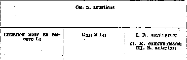
Arising from the brachial plexus-its posterior trunk-immediately after the plexus exits the spatium interscalenium, the nerve very soon separates from other nerves, lies still in the depth of the dome of the axillary fossa on the dorsal side of a. axillaris, descends together with a. profunda brachii along the arm, lying in the sulcus bicipitalis medialis in front of the tendons of musculus teres major and m. latissimus dorsi, here it gives off: 1) n. cutaneus brachii posterior for the skin of the dorsal surface of the arm directly below the tendons of the above-mentioned muscles. Further N. penetrates between caput longum and caput laterale musculus tricipitis brachii along the sulcus nervi radialis, lying in the so-called bag of the triceps muscle on the bone itself (canalis spiralis) and thereby changes its direction, passing from the medial side of the arm to the lateral; still directly before entering this canal, it first gives off 2) rr. musculares for all three heads forming m. triceps brachii and for m. anconaeus; at the border between the middle and lower third of the arm, 4-5 cm. above epicondylus lateralis, the nerve pierces septum intermusculare laterale and lies together with arteria profunda brachii between musculus brachialis and m. brachio-radialis; but still in the spiral canal N. gives off 3) r. cutaneus antibrachii dorsalis, which passes between caput mediale and caput laterale m. tricipitis or between it and musculus brachio-radialis to the dorsal surface of the arm, between processus olecranon and epicondylus lateralis, becomes cutaneous and innervates the skin of the dorsal part of the arm and forearm up to the wrist joint; here the nerve also gives off 4) rr. musculares for the muscles of the dorsal surface of the forearm: a) for musculus brachio-radialis-this nerve may send a branch to the elbow joint, b) for m. extensor carpi radialis longus and c) an inconstant branch for musculus brachialis; in the region of epicondylus lateralis N. divides into both its terminal branches: r. profundus and r. superficialis. 5) R. profundus is first located in canalis supinatorius (in the mass of the homonymous muscle), then on the dorsal surface of the forearm between the superficial and deep layers of extensors and gives a) rr. musculares: a) for musculus extensor carpi radialis brevis, b) for musculus supinator, y) for musculus extensor digitorum communis, <5) for m. extensor digiti V proprius, e) for m. extensor carpi ulnaris, and b) m. interosseus (antibrachii) dorsalis, which is first located between the deep and superficial layers of extensors, then on the dorsal surface of membranae interossea, and gives c) rr. musculares: a) for m. abductor pollicis longus, g) for m. extensor pollicis brevis, y) for m. extensor pollicis longus, h) for m. extensor indicis proprius, d) sensory branches: a) for the interosseous ligament, g) for the periosteum of the radial and ulnar bones, and e) rr. articulares for a) the dorsal surface of the wrist joint and g) for the dorsal surface of the carpal and carpometacarpal joints, c) R. superficialis first passes under musculus brachio-radialis, then between it and m. extensor carpi
Mixed. 1) Innervates the following groups of muscles: a) extensor of the arm at the elbow joint-m. triceps brachii and m. anconaeus; b) flexor and pronator of the forearm-m. brachio-radialis; c) supinator of the forearm-m. supinator; d) extensors of the hand, abducting it to the radial side-ram. extensores carpi radialis longus et brevis; e) extensor of the hand, abducting it to the ulnar side-musculus extensor carpi ulnaris; f) extensor of the fingers-m. extensor digitorum communis; g) extensor in particular of the little finger-musculus extensor digiti quinti proprius, and of the thumb-mm. extensores pollicis brevis et longus; h) abducting and extending the thumb-mm. abductor pollicis longus and extensores pollicis longus et brevis. 2) Transmits sensory irritations from the skin of the dorsal surface of two and a half radial fingers without their terminal phalanges Latin and Russian name. Synonyms Beginning. To which segment does it correspond With which nerves does it anastomose What branches arise from it b) pp. digitales dorsa- a) digiti pollicis radialis, g) digiti pollicis ulnaris, y) digiti indicis radialis, <5) digiti indicis ulnaris, f) digiti tertii radialis Recurrens (r.) (recurrent branch) See n. mandibularis 431; Recurrens (recurrent nerve) N. vagus 1) Ganglion cervicale medium et inferius, 2) n. laryngeus superior 1) Rr. cardiaci inferiores, 2) rr. anastomotici, 3) rr. tracheales, 4) rr. oesophagei superiores, 5) n. laryngeus inferior: a) r. anterior with rr. musculares: a) ramus, g) ramus, y) ramus. <5) ramus, b) ramus posterior gives: a) r. anastomoticus, g) rr. musculares, y) rr. sensitiTi Renales nervi vagi (rr.) (renal branches of the vagus nerve) See n. vagus 433' Renalis nervi splanchnici minoris (r.) (renal branch of the minor splanchnic nerve). Syn.; n. renalis posterior superior See n. splanchnicus minor et minimus 434 Renalis (plexus) (renal plexus) See n. sympathicus Saccularis (nerve of the sac of the hearing apparatus). Syn.: saccularis minor, s. n. vestibuli (Henle), s. n. vestibularis, s. radix posterior, s. vestibularis n. acustici See n. acusticus Saccularis major (large nerve of the sac of the hearing apparatus). Syn.: n. utricularis, s. funiculus superior n. vestibuli, 437! Sacralis primus (first sacral nerve)
What they innervate and where the nerve ends b) a) the skin of the radial edge of the dorsal surface of the thumb to the base of the nail, P) the skin of the ulnar edge of the dorsal surface of the thumb to the base of the nail, y) the skin of the radial edge of the dorsal surface of the index finger to the nail phalanx, <5) the skin of the ulnar edge of the dorsal surface of the index finger to the nail phalanx, e) the skin of the radial edge of the dorsal surface of the third finger to the nail phalanx Topography of the nerve and its branches rad. longus, descends along the palmar surface of the forearm, lying radially from arteria radialis and along m. brachioradialis, passes above the radiocarpal joint onto the dorsal surface, pierces fascia superficialis antibrachii, gives off a) r. anastomoticus ulnaris to ramus dorsalis manus from nervus ulnaris and divides into b) nn. digitales dorsales for the skin of the dorsal surface of the first two fingers and the radial half of the third. 1) Variations in the area of distribution of the cutaneous branches are shown in fig. 67-70 (vol. XX, pp. 769-780). 2) In rare cases, n. thoracodorsalis may branch off from the radial nerve, usually originating directly from fasciculus posterior. 3. Nervus cutaneus antibrachii dorsalis may sometimes be as thick as the trunk of the radial nerve itself. 4. From the radial nerve in the area of the arm, one or two thin branches may branch off to m. brachialis internus (75% according to Frohse) Nature of the nerve and its function. Variations See mandibularis 1) Plexus cardiacus, 2) ganglion cervicale medium et inferius, 3) cervical part of the trachea, 4) cervical part of the esophagus, 5) a) anterior branch to the muscles: a) m. cricoarytenoideus lateralis, P) m. thyreoarytenoideus and m. vocalis, y) m. thyreoepiglotticus, S) m. aryepiglotticus, b) posterior branch: a) n. laryngeus superior, P) to the muscles: m. cricoarytenoideus posterior, m. arytenoideus, y) the mucous membrane below the vocal cords Originating from the vagus nerve and its cervical part on the right side, slightly above the origin of a. subclavia, and on the left side slightly above arcus aortae, the nerve descends, giving off on its way: 1) rr. cardiaci inferiores for the cardiac plexus, 2) rr. anastomotici to the middle and inferior cervical nodes of the sympathetic nerve, 3) rr. tracheales to the cervical part of the respiratory tract and 4) rr. oesophagei superiores for the cervical part of the esophagus; after this it forms a loop on the right around arteria subclavia, and on the left around arcus aortae, and already under the name n. laryngeus inferior ascends along the neck, lying in the groove between the esophagus and the respiratory tract, and divides here into its r. anterior and posterior. Ramus anterior ends in the following muscles of the larynx: a) m. cricoarytenoideus lateralis, P) m. thyreoarytenoideus et m. vocalis, y) m. thyreoepiglotticus and <5) m. aryepiglotticus. Ramus posterior anastomoses with n. laryngeus superior and gives off rr. musculares for a) m. cricoarytenoideus posterior and P) mm. arytenoideus transversus et obliquus and y) rr. sensitivi for the mucous membrane below the vocal cords Mixed. 1. Innervates the following muscle groups: a) abducting the vocal cords and widening the glottis-m. cricoarytenoideus posterior; b) adducting the vocal cords and closing the glottis-m. cricoarytenoideus lateralis, m. arytenoideus transversus et obliquus; c) relaxing the vocal cords-m. vocalis; d) widening the entrance to the larynx and lifting the epiglottis-musculus thyreoepiglotticus, and e) narrowing the entrance to the larynx and lowering the epiglottis-musculus aryepiglotticus. 2. Conducts sensory irritations from the mucous membrane of the larynx below the vocal cords See vagus See splanchnicus minor et minimus See sympathicus See acusticus See acusticus I. Plexus meningeus anterior et posterior; II. N. sympathicus; III. Plexus sacralis and pudendus (partially) Originating from the spinal cord at approximately the level of L1, the nerve gives off 1) r. meningeus, which, remaining in the vertebral canal, participates in the formation of plexus meningeus anterior et posterior; 2) ramus communicans, which originates Mixed. I. Transmits sensory irritations: a) from the skin of almost the entire leg except the area of distribution of lumbar nerves. Latin and Russian names. Synonyms Beginning. Which segment does it correspond to 1 With which nerves does it anastomose What branches arise from it ! 1) n. gluteus superior, 2) n. gluteus inferior, 3) n. piriformis, 4) n. cutaneus femoris posterior, 5) n. ischiadicus, 6) n. pudendus (partially); IV. R. posterior gives: 1) r. medialis, s? 2) r. lateralis nn. clunium mediorum, £ 3) rr. articulares Sacralis quartus (fourth sacral nerve) Spinal cord at the level of L1 Sn and S1 I. R. meningeus; II. R. communicans; III. R. anterior Sacralis quintus (fifth sacral nerve) See Nerve plexuses (plexus coccygeus) Sacralis secundus (second sacral nerve). Syn.: n. bigeminus, s. biradiatus Sacralis tertius (third sacral nerve) In all respects similar to n. sacralis primus Spinal cord at the level slightly below L1 Sn and S1 I. R. meningeus; II. R. communicans; III. R. anterior: 1) n. ischiadicus, 2) n. pudendus; IV. R. posterior Saphenus (major) (hidden N.). Syn.; n. cutaneus femoris internus major N. femoralis l) N. cutaneus femoris posterior, 2) n. cutaneus surae medialis, 3) n. peroneus superficialis l) Ramus infrapatellaris (see), 2) nn. cutanei cruris medii (see) What they innervate and where the nerve ends Topography of the nerve and its branches Nature of the nerve and its function. Variations 1-6) see the corresponding nerves; IV. 1) M. multifidus, the skin over the posterior part of the sacrum and the tip, 2) the skin over the medial part of m. gluteus maximus, 3) articulatio sacroiliaca already in foramen sacrale anterius and enters the anterior surface of the sacrum to join the sympathetic nervous system; 3) r. anterior, which exits foramen sacrale anterius, along the anterior edge of m. piriformis, immediately participates in the formation of plexus sacralis, lying on m. piriformis, and gives off its fibers mainly to nervus ischiadicus, but also to nn. piriformis, gluteus superior et inferior, cutaneus femoris posterior and partially to nervus pudendus; 4) ramus posterior exits foramen sacrale posterius primum and participates in the formation of plexus sacralis posterior, which is located on the posterior surface of the sacrum and on ligamentum sacrotuberosum; from the latter branch off: 1) branches for m. multifidus, for the skin of the posterior part of the sacrum, 2) nn. clunium medii for the skin over the medial part of m. glutei maximi and 3) branches for the sacroiliac joint plexus, b) from the skin of the gluteal region, c) into the leg joint and sacroiliac joint. II. Participates in the innervation of the following muscle groups: a) hip extensors, rotating it outward-mm. glutei, maximus, medius et minimus, b) rotating it outward-mm. piriformis, obturator internus, gemelli, quadratus, c) abducting the thigh in the hip joint-m. tensor fasciae latae, d) hip extensors in the hip joint and leg flexors in the knee joint-mm. biceps femoris, semitendinosus et semimembranosus, e) all muscle groups of the leg and f) the foot. III. Transmits sensory irritations from the membranes of the spinal cord and vertebrae in the sacral region. IV. Participates (to some extent) in the innervation of the muscles and skin of the perineum Plexus meningeus anterior et posterior; II. N. sympathicus; III. Plexus pudendus See pudendus plexus See pudendus plexus See Nerve plexuses (plexus coccygeus) I. Plexus meningeus anterior et posterior; II. N. sympathicus; III. Plexus sacralis et pudendus; See these nerves as well as n. sacralis I In all respects similar to n. sacralis I, but only its r. anterior exits through foramen sacrale anterius III from behind the posterior edge of m. piriformis; the nerve participates in the formation of: a) plexus sacralis, giving off its upper fibers to n. ischiadicus, and b) plexus pudendus, giving off its lower fibers mainly to n. pudendus Mixed. I. Transmits sensory irritations from the area of distribution of nn. ischiadici and pudendi (see). II. Participates in the innervation of the following muscle groups: a) hip extensor in the hip joint-m. gluteus maximus, b) hip extensors in the hip joint and leg flexors in the knee joint-mm. biceps femoris, semitendinosus et semimembranosus, c) all muscle groups of the leg and foot. III. Innervates the skin and muscles of the perineum. IV.
Transmits sensory irritations from the spinal cord and the posterior surface of the sacrum in the sacral region 1) Skin of the medial side of the knee, 2) see corresponding nerve. Beginning from the femoral nerve still very high on the thigh, immediately under the inguinal ligament (Poupart's), it first lies on the lateral, and then on the anterior side of the femoral artery, descends together with it through the adductor canal, passes between the vastus medialis and adductor magnus muscles, and then under the sartorius muscle, reaches under its tendon to the medial side of the knee, gives off its branch - the infrapatellaris for the skin of the medial side of the knee, becomes superficial, descends together with the great saphenous vein on the leg, divides into its medial cutaneous branches of the leg, which run parallel to the branches of this vein in the skin of the medial surface of the leg to the area of the metatarsus. 1. The area of its distribution may vary depending on the degree of development of neighboring cutaneous nerves, as shown in fig. 87-88 (vol. XX, pp. 776-782). 2. Two types are observed: Type I, when there is a single trunk and gradual departure of peripheral branches without significant sensory. Carries sensory irritations from the medial surface of the leg. Latin and Russian names. Synonyms. Beginning. To which segment does it correspond. With which nerves does it anastomose. What branches arise from it.

Saphenus minor (small saphenous nerve). See femoral nerve. Scrotales anteriores (anterior nerves of the scrotum). Syn.: scrotales transversales. Scrotales posteriores (posterior nerves of the scrotum). See ilio-inguinal nerve. See pudendus plexus. Spermaticus externus (external spermatic nerve). Syn.: pudendus externus, genitalis, genito-cruralis. See genito-femoral nerve. Sphenoidalis lateralis (lateral sphenoid nerve). Sphenoidalis medialis (medial sphenoid nerve). Branch connecting the otic ganglion and semilunar ganglion. Branch connecting the otic ganglion with the spheno-palatine ganglion through the pterygoid canal. Spheno-palatini (nervi) (spheno-palatine nerves). Syn.: pterygo-palatini, palatini descendent-tes, plexus spheno-palatinus. Spinales (spinal nerves). N. maxillaris. Several parallel nerves. See cervical dorsal (thoracic), lumbar, sacral and coccygeal nerves. Spinalis accessorius (accessory nerve, cervical part). See accessory nerve. Spinosus (spinal nerve). Syn.: recurrens n. inframaxillaris, rami tertii n. trigemini. N. mandibularis. Splanchnicus imus (lowest splanchnic nerve). Synonyms: splanchnicus tertius, minimus. Splanchnicus major (greater splanchnic nerve). Syn.: splanchnicus superior, supremus. X-XI thoracic nodes of the sympathetic trunk (V). VI-IX thoracic nodes of the sympathetic trunk. What do they innervate and where does the nerve end. Topography of the nerve and its branches connections with neighboring nerves.
Type II, when there is a double or looped trunk with early departure of branches and with a pronounced tendency to form connections between branches and neighboring nerves (obturatorius and peronaeus nerves). Character of the nerve and its function. Variations. See femoral nerve. See ilio-inguinal nerve. See pudendus plexus. See genito-femoral nerve.
Ganglion spheno-palatine-num and through it along the course of palatine nerves. Beginning from the maxillary nerve still in the foramen rotundum, the nerves pass into the pterygo-palatine fossa and enter the spheno-palatine ganglion as its sensory root. See Nerve nodes (ganglion spheno-palatine-num) and palatine nerves. See cervical dorsal (thoracic), lumbar, sacral and coccygeal nerves. See accessory nerve. Dura mater of the base of the brain, greater wing of the sphenoid bone and mucous membrane of the mastoid cells. Separating from the mandibular nerve at the base of the skull, it passes together with the middle meningeal artery through the foramen spinosum into the cranial cavity and spreads in the dura mater, in the mass of the sphenoid bone and in the mastoid cells. Sensory. Carries sensory irritations from the dura mater, from the sphenoid bone (greater wing) and mastoid cells.
Plexus renalis. This inconstant nerve, beginning independently from the X-XI nodes of the sympathetic trunk or communicating branches, passes into the renal plexus. Sympathetic. Carries its fibers to the renal plexus. Ganglion coeliacum and through it to numerous secondary paired and unpaired sympathetic nodes of the abdominal cavity. Beginning with several roots from the (V) VI-IX thoracic nodes of the sympathetic trunk or from the corresponding communicating branches and then merging into one trunk, the nerve descends along the vertebral bodies, penetrates together with the azygos vein through the diaphragm between the intermediate and medial crus into the abdominal cavity, where the celiac ganglion attaches to it, sends fibers to the aortic plexus and participates in the formation of the celiac or solar plexus, from which in turn numerous secondary paired and unpaired plexuses extend to all organs of the abdominal cavity. 1. The major splanchnic nerve may enter the solar plexus directly or after passing through the ganglionic mass near it. 2. Two types can be observed: a) main type, when the number of roots may be 2-4 and they emerge at the level of II-X thoracic vertebrae; b) scattered type - number of roots 4-7, beginning at the level of V-VI thoracic vertebrae. Sympathetic.
Latin and Russian names. Synonyms. Beginning. To which segment does it correspond. With which nerves does it anastomose. What branches arise from it. Splanchnicus minor (lesser splanchnic nerve). Syn.: splanchnicus inferior. X-XI thoracic nodes of the sympathetic trunk. Stapedius (stapedial nerve). Syn.: nervulus stapedius. See facial nerve. Staphylinus n. palatini anterio-ris (r.) (anterior branch of the anterior palatine nerve). One branch from the anterior palatine nerve to the floor of the nasal cavity. Sternales (nn.) (sternal branches). Branches arising from the upper intercostal nerves to the sternum. Stylo-hyoideus (stylo-hyoid nerve). Syn.: stylo-hyoi-deus, styloideus, mastoideus posterior. Branch of the digastric nerve. See facial nerve. Stylo-pharyngeus (stylo-pharyngeal nerve). Syn.: stylo-pharyngeus n. glosso-pharyngei, circumflexus. See glosso-pharyngeus nerve. Subclavius (subclavian). Brachial plexus. Cv 1) R. museularis, 2) ramus. Sublingualis (sublingual gland nerve). See lingual nerve. Submaxillares (nn.) (submaxillary nerves). Syn.: rr. submaxillares ganglii submaxillaris. See Nerve nodes (ganglion submaxillare). Suboccipitalis (suboccipital nerve). Syn.: infraoccipitalis, posterior nervi cervicalis. See first cervical nerve. Subscapulares: superior, medins et inferior (subscapular nerves: superior, middle and inferior). See subscapularis superior, medius, inferior nerves. Subscapularis inferior (inferior subscapular nerve). Syn.: thoraco-dorsalis. Brachial plexus. Cvn-vm Rr. musculares. Subscapularis medius (middle subscapular nerve). Brachial plexus. Cv-vi Rr. musculares. What do they innervate and where does the nerve end. Topography of the nerve and its branches. Character of the nerve and its function. Variations. Ganglion coeliacum and through it to numerous secondary sympathetic nodes of the abdominal cavity. Beginning from the X-XI thoracic nodes of the sympathetic trunk or from the corresponding communicating branches, the nerve descends independently or together with the major splanchnic nerve along the vertebral bodies, passes through the diaphragm laterally to the major splanchnic nerve, reaches the celiac ganglion and passes mainly into the renal plexus. In the solar plexus, the nerve may enter with all its branches or only with part; in this case, the rest enter the nearby ganglionic mass. See facial nerve. Sympathetic.
See p. glosso-pharyngeus 1) M. subclavius, 2) n. pnrenicus Beginning from plexus brachialis, its primary upper trunk. N. goes to fossa supraclavicularis, laterally from n. pnrenicus, to which it sends a branch and ends in m. subclavius Motor. Fixes the clavicle See p. lingualis See Nerve nodes (ganglion submaxillare) See p. cervicali-primus See p. subscapularis superior, medius, inferior M. latissimus dorsi Beginning from the posterior trunk of the brachial plexus, N. is located in the axillary fossa, in its depth, dorsally from other short nerves of the plexus, lies on m. subscapularis in the mass of fatty tissue, reaches here along margo axil lads scapulae to m. latissimus dorsi and ends in the muscle with numerous muscle branches. N. can begin from n. axillaris or more rarely from n. radialp Motor. Innervates the broad muscle of the back, which rotates the shoulder inward, lowers the raised arm downward and throws it on the back with the palm outward M. subscapularis, m. teres major Beginning from the posterior trunk of the brachial plexus, N. in the depth of the axillary fossa is located dorsally from other short nerves, passes along m. subscapularis, sends its branches to the lateral bundles of this muscle and ends in m. teres major Motor. Innervates muscles that rotate the shoulder inward, adduct and lower the raised arm № Latin and Russian name. Synonyms Beginning. To which segment corresponds With which nerves anastomoses What branches depart from it Subscapularis superior (upper subscapular nerve) Plexus bracnialis. Cv-vi Rr. muscu lares Superficialis n. plantaris laterals (g.) (superficial branch of the lateral plantar N.) See p. plantaris lateralis Superficialis n. radialis (g.) (superficial branch of the radial nerve). Syn.: n. cutaneus, s. dorsalis, s. n. radialis superficialis See p. radialis Superior n. laryngei superioris (g.) (upper branch of the superior laryngeal nerve). Syn.: g. exter-nus n. laryngei superioris See p. laryngeus superior 472 Superior n. oculomotorii (upper branch of the oculomotor nerve) See p. oculomotorius Supraclavicu lares: anteriores, medii et posterior (supraclavicular nerves: anterior, middle, and posterior). Syn.: suprasternales ! anteriores, medii et posterior, s. cucuHares (Schwalbe), s. aero-1 miales, s. superficiales scapulae (Langenbeck), s. claviculares, s. sternales Plexus cervicalis. Сш, iv l) Nn. supraclavicu lares anteriores (6-8), 2) nn. supraclaviculares medii (3), 3) nn. supraclavicularis posterior Supraorbitalis (supraorbital nerve). Syn.: frontalis externus, s. major Suprascapularis (suprascapular nerve). Syn.: scapularis medius, s. superior See n. ophthalmicus Plexus bracbialis. Cv-vi 1) Rr. muscu lares.. 2) rr. articulares Supratrochlearis (supratrochlear nerve). Syn.: frontalis interhus, s. minor See n ophthalmicus Suralis (sural nerve). Syn.: communicans surae, s. n. tibialis, s. magnus, s. cutaneus longus cruris et pedis, s. Under this name is meant a small section of the nerve located on the posterior surface of the leg in its lower third, above m. gastro-cnemius; it is a continuation of n. cutanei surae medialis (see), which receives this name after its connection with ramus
What they innervate and where the nerve ends Topography of the nerve and its branches M. subscapularis Beginning from Cv and Cvi, N. lies as the shortest of all subscapular nerves in the depth of the axillary fossa, goes to the most medial bundles of m. subscapularis along margo vertebralis scapulae, where it breaks into its gha-mi musculares Character of the nerve and its function. Variations Motor. Innervates the subscapular muscle, which rotates the shoulder inward, lowers and adducts the raised arm See p. plantaris lateralis See p. radialis See p. laryngeus superior See p. oculomotorius 1) Skin over the sternum in its upper part, articulatio sterno-clavi-cularis, 2) skin of the lateral part of the chest to the fourth rib, 3) skin of the shoulder and over the anterior part of m. delto-idei, gives a branch to n. accessorius Beginning from Сш and Civ near foramen interver-tebrale, the nerves pass along the medial surface of musculi sterno-cleido-mastoidei outward, emerge from under its posterior edge below other cervical plexus cutaneous nerves, descend downward along the posterior edge of the muscle, covered by mus-culus platysma and fascia superficialis colli, and spread as follows: 1) nn. supra-claviculares anteriores cross the sternoclavicular muscle and go to incl-sura sterni, spread in the skin over extre-mitas sternalis clavicnlae, and also give a branch to the sternoclavicular joint; 2) nn. supraclavi-culares medii go downward, cross the clavicle from its middle to processus acromion and spread in the skin of the chest to IV rib; 3) n. supracla-vicularis posterior descends downward along the lateral edge of musculi trapezii and ends in the ko-ze of the upper scapular region and over m. deltoideus Sensory. 1. Carries sensory irritations from the sternoclavicular joint. 2. Carries sensory irritations from the upper part of the chest and shoulder See p. ophtnalmicus 1) M. supraspinatus, m. infraspinatus, 2) articulatio humeri Beginning from the brachial plexus, andv its upper primary trunk, on its dorsal surface, the nerve, being the most lateral and upper (except nervus dorsalis scapulae), goes downward in fossa supraclavicularis along the lateral edge of the plexus, then turns in the lateral side and backward along m. omo-hyoideus, reaches together with it to incisura scapulae, enters under liga-mentum transversum scapulae superius in fossa su-praspinata, lies under musculus supraspinatus. sends its muscle branches to it, goes in the lateral direction to the neck of the scapula, approaches i under ligamentnm transversum scapulae inferius, penetrates into fossa infraspinata and ends in mus- | culus infraspinatus, also sending a branch to the joint- | capsule of the shoulder joint Mixed. I. Sends sensory irritations from the capsule of the shoulder joint. II. Innervates muscles: 1) m. supraspinatus, abducting the shoulder, 2) m. infraspinatus, rotating the shoulder outward, adducting and lowering the raised arm See p. ophtnalmicus Latin and Russian name. Synonyms Beginning. To which segment corresponds With which nerves anastomoses What branches depart from it 477
saphenus brev is inf., s. externus, >
s. minor, s. saphenus tibialis, £
s. r. communicans surae, s.
communicans tibialis anastomoticus peronaeus (see this nerve and n. peronaeus communis); n. suralis gives off rr. calcanei laterales (see) and again receives another name- n. cutaneus dorsalis (pedis) lateralis (see) 478 Sympathicus (nervus, s.truncus) (sympathetic nerve). Syn.: n. gangliosus, s. intercostalis maximus, s. sympathicus magnus, s. maximus, s. sympatheticus, s. trisplanchnicus, s. vasomotorius (Stilling, Henle) The anastomoses of all the communicating nerves of the trunk nerves See in the next column I. Cervical part: A. Ganglion cervicale superius: 1) n. caroticus internus, 2) n. jugularis, 3) n. cardiacus superior, 4) r. anastomoticus, 5) r. anastomoticus, 6) nn. carotici externi, 7) rr. pharyngei, 8) rr. laryngei; B. Ganglion cervicale medium: 9) rr. carotici, 10) n. cardiacus medius; C. Ganglion cervicale inferius: 11) n. cardiacus inferior, 12) n. cardiacimus (not recognized by all); II. Thoracic part: Ganglia thoracalia 13) nn. bronchiales, 14) nn. aortales, 15) n. splanchnicus major, 16) n. splanchnicus minor, 17) n. splanchnicus minimus, s. imus (inconstant); III. Lumbar part: Ganglia lumbalia 18) rr. postganglionares; IV. Pelvic part: Ganglia sacralia 19) rr. postganglionares What they innervate and where the nerve ends Topography of the nerve and its branches Character of the nerve and its function. Variations 1) Plexus caroticus internus, 2) one twig to ganglion jugulare n. vagi, and another-to ganglion petrosum n. glossopharyngei, 3) plexus cardiacus, 4) ganglion nodosum n. vagi, 5) n. hypoglossis, 6) to all branches from arteria carotis externa, 7) plexus pharyngeus, 8) plexus laryngeus, 9) plexus caroticus communis, 10) plexus cardiacus, 11) plexus cardiacus, 12) plexus cardiacus, 13) plexus bronchiales, 14) plexus aortae thoracalis, 15) plexus coeliacus, 16) plexus coeliacus, 17) plexus renalis, 18) plexus coeliacus, mesentericus superior et inferior, 19) plexus hypogastricus (pelvicus) Arising from the rr. communicantes of the spinal nerves, the sympathetic nerve on each side represents: a) the border column- truncus sympathicus, which consists of 20-25 ganglia, of interganglionic fibers-rr. interganglionares, connecting these ganglia of each side with each other, and transverse fibers-rr. transversi, connecting the ganglia of the opposite side with each other; b) from these ganglia go postganglionic fibers-rr. postganglionares, c) to the peripheral ganglia of the sympathetic nervous system: ganglia ciliare, sphenopalatinum, submaxillare and oticum, located in the head, ganglion coeliacum, mesentericum superius et inferius, ganglia renale dextrum et sinistrum, aortico-abdominalis, located in the abdominal cavity, and ganglion pelvicum, located in the pelvis; d) from these ganglia in turn go fibers to the nerve plexuses in all organs, starting from the oral cavity, neck, thoracic and up to the abdominal cavity, to the vessels, to the skin, to the muscles and to the glands. In the cervical part the nerve lies on fascia praevertebralis, with ganglion cervicale superius located on the transverse processes of C6 and C7 and giving off its branches: 1) nervus caroticus internus, which surrounds the internal carotid artery and all its branches and forms plexuses bearing the name of these vessels; in addition plexus caroticus internus gives the following branches: a) radix sympathica ganglii ciliaris, b) rr. orbitales, c) ramus to hypophysis cerebri, d) nervus petrosus profundus major to ganglion sphenopalatinum, e) nn. carotico-tympanici superior et inferior to plexus tympanicus; 2) n. jugularis, which immediately divides into two branches; of them one goes to ganglion jugulare nervi vagi, and the other to ganglion petrosum nervi glossopharyngei; 3) n. cardiacus superior-to the cardiac plexus; 4) r. anastomoticus-to ganglion nodosum n. vagi; 5) r. anastomoticus-to nervus hypoglossus; 6) nn. carotici externi, which surround the external carotid artery and form plexuses along all its branches, and between them also: radix sympathica for ganglion submaxillare from plexus arteriae maxillaris externae and for ganglion oticum radix sympathica from plexus meningeus arteriae meningeae mediae; 7) rr. pharyngei-for plexus pharyngeus and 8) rr. laryngei-for plexus laryngeus. Ganglion cervicale medium-inconstant: sometimes completely absent, and sometimes replaced by two ganglia; lies on truncus thyro-cervicalis at the level of C7 and gives: 9) rr. carotici for plexus caroticus communis and 10) n. cardiacus medius for plexus cardiacus. Ganglion cervicale inferius often fuses with the 1st thoracic, lies above the head of the 1st rib on the transverse process of C7 behind the subclavian artery and gives: 11) n. cardiacus inferior and 12) n. cardiacimus (inconstant) for the cardiac plexus. In the thoracic cavity the nerve lies on the lateral surface of the spine on articulatio costo-vertebralis under the pleura and gives: 13) nn. bronchiales for plexus bronchialis, 14) nn. aortales for the plexus around the thoracic aorta, 15) nn. splanchnicus major, which begins from V-IX ganglia, lies on the anterior surface of the spinal column, descends through the diaphragm and enters plexus coeliacus; to it also approaches 16) n. splanchnicus minor. Plexus coeliacus is located on the anterior surface of the spine at the level of L1 on the sides of a. coeliaca, receives fibers from the vagus nerve and gives secondary plexuses around all abdominal organs both paired and unpaired, namely: a) plexus phrenicus, b) plexus gastricus superior et inferior, c) plexus hepaticus, d) plexus lienalis, e) plexus mesentericus superior, f) plexus mesentericus inferior, g) plexus renalis, h) plexus spermaticus (ovaricus). In the pelvic cavity the nerve also lies on the anterior surface of the spine, receives rr. postganglionares from the corresponding sacral sympathetic ganglia and forms plexus pelvicus with secondary plexuses around the organs of the small pelvis, namely: plexus colicus, vesicalis and vaginalis See Vegetative nervous system № Latin and Russian name. Synonyms Temporales (rr.) (temporal branches). Syn.: rr. temporales n. facialis, s. rr. temporo-frontales, s. nn. faciales temporales Beginning. To which segment corresponds With which nerves anastomoses What branches go from it Temporales superficiales (rr.) (superficial temporal branches of the auriculotemporal nerve). Syn.: rr. temporales subcutanei See n. facialis See n. auriculo-temporalis 481 Temporalis profundus anterior et posterior (anterior and posterior deep temporal nerve) See n. masticatorius Temporo-facialis (temporo-facial nerve) A twig from n. facialis to glandula parotis Tensor vaginae femoris (nerve of the muscle, tightening the fascia of the thigh) A branch from n. glutaeus superior to m. tensor fasciae latae Tensoris tympani (nerve of the muscle, tightening the tympanic membrane) N. pterygoideus internus and ganglion oticum R. muscularis Tensoris veli palatini (nerve of the muscle, tightening the soft palate) N. pterygoideus internus and ganglion oticum R. muscularis Tentorii (nerve of the cerebellar tentorium). Syn.: n. recurrens (Arnoldi), s. tentorii cerebelli, s. r. sinualis, s. n. sinualis Terminalis radialis (terminal radial branch) See n. ophthalmicus See n. medianus Terminalis ulnaris (terminal ulnar branch) See n. medianus Thoracales (thoracic nerves). Syn.: nn. thoracici, s. dorsales, s. costales, s. intercostales, s. spinales dorsales, s. subcostales Spinal cord from approximately the level of the end of the spinous process of D12 to the height of the spinous process of D11. D12-D11 1) Rr. meningei, 2) rr. communicantes, 3) rr. anteriores, 4) rr. posteriores, s. rr. dorsales give: a) rr. mediales, b) rr. laterales 490 i Thoracales anteriores: primus et secunda (anterior thoracic nerves: first and second) Plexus brachialis. C5-C8 and D1 Primus: 1) r. muscularis, 2) r. anastomoticus,- Secundus: 1) r. muscularis, 2) r. muscularis What they innervate and where the nerve ends Topography of the nerve and its branches Character of the nerve and its function. Variations
I. Temporales (гр.) (височные ветви). Син.: rr. temporales n. facialis, s. rr. temporo-frontales, s. nn. faciales temporales Начало. Какому сегменту соответствует C какими нервами анастомозирует Какие ветви от него отходят Temporales superficiales (rr.) (височные ветви ушно-височного нерва). Син.: rr. temporales subcutanei См. п. facialis См. п. auriculo-temporalis 481 Temporalis profundus anterior et posterior (передний и задний глубокий височный нерв) См. п. masticatorius Temporo-facialis (височно-лицевой нерв) Веточка от п. facialis к glandula parotis Tensor vaginae femoris (нерв мышцы, натягивающей фасцию бедра) Ветвь от п. glutaeus superior к m. tensor fasciae latae Tensoris tympani (нерв мышцы, натягивающей барабанную перепонку) N. pterygoideus internus и ganglion oticum R. muscularis Tensoris veli palatini (нерв мышцы, натягивающей мягкое нёбо) N. pterygoideus internus и ganglion oticum R. muscularis Tentorii (нерв мозжечковой палатки). Син.: n. recurrens (Arnoldi), s. tentorii cerebelli, s. r. sinualis, s. n. sinualis Terminalis radialis (конечная лучевая ветвь) См. п. ophthalmicus См. п. medianus Terminalis ulnaris (конечная локтевая ветвь) См. п. medianus Thoracales (грудные нервы). Син.: nn. thoracici, s. dorsales, s. costales, s. intercostales, s. spinales dorsales, s. subcostales Спинной мозг примерно от уровня конца остистого отростка D12 до высоты остистого отростка D11. D12-D11 1) Rr. meningei, 2) rr. communicantes, 3) rr. anteriores, 4) rr. posteriores, s. rr. dorsales дают: a) rr. mediales, b) rr. laterales 490 i Thoracales anteriores: primus et secunda (передние грудные нервы: первый и второй) Plexus brachialis. C5-C8 и D1 Primus: 1) r. muscularis, 2) r. anastomoticus,- Secundus: 1) r. muscularis, 2) r. muscularis Что они иннервируют и где кончается нерв Топография нерва и его ветвей Характер нерва и его
See p. facialis See p. auriculo-temporalis See a. masticatorius M. tensor tympani Beginning with two twigs: 1) from n. pterygoideus internus and 2) from ganglion oticum (the latter receives its motor fibers-radix motoria-mainly from n. pterygoideus internus), the nerve enters the semicanalis tensoris in the pyramid of the temporal bone and ends in m. tensor tympani Motor. Innervates the muscle that tenses the tympanic membrane M. tensor veli palatini Beginning with two twigs: 1) from n. pterygoideus internus and from ganglion oticum-from its anterior edge (the latter receives its radix motoria mainly from the same nerve), the nerve descends from foramen ovale together with n. pterygoideus internus and ends in m. tensor veli palatini Motor. Innervates the muscle that tenses the soft palate See p. ophthalmicus See p. medianus See p. medianus 1) Plexus meningeus anterior et posterior, 2) n. sympathicus (ner-vus splanchnicus major et minor) (see ), 3) a) plexus brachialis-Division, b) n. intercosto-brachialis (Dh), c) nn. intercostales I-XII (see.), 4) a) skin of the back along the midline and muscles: mm. rotatores, multifidus, semispinalis, spinalis dorsi, b) m. longissimus and m. iliocostalis Each of the thoracic nerves still in the spinal canal gives off its branch 1) r. meningeus, which returns back into the spinal canal and participates in the formation of plexus meningeus anterior and posterior, and then the nerve, exiting the intervertebral canal, gives: 2) r. communicans, going to the border column of the sympathetic nervous system; 3) r. anterior, which passes into the intercostal space-n. intercostalis (see.); of these, only the first and partly the second pass into the brachial plexus and participate in the innervation of the skin and muscles of the arm; 4) ramus posterior, which, exiting between the transverse processes of the vertebrae, divides: a) into a medial branch (thicker in the eight upper nerves and thinner in the others) and b) into a lateral branch (thicker in the four lower nerves and thinner in the others) Mixed. 1) Carry sensory irritations from the skin of the trunk and upper limb. 2) Innervate the muscles of the chest and back (see nn. intercostales). 3) Go along the course of n. sympathici to the organs of the abdominal cavity, into muscles and skin. 4) Bring sensory irritations from the membranes of the spinal cord and from the walls of the vertebrae 1) M. pectoralis major, 2) n. thoracalis anterior secundus; 1) m. pectoralis major, 2) m. pectoralis minor Beginning: the first - from the upper secondary trunk of the thoracic plexus, the second - from the lower secondary trunk, from their anterior surface, still in pars supraclavicularis of the brachial plexus, the nerves approach under the clavicle in front of a. and v. subclavia and enter: the first into musculus pectoralis major from its inner surface, and the second into m. pectoralis major and m. pectoralis minor, also from their inner surface Motor. Innervate the pectoral muscles 25v № Latin and Russian name. Synonyms Beginning. To which segment does it correspond With which nerves does it anastomose What branches arise from it Thoracales posteriores (posterior thoracic nerves) Under this name understand n. dorsalis scapulae and n. thoracalis longus 492 Thoracalis longus (long thoracic nerve). Syn.: thoracicus medius, s. posterior, s. lateralis, s. thoracico-dorsalis, s. respiratorius externus inferior Plexus brachialis. Cv-vi (vii) R. muscularis Thoracico-dorsalis (thoraco-dorsal nerve). Syn.: n. marginalis scapulae, s. subscapularis inferior See nn. subscapulares Thyreo-hyoideus (g.) (branch of hypoglossal n. to thyrohyoid muscle) Tibialis (tibial nerve). Syn.: n. popliteus internus, s. posticus Proper branch from ansa hypoglossi, i.e., from n. cervicalis (see n. hypoglossus) N. ischiadicus 1) N. peronaeus, 2) n. femoralis, n. cutaneus femoris posterior, 4) peripheral branches among themselves 3) I. Before division into terminal branches: 1) n. cutaneus surae medialis (see): a) rr. calcanei laterales (see), b) n. cutaneus dorsalis lateralis (see), 2) rr. articulares, 3) rr. musculares, n. canalis musculo-peronaei, 4) r. popliteus, 6) rr. calcanei medialis (see); II. Branches of n. plantaris medialis (see): 6) rr. musculares, r. medialis, 7) r. muscularis, 9) n. plantaris hallucis medialis, 9) nn. digitales plantares communes I-III, 10) nn. lumbricales; III. Branches of n. plantaris lateralis: 11) rr. musculares, r. superficialis, 12) n. plantaris lateralis digiti V: a) rr. musculares, b) rr. lumbricales, 13) n. digitalis plantaris communis IV (see): r. profundus, 14) nn. interossei, 15) rr. musculares 257
HUMAN NERVES
What do they innervate and where does the nerve end Topography of the nerve and its branches Character of the nerve and its function. Variations M. serratus anterior Beginning with two or three roots from the posterior surface of plexus brachialis still in pars supraclavicularis and uniting into one trunk, the nerve descends along the lateral wall of the chest along linea axillaris between musculus subscapularis and musculus serratus anterior, lying tightly on the latter and giving off its twigs to the teeth of musculi serrati anterioris Motor. Innervates m. serratus anterior See nn. subscapulares 1) Skin over the lateral ankle, skin of the lateral edge of the foot, 2) articulatio genu, 3) a) m. gastrocnemius, b) m. plantaris, c) m. tibialis posterior, d) m. soleus, e) mm. flexor digitorum longus, f) mm. flexor hallucis longus, g) for periosteum and foramen nutritura fibulae, 4) a) m. popliteus, b) a. tibialis, c) articulatio talocruralis, d) periosteum of tibia, e) periosteum of bones of the leg, f) membrana interossea (a. interosseus cruris) (see.), 5) skin of the medial surface of the heel, 6) 1 a) m. flexor digitorum
brevis, I б) t. abductor hallucis, \ 7) m. flexor hallucis brevis, i ?JT 8) И 9) v, t кошу подошвенной t стороны пальцев, 10) mm. lumbricales, И) a) m. quadratus plantae, б) m. abductor digiti V, 12) a) m. flexor digit! V, m. opponens digiti V, б) mm. lumbricales Ш, 13) кожу подошвенной стороны пальцев, kH) mm. interossei, ») a) m. adductor hallucis, б) m. flexor hallucis bre- vis, в) m. flexor digiti V, r) m. opponens digiti V Starting from the n. ischiadicus still approximately at the middle of the thigh, but while still remaining with the n. peronaeus communis for some time in a common sheath and giving off on this space a whole series of branches (see nervus ischiadicus), namely: rr. muscu lares for m. obturator internus, mm. gemelli, m. quadratus femoris, m.semiicndinosus, m.semimembrano-sus, m. adductor magnus and t. biceps femoris (ca-p#ut longum), H. in the popliteal fossa occupies a position between the n. peronaeus communis, lying more laterally, and vasa pop Hi aea, lying more medially, gives here a whole series of branches: 1) n. cutaneus surae medialis (see this nerve and n. sura lis), which descends between the common heads of m. gastrocnemii, lies under fascia cruris along v. saphena parva, anastomoses with г. апа-storaoticus peronaeus, receives the name n. sura-lis, pierces fascia cruris, gives its rr. calcanei laterales (see) and then again under another name-nervus cutaneus dorsalls lateralis (see)-spreads in the skin of the lateral edge of the foot; 2) rr, articulares (usually two)-for the knee joint; 8) rr. muscu lares for all muscles of the posterior group of the leg: a) m. gastrocnemius, б) т. plantaris, в) т. tibialis posterior, r) m. soleus, д) т. flexor digitorum longus, e) т. flexor hallucis longus and ж) nervus сапа lis musculo-peronael, which goes along canalis musculo-peronaeus together with vasa peronaea, reaches the fibula and gives nerves to the periosteum and to foramen nutritium fibulae, 4) ramus poplitaeus, к-рая descends along m. poplitaeus on its posterior surface and gives a very many small branches: a) for musculus poplitaeus, б) a. tibialis, в) for the tibia, г) for the periosteum, д) for the ankle joint and e) for the membrana interossea, б) rr. calcanei mediates for the skin of the medial surface of the heel and the posterior part of the foot. Further the N. descends along the leg first between both heads of m. gastrocnemii, then descends under m. soleus, lies between it and musculus tibialis posterior and reaches together with a. tibialis posterior to the medial side of the ankle joint and near malleolus medialis divides into both its terminal branches: n. plantaris medialis and lateralis (their description see respective nerves). 1. Variations in the area of distribution of cutaneous branches are shown in fig. 84-91 (t. X X, p. 775 -782). 2. Both heads of the gastrocnemius muscle usually receive separate branches, but in the mass of the tibial nerve a dorsal bundle can always be distinguished, which includes the n. cutaneus surae medialis, both branches to m. gastrocnemius and to т. soleus. з. The place of origin of ramus articularis genu also varies. 4. R. poplitaeus usually gives off n. diaphysarius tibiae and n.epipliy-sarius tibiae posterior for the respective parts of the tibia and for articulatio tibio-fibularis superior. 5. Ramus articularis pedis (cru-ralis) can depart independently from the trunk or from its branches Mixed. I. Transmits sensory irritations: a) from the skin of the posterior surface of the leg, б) from the sole and plantar surface of the toes, в) from the joints: knee, ankle (from the posterior side) and foot (from its plantar side). II. Innervates muscle groups: a) posterior muscles of the leg, flexing the leg in the knee joint,-т. triceps surae, plantar flexors of the foot,-triceps surae, plantares, flexor hallucis longus and flexor digitorum longus, flexing the toes,-mm. flexor digitorum and flexor hallucis longus, adducting and supinating the foot,-musculus tibialis posterior, б)лсе plantar muscles of the foot № Latin and Russian names. Synonyms Beginning. To which segment does it correspond 1 anastomoses | With which nerves What branches depart from it Tonsillares n. glosso-pharyngei (гг.) (branches of the glossopharyngeal nerve to the tonsil). Syn.: пп. tonsillares ! See n. glosso-pharyngeus | 1 497 Tonetllares г., lingualis (rr.) (branches of the lingual nerve to the tonsil). Syn.: rr. isthmi fau-cium See n. lingualis Tracheales inleriores (rr.) (lower tracheal H.). Syn.: rr. broncbiales anteriores See n. vagus i Tracheales superiores (upper ; See n. laryngeus inferior and n. vagus tracheal nerves). Syn.: гг. i tracheales n. recurrentis ! Trapezius (г.) (trapezius branch) See n. cervioalis quartus Trigeminus: portio major, s. sensitive nportio minor, s.moto-ria (trigeminal-portion sensitive and portion motor). Syn.: quintus, s. par quin-tum nn. encephali Portio major begins in ganglion semiiuna-re; its cells send peripheral collaterals as the nerve itself, and central collaterals end: a) in nucleus sensi-bilis nervi trigemini in tegmentum pontis and ft) in nucleus tractus spina-lis n. trigemini, which continues throughout the entire fossa rhomboi-dea and into the spinal cord. Portio minor begins: a) in nucleus radicis descendentis n. trigemini, lying in the posterior part of the quadrigeminal body on the sides of aquaeductus cerebri (Sylvii), and б) in the nucleus locus coeruleus Besides the anastomoses of its three constituent N., there are macroscopically almost indistinguishable sympathetic anastomotic fibers from plexus саго-tlcus to ganglion semi-lunare Gasseri N. gives three branches: I- n. ophthalmicus, II-п. maxillaris, III-n.man-dibularis, so independent that their descriptions-see respective nerves i i I Trochlearls (trochlear nerve). 1 Begins in nucleus Syn.: n. oculo-muscularis supe- \ nervi trochlearis, pac-rior, s. patheticus, s. par quar- rior, s. par quar-tum nn. encephali tum nn. encephali located in the quadrigeminal body on the sides and at the bottom of aquaeductus cerebri (Sylvii) caudally from nucleus nervi oci-lomo-tori i; its collaterals rise to the dorsal surface of laminae quadrigeminae, cross under co 1 -liculus posterior corpo-ris quadrigemini and вы-i go out from the sides of fre-nulum veil medullaris anterioris 1 i i 1) Plexus cavernosus, S 2) a. ophthalmicus j i j j l. Tubae (rr.) (nerve of the auditory tube). Syn.: tubae Eustachianae, g. auditivae, s. tubae plexus tympanic! Cm. n. glosso-pharyngeus Cm. n. auriculo-temporall j.' Tympani (г.) (nerve of the tympanic membrane). Syn.: г. mem-branae tympani s Tympanicus (tympanic, or Jacobson's nerve). Syn.: n. Ja- cobsoni See n. glosso-pharyngeus 261
HUMAN NERVES
9«2 What they innervate and where the nerve ends. Topography of the nerve and its branches. Character of the nerve and its function. Variations. See p. glosso-pharyngeus See p. lingualis See p. vagus See p. laryngeus inferior and p. vagus See p. cervicalis quartus Beginning with two separate roots: 1) portio sensitive, s. major and 2) portio motoria, s. minor, the nerve emerges as separate roots from the brain on the ventral surface of the pons along the linea trigemino-facialis, which represents the boundary between the pons and crura cerebelli ad pontem, it lies on the anterior surface of the pyramid of the temporal bone in impressio trigemini; here the portio major passes into the ganglion semilunare (Gasseri), being here between two layers of the dura mater in cavum semilunare (Meckeli), in close proximity to processus clinoideus posterior, at the lateral wall of sinus cavernosi. From the semilunar ganglion from its anterior edge depart all three N.: the most medial-nervus ophthalmicus (CM.), the middle-n. maxillaris (see) and the most lateral-n. mandibularis; into the path of the latter nerve enter, without entering the ganglion semilunare and lying ventrally from it, all fibers from portio minor Mixed. Motor fibers enter: a) into nervus mandibularis and in it constitute almost entirely the n. masticatorius (except fibers of n. bucciflaterii) and b) part into nervus lingualis (fibers of n. mylo-hyoidei). Distribution of sensory fibers-see the corresponding nerves obliquus oculi superior Emerging on the dorsal surface of the brain from the quadrigemina on both sides of the frenulum of the anterior sail and passing along the lateral surface of crus cerebri to the ventral surface of the brain, the nerve in the form of a thin thread pierces the dura mater-porus trochlearis behind processus clinoideus posterior, laterally from nervus oculomotorius, and lies adjacent to sinus cavernosus; here it receives sympathetic fibers from plexus cavernosus,- further, the nerve, lying adjacent to nervus ophthalmicus and receiving sensory fibers from it, passes through fissura orbitalis superior into the orbit cavity, bends here over the origin of m. levator palpebrae superioris and enters m. obliquus oculi superior Motor. Rotates, lowers and abducts the eye See p. glosso-pharyngeus See p. auriculo-temporalis See p. glosso-pharyngeus л« Latin and Russian name. Synonyms Beginning. To which segment it corresponds With which nerves it anastomoses What branches depart from it Ulnaris (r.) (ulnar branch) See p. cutaneus antibrachii medialis Ulnaris (ulnar nerve). Syn.: | cubitalis; r. dorsalis manus= ulnaris dorsalis; r. volaris manus=n. ulnaris volaris; r. pro-! fundus-ulnaris volaris profun^ | dus; r. superficialis-ulnaris vo- | laris superficialis Plexus brachialis. Cviii and Di 1) N. musculo-cutaneus, 2) n. medianus, 3) n. radialis, 4) n. cutaneus brachii medialis, 5) peripheral branches between themselves A. Branches of forearm: 1) rr. articulares, 2) rr. musculares, 3) r. cutaneus palmaris; B. Terminal branches: 4) r. dorsalis manus: n. digitalis: a) r. dig. dorsalis digiti V ulnaris, b) r. digitalis dorsalis digiti V radialis, c) n. digitalis dorsalis digiti IV ulnaris, d) r. digitalis dorsalis digiti IV radialis, e) n. digitalis dorsalis digiti III ulnaris, 5) r. volaris manus: r. superficialis: a) r. muscu laris, b) r. lumbricalis, c) n. digitalis volaris digiti V ulnaris, d) n. digitalis volaris communis IV: r. digitalis volaris digiti V radialis, r. digitalis volaris digiti IV ulnaris, r. profundus: a) r. anastomoticus, b) rr. articulares, c) r. interosseus, d) rr. lumbricales, e) rr. musculares, f) rr. perforantes 60S Ulnaris cutanei antibrachii medialis (r.) (medial cutaneous branch of the forearm) See p. cutaneus antibrachii medialis Urethralis nervi perinaei (r.) (urinary branch of the perineal nerve) Branch to bulbus urethrae-see p. pudendus Utricularis (nerve of the auditory sac). Syn.: funiculus superior nervus vestibuli, s. nervus sac-cularis major, s. superior n. vestibuli See n. acusticus 511; Vaginales (nerves of the vagina) See pudendus plexus Vagus (vagus N.). Syn.: pneumo-gastricus, s. par decimum nn. encephali, n.parasympathicus Motor fibers begin from nucleus dorsalis and nucleus ventralis, s. ambiguus, 1) Nervus facialis, 2) n. glossopharyngeus3)n. accessorius, 4) n. hy-poglossus, 5) ganglion I. Head part: 1) r. meningeus, 2) r. auricularis: What they innervate and where the nerve ends Topography of the nerve and its branches Character of the nerve and its function. Variations See p. cutaneus antibrachii medialis 1) Articulatio cubiti, 2) a) m. flexor carpi ulna- ris, b) m. flexor digitorum profundus (caput ulnare), 3) skin of palmar surface of forearm and eminence hypothenaris; 4) skin of dorsal surface of corresponding fingers; 5) r. superficialis: a) m. palmaris, b) m. lumbricalis IV, c) skin of palmar ulnar edge of V finger; d) skin of palmar surface of IV and V fingers facing each other; r. profundus: a) r. dorsalis manus, b) ligaments of wrist, c) mm. interossei volares dorsales, d) mm. lumbricales III and IV, e) m. adductor pollicis, m. flexor digiti V, m. opponens digiti V, m. abductor digiti V, f) for dorsal surface of spatia interossea Beginning from the brachial plexus, its medial (lower) trunk, the nerve descends along the arm, lying in sulcus bicipitalis medialis with the posterior and medial side of a. axillaris, and then a. brachialis and along the anterior surface of m. triceps brachii, its caput mediale; further the N. approaches the elbow joint, superficially bending around the humerus along sulcus n. ulnaris between processus olecranon and epicondylus medialis humeri; here it gives off 1) rr. articulares for the elbow joint. Further the N. passes between both heads of m. flexoris carpi ulnaris already on the palmar surface of the forearm, then goes between the ulnar muscle and musculus flexor digitorum profundus along a. ulnaris, with its medial side and gives off: 2) rr. musculares for the flexors on the forearm: a) m. flexor carpi ulnaris and b) m. flexor digitorum profundus (its caput ulnare) and 3) ramus cutaneus palmaris, which departs in the middle of the forearm as a thin branch, pierces fascia antibrachii between m. flexor carpi ulnaris and musculus flexor digitorum profundus and ends in the skin of the palmar surface of the forearm in its lower third and in the skin of the eminence of the little finger. 4) In this same part begins also r. dorsalis manus, which in the lower third of the forearm passes to the dorsal side of the forearm, pierces fascia antibrachii and above the head of the ulna breaks into nn. digitales dorsales digiti V, IV and III (ulnaris) for the dorsal surface of 21/2 ulnar fingers. As for b) ramus volaris manus, it in the region of os pisiforme under fascia antibrachii divides into r. superficialis and r. profundus. Ramus superficialis gives: a) r. muscularis for m. palmaris brevis, b) r. lumbricalis for musculus lumbricalis and divides into nn. digitales volares V, IV (ulnaris) for the palmar surface of 11/2 ulnar fingers. Ramus profundus gives: a) a thin r. anastomoticus to r. dorsalis manus, located in the region of os pisiforme, b) rr. articulares for ligaments of wrist, c) ramus interosseus for all dorsal and palmar interosseous muscles, d) rr. lumbricales for III and IV lumbrical muscles, e) rr. musculares for all muscles of the eminence of the little finger and for m. adductor pollicis and finally f) rr. perforantes, penetrating to the dorsal surface of interosseous spaces. 1. Areas of distribution of finger skin branches may vary, as shown in fig. 67-70 (t. XX, art. 759-760). 2. Occasionally in the region of the arm there is a Willars anastomosis with n. medianus. 3. N. ulnaris at some distance may go together with n. collateralis ulnaris from n. radialis, and then it may seem that the medial head of the triceps receives branches from n. ulnaris. 4. Although in rare cases, there still occur one or two branches of n. ulnaris for the triceps muscle Mixed. I. Carries sensory irritations: a) from skin of fingers V and IV (partially) from their palmar side and V, IV and III (partially) from their dorsal side, as well as from skin of eminence of the little finger. II. Innervates muscles: a) flexor of wrist, abducting it to the ulnar side (m. flexor carpi ulnaris); b) flexor of fingers (flexor digitorum profundus); c) muscles of hand: adductor of thumb (musculus adductor pollicis), all three muscles of little finger, short palmar muscle, all interosseous and one lumbrical See p. cutaneus antibrachii medialis See p. acusticus See pudendus plexus 1) Dura mater, Beginning from the medulla oblongata in sulcus lat er alis behind oliva 12-16 separate root fibers, going through foramen jugulare to ganglion Jugulare, the nerve already goes as one Mixed: sensory, secretory, visceromotor, motor. I. Transmits sensory i«7 № Latin and Russian name. Synonyms Beginning.
To which segment does it correspond? With which nerves does it anastomose? What branches arise from it? The sensory fibers begin in the jugular cervical ganglion superior and inferior. I. Bulbar part: a) r. anastomoticus, b) r. cutaneus, c) r. anastomoticus, d) r. anastomoticus superior, e) r. anastomoticus; II. Cervical part: f) r. anastomoticus inferior, g) rr. anastomotici, h) rr. pharyngei superior et inferior: a) rr. musculares, b) r. sensitivus, c) r. lingualis, i) n. laryngeus superior: a) r. anastomoticus, b) r. anastomoticus, c) r. anastomoticus, d) r. externus: rr. musculares, r. sensitivus, r. cardiacus superior, e) r. internus: rr. epiglottici, rr. inferiores, rr. pharyngei, r. anastomoticus, j) rr. cardiaci superiores, among them n. depressor cordis, k) n. recurrens: a) rr. cardiaci inferiores, b) rr. anastomotici, c) rr. tracheales superiores, d) rr. oesophagei superiores, l) n. laryngeus inferior: a) r. anterior: rr. musculares, b) r. posterior, r. sensitivus; III. Thoracic part: m) rr. tracheales inferiores, n) rr. bronchiales anteriores, o) rr. bronchiales posteriores, p) rr. oesophagei, q) rr. pericardiaci; IV. Abdominal part: r) rr. gastrici (vagus sinister), rr. gastrici (vagus dexter), s) rr. hepatici, t) rr. coeliaci (vagus dexter). What do they innervate and where does the nerve begin? Topography of the nerve and its branches. Character of the nerve and its function.
a) r. auricularis n. facialis, b) the skin of the convex surface of the auricle, c) n. glosso-pharyngeus, d) ganglion cervicale superius, e) n. accessorius, f) ganglion cervicale superius, g) n. hypoglossus, h) plexus pharyngeus: a) mm. constrictores pharyngis: superior, medius et inferior, m. levator veli palatini, m. uvulae, m. pharyngo-palatinus, m. stylo-pharyngeus, b) the tongue together with n. hypoglossus and plexus caroticus externus, i) a) plexus pharyngeus, b) plexus caroticus externus, c) ganglion cervicale superius, d) m. constrictor pharyngis superior, m. crico-thyreoideus, the mucous membrane above the vocal cords, glandula thyreoidea, depressor cordis, e) epiglottis, the posterior wall of the larynx, n. laryngeus superior, j) plexus cardiacus, k) a) plexus cardiacus, b) ganglion cervicale inferius, c) plexus trachealis, d) plexus oesophageus, l) a) n. laryngeus superior: m. crico-arytaenoideus lateralis, m. thyreo-arytaenoideus, m. vocalis, m. thyreo-epiglotticus, m. ary-epiglotticus, b) m. crico-arytaenoideus posticus, mm. arytaenoidei, the mucous membrane below the vocal cords, m) plexus trachealis, n) plexus bronchialis anterior, o) plexus bronchialis posterior, p) plexus oesophageus anterior (vagus sinister) et posterior (vagus dexter), q) plexus pericardiacus, r) plexus gastricus anterior, plexus gastricus posterior, s) plexus hepaticus, t) plexus coeliacus for the spleen (r. lienalis).
The trunk to the ganglion nodosum, having in front of it the n. glosso-pharyngeus, behind it the n. accessorius and vena jugularis interna, and medially the nervus hypoglossus; in this space between both nuclei (about 1 cm) the nerve gives off its branches: 1) ramus meningeus, which returns to the brain and spreads in the dura mater along the sinus transversus and occipitalis. 2) Ramus auricularis, which, receiving a twig from the ganglion petrosum nervi glosso-pharyngei, goes into the fossa jugularis in front of the bulbus superior venae jugularis internae and enters the canaliculus mastoideus, crosses with the nervus facialis, reaches the fissura tympano-mastoidea and divides here into its: a) ramus anastomoticus for the ramus auricularis posterior n. facialis and b) ramus cutaneus for the skin of the convex surface of the auricle and the lower posterior wall of the external auditory canal; 3) ramus anastomoticus to the n. glosso-pharyngeus, which lies next to it; 4) r. anastomoticus superior to the ganglion cervicale superius and 5) ramus anastomoticus to the nervus accessorius, which also lies next to it.
Then in the region of the ganglion nodosum the nerve receives the ramus internus nervi accessorii and now gives off in the cervical region - first of all from the ganglion nodosum itself - the following branches: 6) ramus anastomoticus inferior to the ganglion cervicalis superius and 7) rr. anastomotici to the nervus hypoglossus, and further: 8) rr. pharyngei superior et inferior to the pharyngeal plexus, which is located on the lateral wall of the pharynx, on the m. constrictor pharyngis medius, receives twigs from the n. glosso-pharyngeus (rr. pharyngei) and from the nervus sympathicus (rr. pharyngei); the plexus itself gives twigs to the muscles of the pharynx, its mucous membrane and to the path of the n. hypoglossus (r. lingualis n. vagi).
9) N. laryngeus superior also begins in the ganglion nodosum, descends along the medial wall of the a. carotis interna downward and forward, receives on this path its anastomotic twigs from a) plexus pharyngeus, b) ganglion cervicale superius and gives branches to the plexus caroticus internus, and then near the membrana thyreoidea divides into two branches: c) r. externus, which descends downward, gives rr. musculares for the m. constrictor pharyngis inferior and m. crico-thyreoideus, a twig to the mucous membrane, a twig to the thyroid gland and the nervus depressor cordis and d) r. internus, which together with a twig (r. laryngeus) from the arteria thyreoidea superior pierces the membrana thyreo-hyoidea, penetrates into the larynx and gives twigs: rr. epiglottici to the mucous membrane of the epiglottis and to the root of the tongue (ramus lingualis), rr. inferiores to the mucous membrane of the larynx below the vocal cords, rr. pharyngei to the posterior wall of the larynx and r. anastomoticus to the n. laryngeus inferior - Galloper's anastomosis.
The nerve itself descends along the neck in the groove between the a. carotis interna, and below the a. carotis communis and v. jugularis interna and in front of the nervus sympathicus and gives off in this space: 10) the n. depressor cordis and other rr. cardiaci superiores to the cardiac plexus, 11) nervus recurrens; these nerves descend into the thoracic cavity, lying on the right in front of the a. subclavia, and on the left in front of the arcus aortae, form a loop around them, return upward along the neck in the groove between the esophagus and the anterior wall of the spine and give off here: a) rr. cardiaci inferiores to the cardiac plexus, b) rr. anastomotici to the ganglion cervicale inferius, c) rr. tracheales superiores to the trachea, d) rr. oesophagei superiores to the esophagus.
Then the N. receives a new name - 12) the n. laryngeus inferior, ascends in the groove between the spine and the esophagus and gives: e) r. anterior for all muscles of the larynx except the m. crico-thyreoideus to the crico-arytaenoideus posticus and f) r. posterior for this muscle and mm. arytaenoidei, for the mucous membrane below the vocal cords, and Galloper's anastomosis. In the thoracic region the nerve lies adjacent to the esophagus and with it descends into the abdominal cavity; in the thoracic region it gives: 13) rr. tracheales inferiores, 14) rr. bronchiales anteriores to the plexus bronchialis anterior, 15) rr. bronchiales posteriores to the plexus bronchialis posterior, 16) rr. oesophagei and 17) rr. pericardiaci.
Finally in the abdominal region the left N. forms: 18) rr. gastrici for the plexus gastricus anterior, 19) rr. hepatici; the right N. 18) rr. gastrici for the plexus gastricus posterior and 20) rr. coeliaci for the plexus coeliacus, and through it for the spleen, in some cases for the pancreas (for its irritation: a) from the external auditory canal and auricle, b) the root of the tongue and epiglottis, c) the larynx, pharynx and internal organs, d) from the dura mater).
II. It sends secretory fibers for the mucous membrane of the stomach and for the pancreas. III. It sends vasoconstrictors for the blood vessels. IV. Inhibitory fibers for the heart. V. Motor fibers to the muscles of the palate, pharynx, larynx.
Latin and Russian names. Synonyms. Beginning. To which segment does it correspond? With which nerves does it anastomose? What branches arise from it?
Vesicales: inferiores et superiores plexus vesicalis (lower and upper nerves of the vesical plexus to the urinary bladder) See Nerve plexuses (plexus vesicalis n. sympathici)
Vestibularis (vestibular nerve). Syn.: n. vestibuli, radix posterior, vestibularis nervi acustici See n. acusticus
Vidianus (Vidian nerve). Syn.: n. canalis pterygoidei
See n. canalis pterygoidei Volares: communes et proprii (rr.) (palmar nerves common and proper) See pp. digitales volares: communes et proprii Volaris (g.) (palmar) See p. cutaneus antibrachii medialis Volaris manus (g.) (palmar branch of the hand) See p. ulnaris Volaris n. cutanei antibrachii medialis (palmar branch of the cutaneous nerve of the forearm) See p. cutaneus antibracliii medialis Zygomatici (rr.) (zygomatic branches). Syn.: orbitales, s. palpebrales, s. nn. faciales superiors, v. nn. malares See n. facialis Zygomatico-facialis (g.) (zygomatic-facial nerve). Syn.: g. facialis n. subcutanei malae, s. malae n. orbitalis See p. zygomaticus Zygomatico-temporalis (g.) (zygomatic-temporal nerve). Syn.: ramus temporalis n. subcutanei malae See p. zygomaticus Zygomaticus (zygomatic nerve). Syn.: subcutaneus malae, s. orbitalis (Henle), s. malaris, s. orbitarius, s. temporo-malaris (Quain) N. maxillaris N. lacrimalis 1) R. anastomoticus, 2) r. zygomatico-temporalis, 3) r. zygomatico-facialis What they innervate and where the nerve ends Topography of the nerve and its branches Character of the nerve and its function. Variations of the pancreas, small intestines, adrenal glands, kidneys (g. genitales) may have separate branches from n. vagi-ri': pancreatici n. vagi), small intestines, adrenal glands and kidneys. 1. Nervus vagus may not enter directly into plexus Solaris, but, bypassing it, enter plexus mesentericus superior. 2. It can also in rarest cases, without entering plexus Solaris, end in the walls of the stomach. 3. Both vagus nerves can enter the solar plexus only from one side, most often the left See pudendus plexus See Nerve plexuses (plexus vesicalis n. sympatbici) See p. acusticus See p. canalis pterygoidei See pp. digitales volares: communes et proprii See n. cutaneus antibrachii medians See n. ulnaris See n. cutaneus antibrachii medialis See n. facialis See n. zygomaticus See n. zygomaticus 1) N. lacrimalis, 2) skin of the anterior region of the temple, 3) skin of the cheek and outer corner of the eye Originating from the trunk of n. maxillaris still in the depth of fossa pterygo-palatina, the nerve passes through fissura orbitalis inferior into the orbit, and here receives: 1) ramus anastomoticus cum n. zygomatico from n. lacrimalis, approaches the lateral wall of the orbit and enters canalis zygomaticus, dividing there into both its branches: r. zygomatico-temporalis and r. zygomatico-facialis. 2) Ramus zygomatico-temporalis emerges from canalis zygomatico-temporalis into the temporal fossa, passes through the temporal muscle and temporal fascia and branches in the skin of the temple. 3) R. zygomatico-facialis passes through canalis zygomatico-facialis, emerges onto the zygomatic bone and branches in the skin of the cheek and outer corner of the eye Sensory, Carries sensory irritations from the skin of the temple to the cheek In the center, NEB1UM OLEAxXDER L,
Related articles
Cite this page
“Human Nerves ([For anatomy, physiology and pathology of).” Soviet Medical Encyclopedia. English translation of Bolshaya Meditsinskaya Entsiklopediya, 1st ed. (Moscow, 1928–1936), ed. N. A. Semashko. https://sovietmedicalencyclopedia.pages.dev/article/human-nerves-2/