Hanko and Nerve Ganglia
Historical document, translated for reference. It reflects medical knowledge of the 1920s–30s and is not medical advice.
Summary
This article provides an overview of the Finnish seaside resort of Hanko and a detailed anatomical description of nerve ganglia, including their classification into spinal and sympathetic types, cellular structure, and functional characteristics as understood in the 1930s.
Encyclopedia article (1928–1936)
HANKO (in Finnish: Hanko, in Swedish: Hangö), a city (about 7,500 inhabitants), a seaside resort and climatic station in Finland, on the shore of the Baltic Sea. The resort adjoins the city from the southeast and possesses a luxurious park, a hydrotherapy establishment, a Kursaal, and a seaside beach. The climate is moderately humid; the average temperature in May is 6.6°, in June 12.8°, in July 15.4°, in August 10°, and in September 11.5°; the number of rainy days ranges from 33 to 40%. The water temperature fluctuates significantly (in June, July, and August—from 8.5 to 17.6°); the air is very clean. A special feature of Hanko is the use of closed swimming pools with seawater, which have a constant inflow of water and a specific temperature. Other therapeutic facilities include: carbon dioxide, pine, steam, hot-air, electric, and light baths; showers, mud baths, and sand baths. Indications: nervous diseases, metabolic diseases (gout, diabetes, obesity), heart, stomach, and intestinal diseases. There are many villas. The season lasts from June 1 to September 15. NERVE GANGLIA (from Greek ganglion—knot), nerve nodes. Nerve ganglia are defined as delimited accumulations of nerve cells of various sizes (down to microscopic) located along the tract of nerve trunks. According to their structure and location, nerve ganglia are divided into spinal ganglia (intervertebral nodes) and sympathetic ganglia (nodes). (For ganglia of the central nervous system, see the corresponding cranial nerves.) Nerve ganglia are covered on the outside by a connective tissue sheath, a continuation of the perineurium (see Nerves); from it, connective tissue strands and layers extend into the interior of the ganglion, surrounding nerve cells and fibers with their ramifications. Cells are usually located at the periphery, fibers along the axis of the node. Inside the ganglion, there is a dense network of blood vessels, the capillary ramifications of which entwine individual nerve cells and bundles of fibers. Spinal ganglia. The cells of the spinal ganglia have all the typical structural features of nerve cells: the cell contains a rounded or oval, large, chromatin-poor (vesicular) nucleus with a clearly protruding, rounded, well-staining nucleolus; in the cell protoplasm, chondrioma is noted in the form of granules (mitochondria) and short threads (chondriomites); a significant structural feature is the neurofibrils, which usually form a dense network of thin fibers in the inner part of the cell body and a sparser interweaving in its peripheral parts; thicker bundles of fibrils, which head into the processes, are formed from the finest fibers; between the neurofibrils are irregularly angular clumps of tigroid substance (Nissl bodies), which are usually absent at the site where the axial process originates; also noted are granules of light, yellowish, or darker, brown pigment, which accumulate at the site of origin of the axon (see); apparently, the quantity and color of the pigment correspond to the darker or lighter coloration of the animal. Cells of the spinal ganglia, unlike cells of the central nervous system, are usually surrounded by a connective tissue capsule consisting of collagen fibers with a large number of cellular elements; inside such a capsule, near the very surface of the nerve cells, special cells are arranged in a single row—"satellites" (mantle cells, amphicytes), which most authors do not classify as connective tissue cells, but equate to the cells of the Schwann sheath of the nerve fiber (see table, fig. 2 c). By shape, the nerve cells of nerve ganglia can be unipolar, bipolar, and multipolar. The cells of the intervertebral nodes in the vast majority belong to unipolar cells and are very diverse in their shape and the course of their processes. Dogiel distinguishes 11 types; however, they can be reduced to three main types: 1st type—unipolar cells with a T-shaped process, which gives a central fiber into the substance of the spinal cord (posterior roots) and a peripheral, sensory fiber; they can in turn be subdivided into two kinds: a) Large round cells, the axial process of which forms several turns either inside the capsule or already after emerging from it, and then, becoming covered with a medullary sheath at the level of one of the Ranvier nodes, divides into two branches, of which one goes to the center through the posterior root of the spinal cord, and the other heads to the periphery, where it provides free or encapsulated receptor endings (see table, fig. 2 a). b) Small pear-shaped cells, without a coil on the nerve process, are distinguished by the absence of a myelin covering on the nerve fiber (see table, fig. 2 b). 2nd type—rounded unipolar cells, the axon of which is surrounded by a myelin sheath and repeatedly divides into a large number of branchlets, which head toward the cells of the 1st type and form pericapsular networks around them, from which thin nerve fibers penetrate to the nerve cells themselves, forming pericellular plexuses around them (see table, figure 1 v). Each cell of the 1st type receives branchlets from several cells of the 2nd type. Thus, these cells undoubtedly perform an associative function. Cells of the 3rd type are similar to cells of the 1st type, unipolar, with a T-shaped process, one branch of which, thinner, goes into the central nervous system, and the other, thicker, covered with a myelin sheath, branches inside the spinal ganglion and forms the sensory terminal apparatus of the nerve node itself (see table, fig. 1 g). In addition to the indicated cellular forms, one should also note multipolar nerve cells, the axial process of which gives two branches—one going to the periphery, and the other providing its ramifications inside the ganglion; these cells should be viewed, according to Dogiel, as sympathetic nerve cells mixing with the elements of the spinal ganglion (see table, fig. 1 d); this is why the number of nerve fibers of the posterior root of the spinal cord is significantly less than the number of nerve cells. Besides the nerve fibers and processes of the cells of the spinal ganglion itself, one can undoubtedly also note non-medullated sympathetic fibers penetrating here from the outside through the rami communicantes. The ganglia g. Gasseri, g. jugulare, and g. nodosum n. vagi, g. petrosum n. glossopharyngei, and g. geniculi n. facialis have the same structure as the spinal ganglia. The ganglia of the auditory nerve, just like those of the lower spinal nerves, contain bipolar nerve cells. Sympathetic ganglia consist of relatively small cells, mono- or binuclear, multipolar, often pigmented, and of nerve fibers (see table, fig. 4). They can be divided into three types: 1st type (see table, fig. 5a)—cells have numerous, often flattened dendrites, which give a large number of branches forming a dense plexus within the given node; from the body or from one of the dendrites.
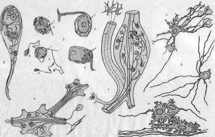
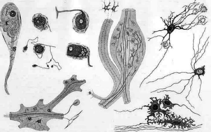
Fig. 1. Degenerative changes in the nerve cells of the sympathetic ganglion during chronic poisoning. Cells are swollen and vacuolated. Nerve fibers of the cervical sympathetic ganglion during chronic poisoning. (According to Ter-Pogosyan and Molchanov).
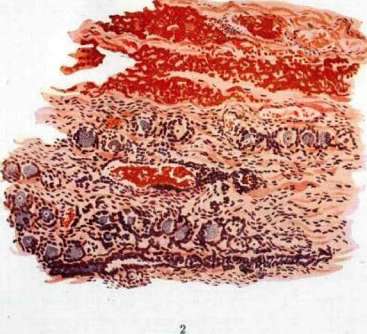
Fig. 2. Ganglion nodosum n. vagi in typhus (typhus exanthematicus). Hemorrhage in the stroma of the ganglion; hyperemia of vessels, perivascular infiltration, proliferation of glia; disappearance of Nissl bodies; vacuolization, cloudy swelling of protoplasm, neuronophagia, neuronolysis. (According to Tardykovsky and Mogilnitsky). To the article Nerve Ganglia. An axon departs in the form of a thin varicose thread, which, without being covered by myelin, passes through a whole series of sympathetic ganglia; its terminal branches end on smooth muscle cells (motor cells of the sympathetic ganglion). Cells of the 2nd type (see table, fig. 5 b) have thin dendrites, similar to axis-cylinder processes, which go beyond the limits of the given sympathetic ganglion (the method of their termination has not been sufficiently clarified); the axon gives off a number of collaterals on its path and, apparently, passes to glands as a secretory fiber. The 3rd type (see table, fig. 5 c) gives off thin dendrites that do not go beyond the limits of the given ganglion, while the axial process in the form of a thicker fiber passes into other ganglia and, apparently, is an association fiber. The number of cells of the 3rd type is small, and in smaller sympathetic ganglia they are not found at all. The structure of sympathetic ganglia was developed, mainly, by Dogiel. Recently, Stöhr Jr. rejects his data and asserts that the processes of sympathetic cells, connecting with each other, form a continuous network in which it is impossible to distinguish axons from dendrites, and that, thus, there are no isolated neurons in sympathetic ganglia. As in spinal ganglia, sympathetic cells are surrounded by a connective tissue capsule and satellite cells. A peculiarity of sympathetic cells is the presence of special flask-shaped outgrowths of the body, which are usually located inside the capsule; with age, they increase, their significance has not been clarified. As for nerve fibers, the sympathetic ganglion, in addition to its own fibers, also contains an admixture of myelinated fibers, originating, apparently, from the cerebrospinal system and forming with their terminal branches networks around the cells of the ganglion; the fibers of the sympathetic ganglion itself (processes of sympathetic ganglionic cells) also give rise to dense networks around the nerve cells of the ganglion (see table, fig. 3). The development of nerve ganglia occurs as follows: at the moment when the neural tube is still open, a cellular ridge forms at the place of transition of the skin layer into the primordium of the spinal cord, which grows out to the sides; after the closure of the neural tube, the detached cellular ridge lies between the neural tube and the skin layer. Subsequently, the cord grows laterally and segments. Individual groups of its cells give rise to spinal ganglia; their connection with the spinal cord is established secondarily by the ingrowth of fibers from the ganglion into the spinal cord; at the periphery, another process grows out, giving rise to a peripheral receptor fiber. Initially, the cells of the spinal ganglion appear bipolar, and then the places of origin of the processes move closer together, and thus the cell becomes unipolar. Sympathetic ganglia are formed as outgrowths of the ventral side of the spinal ganglion and subsequently actively move to the place of their final location. At a certain stage of development, in the primordium of the intervertebral ganglion, one can see two groups of cells: dorsal (spinal ganglion) and ventral (sympathetic ganglion), connected by a bridge of fibers (ramus communicans).
V. Fomin. Pathological anatomy of nerve ganglia. The study of changes in nerve ganglia in various diseases and the assessment of these changes in terms of their significance for the organism began to attract the attention of researchers relatively recently. The first works of this kind, dating back to the 70s of the 19th century, belong to Russian scientists (Popov, Lyubimov) and concern changes in sympathetic ganglia in infectious diseases. Subsequent studies by numerous scientists from various countries, among whom Russian scientists again played a major role (Abrikosov, Vinogradov, Vetvinsky, Muravyov, Rumyantsev, Davydovsky, Mogilnitsky, and many others), relate, mainly, to the study of pathological-anatomical changes occurring in a wide variety of diseases in the cervical and abdominal sympathetic ganglia, cardiac ganglia, ganglia of the vagus nerve, and in the Auerbach and Meissner plexuses. On the basis of these works, the following general ideas about the pathological anatomy of nerve ganglia can be given. Age-related changes in nerve ganglia consist of the appearance in the protoplasm of nerve cells of a fine-grained brown pigment, lipofuscin, which, beginning to be deposited even in childhood, gradually increases in quantity; simultaneously, atrophic changes on the part of the nerve cells and thickening, sclerosis of the interstitial connective tissue are noted. These age-related changes are observed as something "normal" in sympathetic nerve ganglia and in intervertebral ganglia, whereas they are not characteristic of cardiac ganglia. Changes similar to age-related ones take place in nerve ganglia in cachexia caused by various debilitating chronic diseases (cancer, starvation, tuberculosis, etc.). Degenerative changes in nerve cells, manifested in tigrolysis (see), changes in the neurofibrillar apparatus, vacuolization, degenerative fatty degeneration (see color table, fig. 1), as well as necrosis of cells with degeneration of nerve fibers, are observed to one degree or another in all infectious diseases and in many poisonings; of the infections, diphtheria gives the strongest degree of degeneration of nerve cells, mainly of cardiac ganglia. In many infectious diseases, in addition to degenerative changes in nerve cells, inflammatory manifestations on the part of the interstitial tissue are also observed in nerve ganglia in the form of hyperemia, edema, and inflammatory infiltration (see color table, fig. 2); this is most sharply expressed in typhus, epidemic influenza, rabies, and lobar pneumonia. In septicopyemia, abscesses are encountered in nerve ganglia. Among circulatory disorders in nerve ganglia, there may be hemorrhages, for example, in infections, as well as due to atherosclerosis of vessels or as a partial manifestation of hemorrhagic diathesis. These changes leave phenomena of sclerosis and scars in the ganglia along with one or another loss of nerve cells and fibers. From nerve ganglia, mainly sympathetic ones, tumors can originate, relating to mature or immature ganglioneuromas (see). The above-mentioned pathological-anatomical changes in nerve ganglia lie at the basis of those disturbances in various functions of the autonomic nervous system (see) which are observed in corresponding diseases.
Related articles
Mentioned in
Cite this page
“Hanko and Nerve Ganglia.” Soviet Medical Encyclopedia. English translation of Bolshaya Meditsinskaya Entsiklopediya, 1st ed. (Moscow, 1928–1936), ed. N. A. Semashko. https://sovietmedicalencyclopedia.pages.dev/article/ganglion/