Metamerism
Historical document, translated for reference. It reflects medical knowledge of the 1920s–30s and is not medical advice.
Summary
Metamerism is a type of body structure characterized by the sequential arrangement of homologous segments called metameres. This fundamental organizational principle is found in all vertebrates and the majority of invertebrates, though it becomes modified in more complex organisms.
Encyclopedia article (1928–1936)
METAMERISM (from Greek meta- after and meros-part), a type of body structure characterized by the sequential arrangement of a series of homologous segments, or metameres. Metameric body structure is characteristic of all vertebrates and the overwhelming majority of invertebrate animals. In each metamere, corresponding elements can be distinguished: of the skin (dermatome), muscles (myotome), skeleton (sclerotome), nerves (neuromere), and the center of the nervous apparatus (myelotome). In lower (chordate) animals, metamerism is observed in the connective tissue skeleton. Typical metamers of connective tissue are myocommata—the connective tissue septa that separate the metameric muscles from each other. Myocommata serve as the framework for the attachment of metameric muscles and can be considered homologous to the ribs of vertebrates. - The question of the origin of metamerism cannot yet be considered definitively resolved. According to Haeckel, metamerism can be considered as a phenomenon of colonialism, the combination of separate individuals into a single whole. The process of formation of metameric segments can be viewed as the result of asexual reproduction: each metamere represents a new individual, which however does not separate and does not transform into an independent organism, but remains connected with the other metameres, combining with them into a single individuality. According to another theory, metamerism originated from the repetition of organs: in some lower invertebrates that do not have a metameric structure (flatworms), symmetrical branches are observed, extending in pairs from both sides of longitudinally located organs; such metameric branching is observed in the intestine, sexual organs, and excretory organs. If one assumes that such repetition of parts spread to all organs and became even more regular, one can imagine the evolution of a premetameric organism into a metameric one. Ideal metamerism, i.e., an animal whose entire body would be built from completely identical segments, does not in fact exist; this ideal metamerism is primarily violated by the circumstance that the most anterior metameres, or segments, with which animals first encounter surrounding objects, become the leading (head) department of the body; in this head department, special anatomophysiological apparatuses develop for perceiving irritations at a distance (distance receptors: olfactory, visual, auditory) and for carrying out general reactions of the organism as a whole. In addition, part of the anterior metameres in lower animals (fish) is closely connected with the respiratory apparatus (gills), which undergoes significant transformations in the course of evolution. These anterior metameres connected with the gills are designated by the term branchiomeres, i.e., gill metameres. Since in the region of branchiomeres there is a close connection between somatic elements and visceral elements that disappear in the further course of evolution, the innervation of the branchiomere region has certain peculiarities depending on the transformation of visceral elements into somatic ones [for example, the n. accessorius, which in humans is a somatic nerve, does not arise from the anterior horns, like all other somatic motor nerves, but from the lateral horns, which is explained by its origin from the sympathetic nerve that innervated the gills in the past (n. branchiomotorius)]. However, despite all these transformations, traces of metamerism can be found in the skull and brain of humans. Although the view once expressed by Goethe and Oken regarding the origin of the skull from vertebrae is now refuted, the metamerism type of structure of the head of vertebrate animals is beyond doubt (Wiedersheim). In addition to the violation of metamerism in connection with the formation of the head end or the so-called process of cephalization in the course of evolution, other factors also arose that contributed to the transformation of metameres. Among these factors, the development of limbs should be mentioned first. The formation of limbs occurred through the growth from the body trunk of dermatomic, myotomic, sclerotomic, and neuromeric elements
elements. The development of limbs led to the modification of the metameric structure in the regions where they appeared. The limbs themselves represent a complication of the metameric plan, as they develop from specific metameres and acquire their own structural organization. In the course of evolution, the metameric structure became increasingly modified, especially in the anterior part of the body, which specialized for various functions. The process of cephalization, or the formation of a head, resulted in the concentration of sensory organs and nervous tissue in the anterior metameres, leading to their specialization and deviation from the basic metameric pattern. Despite these modifications, the fundamental metameric plan remains evident in the organization of the nervous system, the arrangement of certain muscles, and the skeletal structure of vertebrates. The study of metamerism provides important insights into the evolutionary development of animal organisms and the basic principles of their organization.
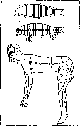
elements corresponding to a specific metameric level; myelomers, i.e., elements corresponding to spinal reflex centers, did not participate in this outgrowth. As a result of such gradual outgrowth of dermato-, myo-, scleroto-, and neurameric elements for the formation of limbs, a significant disruption of metamerism occurred, consisting in the fact that the correct sequence of metamers on the trunk was interrupted at the levels of limb formation (see figure). Thus, while the metameric spinal centers (myelomers) retained their correct sequential arrangement, the metamers of the trunk were disrupted in their sequence due to the formation of limbs. In humans, an apparent disruption of the correct sequence of metamers also occurred due to the adoption of an upright position; in connection with this, the upper limbs are positioned parallel to the trunk instead of having a perpendicular position to it, as is the case in all quadruped mammals; similarly, the lower limbs instead of the perpendicular position to the trunk found in quadrupeds, in humans due to the adoption of an upright position, are as if an extension of the trunk. All this leads to an apparent sharp disruption of the sequential arrangement of metamers; this apparent irregularity can be eliminated if one imagines a human standing on all four limbs (see figure); in this way, those disruptions in the sequential arrangement of metamers (dermatomers are shown in the figure) that are caused by the upright position of the body are eliminated. In general, it should be borne in mind that in humans, features of metamerism have been preserved most of all in the spinal reflex centers and the roots of the spinal nerves; in the somatic division, features of metamerism have been preserved more or less clearly in the thoracic region: the correct alternation of ribs, intercostal muscles, intercostal nerves; in the visceral organs, in humans as in all vertebrates, there are no features of metamerism. The clinical significance of metamerism lies mainly in neuropathology; in this respect, an especially important role belongs to the data of dermatomerism, i.e., the metamerism of skin sensitivity. The role of these data is as follows: there is a certain exact relationship between the sensory innervation of the skin and the segments of the spinal cord, shown in the figure; therefore, on the basis of sensory disturbances, it is possible to conclude the level of spinal cord damage; since the distribution of skin sensitivity by segments does not coincide with its distribution by peripheral sensory nerves, the form of the spread of sensory disturbances can generally differentiate segmental lesions (spinal and root) from lesions of peripheral nerves. The above applies not only to the area of the trunk and limbs, but also to the area of the face: the nucleus of the trigeminal nerve, which is a complete analog of the posterior horns of the spinal cord, innervates the face according to the type of primary metamerism, as indicated in the figure. Disorders of sensitivity distributed according to the concentric lines marked in the figure indicate a nuclear lesion; with damage to individual branches of the trigeminal nerve, as is known, the disorders of sensitivity have a completely different distribution. It should be noted that the areas of metameric innervation indicated in the diagrams refer only to the predominant connection of a given area of skin with a specific segment of the spinal cord; in reality, each given area of skin is innervated by at least three segments: mainly by the one indicated in the diagram, but also partly by adjacent higher and lower ones. For example, in the figure, the innervation of the level corresponding to the umbilical line is indicated from the X thoracic segment; in reality, this area is mainly innervated from the X thoracic segment, but also partly from the IX and XI. This circumstance is of great importance for the precise determination of the upper level of spinal cord damage, as is required, for example, in surgical interventions for spinal cord tumors; with complete loss of sensitivity in a certain area of skin, the segment corresponding to it in the diagram should be considered damaged, as well as the one above it. The data on metamerism also play a certain role in the diagnosis of diseases of internal organs. Although, as indicated above, the internal organs are completely devoid of metamerism features, and some of them even appear asymmetrical (spleen, liver, loops of intestines), however, the sympathetic innervation of internal organs has metameric centers in the lateral horns of the spinal cord. In these centers, afferent sympathetic fibers carrying impulses from various internal organs end; here, in the lateral horns, efferent sympathetic fibers arise, conducting motor impulses to the smooth muscles of internal organs. As is known, the pains observed in diseases of internal organs are the result of the spread of impulses from sympathetic afferent nerves to the cells of somatic nerves in the posterior horns and the subsequent projection of these impulses into areas corresponding to these somatic nerves. In other words, pains in diseases of internal organs are the reflection of impulses from sympathetic nerves into the area of somatic sensory nerves, which have centers in the spinal cord at the same level as these sympathetic nerves (see Head zones). Thus, guided by the data of sympathetic and somatic metamerism of the spinal cord, it is possible to recognize diseases of various internal organs on the basis of the localization of pains and hyperesthesia of the skin.
M. Astvatsaturov.
Related articles
Mentioned in
Cite this page
“Metamerism.” Soviet Medical Encyclopedia. English translation of Bolshaya Meditsinskaya Entsiklopediya, 1st ed. (Moscow, 1928–1936), ed. N. A. Semashko. https://sovietmedicalencyclopedia.pages.dev/article/metamerism/