Head's Zones
Historical document, translated for reference. It reflects medical knowledge of the 1920s–30s and is not medical advice.
Summary
This article from the 1928-1936 Great Medical Encyclopedia describes Head's zones (areas of cutaneous hyperesthesia and referred pain associated with visceral diseases) and details their neuroanatomical mapping and diagnostic utility as proposed by Henry Head.
Encyclopedia article (1928–1936)
HEAD'S ZONES (Head), established by Head, are specific areas of the skin in which referred pains, as well as pain and temperature hyperesthesia, appear during diseases of internal organs. The boundaries of these "zones" correspond to dermatomes, the radicular distribution of cutaneous sensibility, and, at the same time, correspond to the zones along which herpes zoster spreads. The emergence of Head's zones finds its explanation, according to the author himself, in the irradiation of excitations received from a diseased internal organ and conducted via sympathetic nerve fibers running from it to the spinal centers in which these sympathetic fibers terminate. The excitation of the spinal centers arising in this manner manifests itself by projecting pains (and hyperesthesia) into those skin areas that are innervated by the roots corresponding to these centers. Thus, for example, a disease of the rectum causes irritation in its sympathetic fibers terminating in the region of the II-III-IV sacral segments of the spinal cord; the gray matter of the specified segments manifests its irritation arising in this way by the projection of pains (and hyperesthesia) into the skin areas innervated by the II-III-IV sacral roots, i.e., into the perianal and scrotal zones. To discover cutaneous hyperesthesias accompanying diseases of internal organs and to establish their boundaries in each individual case, Head used several methods. 1st method: the skin and subcutaneous tissue in the investigated area are slightly pinched by the investigator between the thumb and index finger and slightly lifted upward; under normal conditions, this manipulation is painless, whereas in cases with the presence of irradiated hyperesthesia, it produces more or less severe pain. 2nd method: the skin is touched with the head of a pin—as soon as one enters the region of Head's zones, a pain sensation is obtained that is completely absent in normal areas. 3rd method: the skin is touched with a warm wet sponge or a test tube filled with lukewarm water—such a touch, indifferent in other places, produces a sensation of pain and burning in Head's zones. Using these methods and having studied a large amount of material, Head established precise correlations between internal organs and radicular cutaneous innervation. These correlations found expression in his well-known diagram, which is presented here (see figure 1). Heart: III-IV cervical roots, II-VIII and, possibly, also I and IX thoracic. Lungs: III-IV cervical roots, III-IX and, possibly, II thoracic. Stomach: IV and, possibly, III cervical roots and VII-IX and, perhaps, VI and X thoracic. Intestines: IX-XII thoracic roots. Rectum: II-IV sacral roots. Liver: III and IV cervical
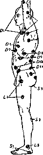
Figure 1. Head's zones on the trunk and limbs and their relation to the segments of the spinal cord.
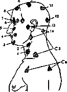
roots, VII-X and, possibly, VI thoracic. Gallbladder: VIII, IX and, possibly, VII thoracic roots. Kidney and ureter: X-XII thoracic roots, I and, possibly, II lumbar. Urinary bladder: XI and XII thoracic roots, I and II lumbar, III and IV sacral. Prostate gland: X-XII thoracic roots, possibly V lumbar, I-III sacral. Testes: X thoracic root. Epididymis: XI and XII thoracic roots and, possibly, I lumbar. Ovary: X thoracic root. Fallopian tube: XI and XII thoracic roots, I and, possibly, II lumbar. Uterus: X-XII thoracic roots, I and, possibly, II lumbar. Orificium uteri: II-IV and, possibly, also I sacral roots. Mamma: IV and V thoracic roots. As can be seen from this diagram, two radicular zones are not affected by the irradiation of excitations from internal organs: first, the lower cervical segments (V, VI, VII, and VIII) and, second, the middle lumbar segments (III and IV). Head's zones have also been discovered in disease of internal organs in the region of the head (see figure 2). Pains in the frontonasal region correspond to lesions of the apices of the lungs, stomach, liver, and mouth of the aorta (corresponding spinal zones: III and IV cervical segments). Pains in the middle orbital region correspond to lesions of the lungs, heart, and ascending aorta (II, III, and IV thoracic segments). Pains in the frontotemporal region correspond to lesions of the lungs and, possibly, the heart (V-VI thoracic segments). Pains in the temporal region correspond to lesions of the lower lobes of the lungs, heart, and cardia (VII thoracic segment). Pains in the parietal region correspond to lesions of the pylorus and the upper part of the intestines (IX thoracic segment). Pains in the occipital region correspond to lesions of the liver, large intestines, ovaries, testes, Fallopian tubes, uterus, and urinary bladder (X, XI, XII thoracic roots). The diagnostic significance of Head's zones is understandable: by establishing the zones of pain and hyperesthesia and comparing their boundaries with Head's given diagram, one can make diagnostic conclusions about which internal organ is affected in a given case. However, these conclusions encounter significant difficulties in practice. First of all, they are based on the subjective testimonies of the patient himself. Figure 2. Head's zones on the head and neck and their relation to Head's zones on the trunk in certain diseases of the internal organs of the chest and abdominal cavities: 1 - zona laryngei inf.; 2 - z. laryngei sup.; 3 - z. mentalis; 4 - z. naso-labialis; 5 - z. maxillaris; 6 - z. fronto-temporalis (D5-6); 7 - z. fronto-nasalis (C3-4); 8 - z. orbitalis med. (D2-3); 9 - z. temporalis (D4); 10 - z. verticalis (D5); 11 - z. parietalis (D9); 12 - z. occipitalis (D10-12); 13 - z. mandibularis; 14 - z. hyoidea; C1-C4 - cervical segments of the spinal cord. Then, as the diagram shows, hyperesthesia of the same zones can be encountered in diseases of very different organs: thus, the zones of the lungs and heart almost completely coincide, the zones of the stomach and liver, of the urinary bladder and prostate, etc., almost coincide. Above all, however, inconveniences are caused by the so-called "generalization" of visceral excitations, noted already by Head himself and often disrupting the strictness of his diagram: pain, as a result of a disease of a given internal organ, may under certain conditions localize in a zone corresponding to a completely different organ. Head finds an explanation for such generalization and for its direction in the specific resistance of individual spinal zones. It is very easy, for example, in women for the zone of the X thoracic segment to be secondarily captured, then the zone of the VI thoracic segment follows, and so on, provided that the specified specific resistance is lowered. And many conditions lead to such a lowering, among them also such frequent ones as anemia and infectious diseases. The possibility of generalization makes one treat the diagnostic evaluation of each individual case with caution and take into consideration many extraneous factors in such an evaluation most seriously. Nevertheless, Head's method is of great diagnostic value, especially as a method of auxiliary significance.
I. Filimonov
Related articles
Mentioned in
Cite this page
“Head's Zones.” Soviet Medical Encyclopedia. English translation of Bolshaya Meditsinskaya Entsiklopediya, 1st ed. (Moscow, 1928–1936), ed. N. A. Semashko. https://sovietmedicalencyclopedia.pages.dev/article/ged-zones/