Herpes
Historical document, translated for reference. It reflects medical knowledge of the 1920s–30s and is not medical advice.
Summary
Herpes refers to dermatoses with acute and cyclic courses characterized by the eruption of closely grouped blisters on a limited area of edematous, hyperemic skin. The article distinguishes between herpes zoster (shingles), herpes simplex, and herpes febrilis, with detailed descriptions of their clinical manifestations.
Encyclopedia article (1928–1936)
HERPES. Contents: Herpes zoster...................682 Herpes simplex..................688 Herpes febrills..................691 Herpes (from Greek herpo- I crawl), from a morphological point of view - dermatoses with acute and cyclic courses, characterized by the eruption on a limited area of edematous, hyperemic skin of groups of closely located blisters. Distinguished are: H. zoster (zoster, zona, shingles), H. simplex (blight, H. genitalis, labialis, etc.) and H. febrilis. Herpes zoster. The eruption develops acutely, either without prodromal phenomena, or with some disturbances of the general condition (malaise, headache, fever, usually moderate, gastrointestinal disturbances, etc.). Particularly characteristic are neuralgic pains of varying intensity, localized in the area of distribution in the skin of this or that nerve - either over the entire area of the future eruption, or at separate points corresponding to the places of exit and division of the nerves. Then on the reddened and slightly swollen skin appear separate groups of papules, the size of a millet grain to a small lentil. After several hours, sometimes after 1-2 days, they transform into tense blisters filled with transparent fluid. The eruption is accompanied by more or less severe burning or tingling. The blisters may remain completely isolated until the end or merge with neighboring ones into larger blisterous efflorescences with festooned outlines. After 2-4 days, the transparent content of the blisters becomes cloudy, turning purulent. At the same time, the tension of the blisters weakens, their cover shrivels and then soon dries together with the contents into a yellow-brown crust. After its falling off, either a pale spot or a superficial scar remains. This evolution of the eruptive element takes 7-10 days. The efflorescences of a separate group are uniform, as they erupt at the same time. But in the following days new groups of eruptive elements may appear nearby; the rash by repeated flashes can develop over 2-3 weeks. Then the general picture of the disease becomes polymorphic: alongside the newly appeared papules, all the already described stages of their further evolution are visible. With repeated eruptions, the general course of the dermatosis takes 4-5 or more weeks. From this, the most common picture of herpes zoster, various deviations are observed. In H. zoster abortivus, the inflammatory phenomena quickly subside, the few papules are covered with scales, and the blisters with superficial crusts, which soon fall off, and the rash disappears without a trace. The number of eruptive groups can also vary: from single ones, even a single one, to an almost continuous eruption formed by the merging of numerous groups of eruptive elements and occupying, for example, an area of skin 2-3 intercostal spaces wide, from the spine to the anterior median line of the trunk. Further, 68S
HERP
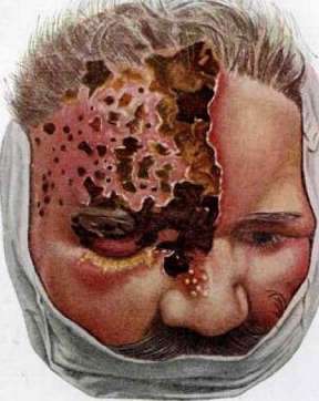
Herpes zoster occurs with very large blisters, up to the size of a pigeon's egg (N. zoster bullosus). Herpes zoster with hemorrhagic-serous blister contents occurs—N. zoster haemorrhagicus; in this form, in addition to the bluish-black color of the blisters, there are sometimes very severe pains and as a rule a deeper destruction of the skin, as a result of which depressed scars remain. The most severe form is represented by N. zoster gangraenosus [see separate table (pp. 687-688), Fig. 1], in which closely grouped blisters form, the size of a lentil, partly merging. Their contents soon become purulent, often purulent-hemorrhagic. The surrounding skin takes on a bluish-purple color. The blisters soon break down and form more or less deep ulcers covered with a dirty-brown scab. The necrosis of the skin can have a small-nest character, and then isolated scars remain as a result, or else in dense, confluent rashes, more or less large areas of skin undergo necrosis; in such cases the picture of the disease resembles burns from caustic substances. Sometimes N. zoster gangraenosus develops without a noticeable blister stage, immediately in the form of necrosis of more or less extensive areas of skin, which becomes black, mummified; in such cases the diagnosis is based on the localization of the necrosis in the area of distribution of a certain nerve and the scalloped character of its outlines. N. zoster gangraenosus is distinguished by especially painful pains, a course of two or more months, and the formation of scars. Atypical forms of N. zoster should include cases of rash developing immediately or successively in the area of distribution of several nerves. Its highest form is N. zoster generalisatus, in which, in addition to typical unilateral belt-like rashes, there are so-called aberrant blisters scattered over the entire surface of the skin. In moderate and severe forms of N. zoster, painful swelling of regional lymphatic glands is observed. These adenitis do not suppurate and disappear without a trace. The subjective symptoms in N. zoster consist, mainly, of the mentioned neuralgic pains of varying intensity, sometimes extremely severe; in some cases these pains do not disappear, as is usually the case, with the resolution of the rash, but remain for a long time, especially when N. zoster is localized in the area of distribution of the trigeminal nerve. Sometimes the pains have a rheumatoid character (in the form of lumbago, ischias), sensitivity to pressure appears and with movements of the long muscles, etc.; sometimes the pains simulate pleuritic pains. Along with this, other disturbances of sensitivity occur—hyperesthesias, hyperalgesias, paresthesias (feeling of numbness, crawling of ants, itching, etc.). Rarely, after the resolution of N. zoster, paralysis of the eye muscles, facial nerve, more rarely—muscles of the upper extremities and others remain. Sometimes various trophic and vascular disorders, disturbances of secretion (e.g., increased or absence of sweating, loss or graying of hair, loss of teeth, etc.) are observed. Some forms of herpes zoster are accompanied by fever and in their course and general picture have the character of an acute infection. Increases in temperature are observed, according to Blaschko, in ⅛-⅜ of cases, not exceeding as a rule 39° and usually ceasing with the appearance of the blisters. Localization. Herpes zoster can develop in the area of distribution of any nerve; in 60% of cases it is observed on the trunk, especially on the chest and back (herpes dorso-pectoralis), along the course of the intercostal nerves [see separate table (pp. 631-632), Fig. 2]. The second most frequent location is herpes zoster of the face and head, especially in the area of the trigeminal nerve, on the neck, occiput [see separate table (pp. 687-688), Fig. 2)]; least often on the extremities. On the head, N. zoster ophthalmicus, developing in the area of the frontal, ciliary and other branches of the trigeminal nerve, is distinguished by an especially severe course. Following the prodromal intense neuralgic pains, redness and swelling of the skin of the forehead, eyelids, nose, temporal region and conjunctiva, tearing and photophobia appear. Subsequently, in the area of distribution of the n. frontalis, supratrochlearis and sometimes lacrimalis, groups of blisters develop; they can also appear on the skin of the nose and cheeks, in the area of distribution of the n. nasociliarium and supramaxillaris. The eyeball is often affected: small blisters appear along the edge of the cornea, leaving scars behind. The development of iritis, hypopyon, deep corneal ulcers and even panophthalmitis is possible. As a result of the scarring of deeper ulcers, in severe forms of N. zoster, ectropion or entropion may develop on the eyelids. Sometimes, due to paralysis of the eye muscles, ptosis, strabismus, etc. appear. On the trunk, N. zoster is especially typical. Usually the rash is distributed on one half of the trunk, in the area of one or another intercostal space or immediately two to three, in the form of a half-belt; it starts from the spine and ends at the anterior median line (sometimes, however, individual elements go beyond these limiting borders). Much less frequently N. zoster is bilateral. N. zoster can also develop on mucous membranes, e.g., oral, nasal cavity, etc. Here the blisters quickly lose their covering and turn into erosions or ulcers of a round shape (sometimes scalloped), with sharp, as if cut-out edges and a bottom covered with a dirty-yellowish coating, surrounded by a bright red inflammatory halo. N. zoster of the mucous membranes is accompanied by severe pains. Pathological anatomy. The works of Hoffmann, Frieboes (E. Hoffmann, Frieboes) and others established that the histopathological changes in N. zoster begin with increased proliferation of the cells of the Malpighian layer, as evidenced by numerous mitoses (status proliferationis). Soon a series of further changes occur in the structure of the Malpighian layer: 1) the prickle cells undergo a peculiar degeneration, and 2) a more or less strong edema develops in the intercellular spaces. The degeneration of the cells manifests itself as follows: a) the nuclei of the prickle cells amitotically divide; their number reaches 10-20-30 or more; b) the protoplasm of the cells, losing its fibrillar structure, swells, becomes homogeneous; c) the cells significantly increase in size, turning into a ball—the 'cell balloon' (Unna); d) due to the disappearance of 'intercellular bridges' the cell loses its connection with neighboring elements. Several cells are subjected to the described changes at the same time. At the same time, the intercellular spaces of the Malpighian layer are filled with edematous fluid—the 'spongy state' (Unna). The serous fluid, accumulating mainly in areas where 'cell balloons' have formed, pushes apart the rows of epithelial cells, as a result of which a more or less large cavity—a blister—is formed; the 'cell balloons' due to their weight sink to its bottom; in the contents of the blisters that have arisen, in addition to these elements, in small quantities: lymphocytes, polymorphonuclear cells, sometimes eosinophils, erythrocytes, and also remnants of degenerating epithelial cells are found. The walls of the blister are formed by prickle cells that have already undergone the above-described changes. Unna gave the name 'ballonierende Degeneration' to the complex of all these phenomena. Thus, in the Malpighian layer, single-chamber blisters of various sizes arise, but along with them, multi-chamber blisters filled (at first) with a gelatinous, detritus-like mass, due to their origin to a special type of degeneration called by Unna 'reticulierende Degeneration', in which vacuoles arise in the protoplasm of groups of prickle cells; they gradually increase, and in the end only a thin peripheral rim remains from the protoplasm. The nucleus quickly disintegrates, and the entire cell turns into a blister. A conglomerate of thus changed cells forms a multi-chamber blister of larger or smaller sizes, the partitions of which are the remains of the protoplasm of vacuolized prickle cells. The first type of blister formation—ballonierende Degeneration—is found predominantly in the deeper layers of the epithelium, while the second—reticulierende Degeneration—in its superficial parts, closer to the horny layer. Sometimes both varieties of blisters can be seen on one preparation. In the papillary and subpapillary layers of the skin, all transitions from barely noticeable inflammation (e.g., only hyperemia) to highly developed inflammatory infiltration, sometimes with phenomena of thrombosis and destruction of the vessel walls with their contents, are encountered. In the deeper parts the infiltrates consist mainly of lymphocytes and plasma cells, sometimes also giant cells; near the epithelium a large admixture of polymorphonuclear cells. The latter often extend into the dilated interepithelial canals, and then the boundary between the papillae and the epithelium becomes blurred. The papillae and subpapillary layer in the immediate vicinity of the blister are always more or less edematous, the collagen tissue swells, the elastic tissue is usually preserved. The skin appendages are usually not affected, but if, for example, a blister develops near a hair follicle, 'ballooning' degeneration can develop in the cells of its sheath.
Kopitowsky, Lip-schütz and others have described in H. zoster in the degenerated prickle cells, in the nuclei of connective tissue cells, and in the endothelial cells of the intima of small vessels special inclusion bodies, according to Lip-schütz, Zoster-Körperchen. These are round or oval, of various sizes, compact, sharply delineated eosinophilic formations, occurring singly or in groups of 2-3 or more, mainly in the nuclei, and sometimes in the protoplasm of the cells. Since they react differently to staining substances than nucleoli, Lip-schütz considers them as 'the product of the reaction of cells to the virus parasitizing in them.' In some forms of H. zoster, inflammatory infiltration is observed in the spinal ganglia (resp. gangl. Gasseri), in the roots and adjacent segments of the spinal cord, along with hemorrhages. The more or less dense cellular infiltrate consists mainly of lymphocytes, sometimes with an admixture of plasma cells. This process is accompanied by neuronophagia with the death of many ganglion cells. Etiology and pathogenesis. H. zoster affects both sexes equally and any age. In some cases, it develops during other infectious diseases, both acute (for example, pneumonia, pleurisy, cerebrospinal meningitis, etc.) and chronic (tuberculosis, syphilis, etc.). In other cases, it is apparently associated with endogenous or exogenous intoxication: it has been observed in uremia, poisoning by carbon monoxide, morphine, and especially arsenic. H. arsenicalis is more frequently observed—either during treatment with arsenic preparations, especially arsenobenzenes, or from consumption of food products containing arsenic. An example of the latter is the so-called Manchester 'epidemic' of 1900 from the consumption of beer in the production of which glucose containing arsenic was used. In a number of cases, the development of H. zoster is associated with trauma to the central nervous system, skull, spine, or peripheral nerves. This H. traumaticus arises at the site of injury, nearby, or at a greater or lesser distance from it. More frequently observed is idiopathic herpes zoster, which gives the impression of a special infection (cyclic course, elevated temperature, prodromal phenomena, swelling of regional glands, usual absence of recurrences, occurrence of small 'epidemics', increased frequency in autumn and spring, etc.). A number of authors have noted pleocytosis of the spinal fluid in herpes zoster; others have noted leukocytosis of the blood at the onset of the attack and eosinophilia during the recovery period. All this taken together, in connection with the localization of H. zoster along the course of the nerves and the histological findings in the central and peripheral nervous system, gives the impression of an infection by some neurotropic virus, perhaps invisible and filterable, which is why many dermatologists consider herpes zoster the result of a vasomotor-trophic reflex caused by disease of some part of the neuron. There are serious data for recognizing the identity of the viruses of herpes zoster and varicella, first pointed out in 1892 by Bokai: 1. Sometimes H. zoster and varicella are observed together in a patient. 2. Sometimes varicella develops following H. zoster or vice versa. 3. Sometimes H. zoster develops in a person who has been in contact with a varicella patient, or vice versa, with an interval of 12-17 days, corresponding to the incubation period of varicella. 4. H. zoster in adults sometimes gives rise to an epidemic of varicella in children. Particularly convincing in this respect are the observations of François-Dainville and Reynaud in the children's day nurseries of the Seine department (France), where following H. zoster in a nurse, an epidemic developed in a number of infants, starting with her own child. 5. The histological elements of herpes zoster and varicella are very similar. 6. In the serum of patients with H. zoster, the presence of varicella antibodies has been established (Netter, Urbain and others). 7. Inoculation into the skin or under the skin of the serous fluid of H. zoster vesicles can give immunity to varicella if vesicles develop at the site of inoculation (experiments of Kundratitz on children). Convincing evidence of the inoculability of H. zoster on the rabbit cornea (which has been proven in relation to H. simplex) has not been presented, and this is considered a fundamental difference between the two species of the herpes group. Diagnosis in most cases is not difficult: the characteristic localization strictly in the area of distribution of one or another nerve, the one-sided arrangement, disturbances of sensitivity, neuralgic pains, the acute development of the disease and the cyclic course sharply distinguish H. zoster from other vesicular rashes—eczema, trichophytosis, H. simplex, etc. Doubts in diagnosis may arise only in abortive forms of H. zoster or initial forms of H. of the hairy scalp or its ophthalmic form, when hyperemia and edema come to the fore, which may simulate erysipelas, but the further course and picture of the disease usually quickly clarify the matter. - The prognosis in the vast majority of cases is favorable: the rash disappears without a trace. The prognosis is worse for hemorrhagic and especially gangrenous H. zoster: in the former, scars often remain, in the latter always, sometimes deep and extensive. Caution is needed in prognosis in elderly individuals: in them, sequential neuralgias and other nervous disorders often remain for a long time, for example, pareses. The prognosis is always doubtful in H. zoster ophthalmicus (see description of its clinical picture) and zona otica with a rash on the cheek and auricle, in which paralysis of the VII or VIII pair is not uncommon and sometimes even disturbance of balance.-Treatment. Various methods have been repeatedly proposed for stopping H. zoster or, at least, alleviating and shortening its course. Among them, large doses of sodium salicylate and local application of red light deserve attention. For neuralgic pains: antipyrin-0.5, three times a day, pyramidon-0.3-0.5, three times, and other anti-neuralgic drugs. For very severe pains with insomnia-morphine. For sequential neuralgias, diathermy or ultraviolet rays help; sometimes pilocarpin (1% sol. 10-15 drops, 2 times) and potassium iodide 0.5-3 times have proven useful.-Local treatment of the rash. Protect the area of the rash from any kind of trauma with a bandage (not too warm). Preserve the integrity of the vesicle cover; efflorescences deprived of it are usually painful. With intact vesicles—abundant dusting with indifferent powders (starch, zinc oxide, talc). When the vesicles subside and are covered with crusts—indifferent or slightly disinfecting pastes, such as zinc, 5% dermatol, 3-5% viioform. For the gangrenous-ulcerative form—dusting with iodoform, iodoform ointment, etc.-H. zoster varicellosa—see Chickenpox.
Herpes simplex (vesicular herpes), an acute eruption, usually of short duration, consisting of one or more groups of small vesicles, located on slightly edematous and reddened skin and initially filled with clear fluid. The favorite localization is the area around the mouth, especially the lips (H. labialis), the nose (H. nasalis), the genital organs (H. genitalis); less frequently—the auricle (H. auricularis), eyelids, cheeks, etc. It also occurs on the mucous membranes of the oral cavity, pharynx, larynx, urethra, nose, etc. The eruption is usually preceded by a feeling of itching, less frequently—tingling, burning. The vesicles are as a rule of the same size, from pinhead to millet seed, appear at once in a close group, located either isolated or partially merging. Their contents, initially serous, within the next two to three days becomes cloudy, turns purulent and dries into a loosely adherent crust. Often the vesicles burst, giving a superficial erosion surrounded by a red halo. An erosion arising from an isolated vesicle is round in shape; erosions that developed from merged vesicles have finely scalloped outlines. The eroded efflorescences of herpes simplex are also covered with thin crusts. After the crusts fall off, in place of the former eruption either no trace remains or a brief, reddish spot; never are there scars. H. simplex is in most cases an ephemeral disease, lasting several days, at most 1-2 weeks, but often recurrent.-H. genitalis (see figure 3) occurs in the male in the coronal sulcus, on the glans, prepuce, shaft of the penis, scrotum, on the mucous membrane of the urethra. Usually
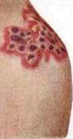
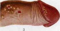
Fig. 1. Herpes simplex (herpes febrilis). Fig. 2. Herpes zoster (shingles). Fig. 3. Herpes simplex (herpes febrilis). Fig. 4. Herpes zoster (shingles). The course and picture of it differ in no way from those in any other localization; sometimes, with localization in the prepuce sac, under irritating influence, e.g., of decomposing smegma, the base of its erosions may be covered with a diphtheritic coating, deepen and give a purulent discharge; regional glands swell and become painful. Such complicated cases of herpes genitalis can create significant diagnostic difficulties. When developing on the urethral mucosa, H. genitalis can simulate a sluggishly developing gonorrhea. With irrational treatment of herpes genitalis erosions with cauterizing substances (e.g., lunar caustic), their base becomes slightly indurated, which can simulate an erosive primary syphilis. In women, H. genitalis is located mainly on the external genital organs and occasionally on the vaginal or cervical mucosa. When localized on the labia minora, the latter may swell. A special form is represented by the so-called H. vulvaris profusus: with moderate fever and intense burning, a sharp swelling of the vulva develops. Then groups of vesicles quickly appear on the external genital organs, inner surface of the thighs, pubis, and perineum, often merging into large efflorescences. The vesicles quickly burst, giving erosions. Their base is covered with diphtheritic decay, excreting a mucopurulent fluid with a foul odor. Regional lymphatic glands swell and are painful. With appropriate treatment, recovery occurs in 3-4 weeks. The appearance of H. on the tonsils—angina herpetica—develops suddenly, with fever and intense pain in the pharynx; upon examination, on the reddened and swollen tonsils, there are groups of small vesicles, and more often erosions with polycyclic edges and base, often covered with diphtheroid decay. The etiology of H. simplex cannot be considered finally established. It is possible that not for all cases of this dermatosis is it the same. It is highly probable that sometimes the etiological factor is trauma: H. labialis after dental operations, recurrent H. genitalis post coitum. The connection between H. simplex and the endocrine system cannot be excluded (e.g., recurrent herpes during menstruations—H. menstrualis s. sexualis). Vesicular herpes often appears during or after some infectious diseases: lobar pneumonia, malaria, influenza; conversely, it is extremely rare with streptococcal sepsis, typhoid fever, and lethargic encephalitis. It is worth mentioning that sometimes the appearance of a hard chancre is preceded by an eruption of H., and that infection with syphilis serves as a trigger for recurrence of H. of the genital parts. An intoxication H. is also described in carbon monoxide poisoning, poisoning with benzenes, cocaine, etc. It is not uncommon in the treatment with arsenic, especially arsphenamines. Finally, it can apparently be an independent infectious disease, the so-called febris herpetica. Since 1913, the etiology of dermatoses of the H. group has been thoroughly studied by numerous researchers. Inoculation of the contents of herpes vesicles of any localization into the rabbit's cornea can cause a special keratitis, which can be experimentally reinoculated to a whole series of rabbits and which sometimes even ends in fatal encephalitis. Gilde-meister and Herzberg succeeded in inoculation on the guinea pig's sole, and Teissier and others showed the possibility of inoculating H. on the skin of both carriers and healthy individuals. It is assumed that the causative agent of H. is a filterable virus (see Virus), close, and according to Levaditi, apparently identical to the virus of epidemic encephalitis. Apparently, this virus (or viruses) is located not only in the skin, in the contents of vesicles, but also in other parts of the body, particularly in the central nervous system: 1) in animals infected in the cornea with the contents of H. vesicles, encephalitic changes have been found; 2) positive inoculations on rabbits have been obtained with lumbar fluid from patients with herpes (Bastai, Rouillard); it has a special affinity for the nervous system and spreads in the body along the nerves (Grutter), like the rabies virus. Apparently, there are also "virus carriers," since Flexner and Moss caused herpetic keratitis by inoculating the saliva of healthy people. There is reason to believe that the skin manifestations in H. are the result of the combined action of the virus a) on the skin directly and b) on the nervous system. Thus, the skin changes are only one of the symptoms of the general disease, in which the central nervous system also plays a prominent role, the symptom of which is the not infrequent pleocytosis of the cerebrospinal fluid. Histologically, H. simplex gives changes essentially identical to those in herpes zoster. Diagnosis. Cardinal symptoms: 1) grouped arrangement of small vesicles; 2) favorite localization, mainly on the transition sites of skin to mucous membrane; 3) micropolycyclic edge of erosions at the site of merged vesicles; 4) absence of induration in the base of erosions, its evenness and undercut edges; 5) absence of severe subjective disorders; 6) rapid and traceless resolution. From the soft chancre, the erosion in H. genitalis is distinguished, besides, by the inability to autoinoculate with subsequent typical evolution for a venereal ulcer, absence of Ducrey's streptobacilli. Main differences from erosive primary syphilis: absence of induration of the base (cave confusion with induration after cauterizing substances!); absence of the pale spirochete; absence of typical accompanying adenitis; presence of micropolycyclic edge. - Treatment. Observance of cleanliness; in the vesicular stage—powderings with indifferent drying powders: talc, starch, zinc oxide, etc., or zinc paste; in the erosive stage—compresses with weak (1-2 percent) solutions of lunar caustic.
P. Grigoriev; Herpes febrilis (see pathogen above), an acute general infectious disease. Until recently, herpes febrilis was not regarded as a disease sui generis, but only as a symptom that can occur in various infections, and only in recent years has this disease been identified as a specific nosological unit. It occurs predominantly sporadically, rarely in the form of small epidemics, more often in the autumn and winter months. Herpes febrilis affects younger age groups, with women becoming ill more often than men. Factors that weaken the body's resistance serve as predisposing factors: fatigue, inadequate nutrition, cooling, all kinds of excesses, in women-menses, etc. The virus can be found in the serous fluid of herpetic skin vesicles, then in the cerebrospinal fluid, in the blood, in extracts from the brain (in experimental infection of rabbits), and not only during the acute period of infection, but even in persons who have long since had N.; the virus is in a latent state in them, and then, under the influence of various factors that weaken the body (cooling, fatigue, gastrointestinal disorders, certain infections, parenteral administration of foreign protein, etc.), it can be activated and give the characteristic skin rash. This is probably the way to explain the appearance of herpetic rash at the beginning of some infections. Clinical picture. The disease sometimes begins with brief prodromal phenomena: malaise, lassitude, loss of appetite; however, more often without these precursors, after a chill, t° quickly rises, sometimes to 40°, the pulse and breathing become more frequent accordingly, headache, general weakness, redness of the face, coated tongue, often quite severe pains in the limbs, chills in the chest, sometimes agitation and even delirium. These phenomena can be so violent that they lead to suspicion of the beginning of some severe infection. However, on the 2nd-4th day, t° falls with profuse sweating, often critically, sometimes below normal, the general condition of the patient quickly improves, and a characteristic vesicular rash appears, mostly of a hemorrhagic nature. The favorite localization of this rash is the skin of the face near the mucous membranes (on the lips, around the nostrils). However, it can also be on other parts of the facial skin-on the chin, cheeks, eyelids, sometimes even on the ears, neck, chest, very rarely on the extremities; in women, often on the genital organs. The rash can also be localized on the mucous membranes of the lips, cheeks, in the throat, on the tonsils (angina herpetica). The cycle of development of this rash on the skin is as follows: on certain, above-mentioned places on the skin, burning, redness and one or more groups of small elevations of the epideris-nodules-appear acutely, which quickly turn into vesicles due to the accumulation of serous fluid under the epidermis, often of a hemorrhagic nature. The vesicles, after several hours or 1-2 days, dry into a crust due to absorption of the fluid. Under the crust, due to the reverse development of inflammation and cessation of fluid exudation, epidermis forms; the crust falls off. For some time after the rash, a brownish pigmentation of the skin remains at the site. The number of groups of vesicles is extremely variable. When a herpetic rash appears on the mucous membranes, edema forms in the surrounding area of the mucous membrane, then the vesicles quickly burst and give rise to superficial, quickly healing ulcers. From the side of internal organs, few changes are found: sometimes enlargement of the spleen, bronchitis, and slight albuminuria can be determined. The disease lasts 4-5, more often 1-2 days and has a tendency to recur. The prognosis is always favorable. When making a diagnosis, great caution must always be exercised, as the clinical manifestations of the disease in the first days of the illness are common to the initial symptoms of many other infectious diseases. And even when a herpetic rash appears, it is not always possible to exclude the presence of another infection that can exacerbate the herpes virus, which is in a latent state in the body. Mostly the diagnosis has to be made by exclusion. If there is suspicion of central pneumonia, a diagnosis can be helped by X-ray examination. Therapy is symptomatic. Bed rest, a fever diet, and cleansing of the bowels are prescribed. For high t°-antipyretics (quinine, sodium salicylate), for rheumatoid pains-antineuralgic agents. If the rash is painful, it can be smeared with zinc and anesthetic ointments and dusted with INDIfferent powder. K. Georgievsky. H. circinatus, H. Iris, see Erythema (Erythema exsudativum multiforme). H. circinatus bullosus, H. gestationis, H. phlyctaenodes, see Duhring's disease. H. pjaemicus, see Impetigo herpetiformis. H. tonsurans, see Trichophytosis. H. tonsurans maculosus, see Pityriasis rosea. H. vegetans, see Pemphigus vegetans. Lit.: Ustinovsky A., On the etiology of Erythema exsudativum multiforme, zona and pityriasis rosea, 'Moskovsky med. zhurnal', 1926, no. 11; Zeleny I., Mercury herpes zoster (Herpes zoster), 'Russian journal of dermatological and venereal diseases', vol. XII, no. 9. 1906; Doerr R., Encephalitis- u. Herpesvirus (Handbuch der pathogenen Mikroorganismen, hrsg. v. W. Kolle, R. Kraus u. P. Uhlenhuth. B. VIII, Jena-B.- Wien-print); Schönfeld W., Zoster u. Herpes simplex (Handbuch der Haut- u. Geschlechtskrankheiten, herausgegeben von J. Jadassohn, Band VII, T. 1, Berlin, 1927); Doerr R. u. Schnabel A., Weitere experimentelle Beiträge zur Aetiologie und Verbreitungsart des Herpes febrilis beim Menschen, Schweizerische med. Wochenschr., 1921, no. 20; Doerr R. u. Zdansky E., Kritisches u. Experimentelles zur aetiologischen Erforschung des Herpes febrilis u. der Encephalitis lethargica, Zeitschr. f. Hygiene und Infektionskrankheiten, B. CI, 1924; Gold E., Anatomische Untersuchungen eines Falles von Herpes zoster, Dermatologische Zeitschrift, B. XXIV, 1917; Guszmann D., Beiträge zur Herpes-zoster-Varizellenfrage, Dermatologische Wochenschrift, B. LXXIX, 1924; Hoffmann E. u. Friebocs W., Beitrag zur Histopathologie des Herpes zoster, Arch. f. Dermatologie u. Syphilis, B. CXIII, 1912; Lauda E. u. Rezek Ph., Zur Histopathologie des Herpes simplex, Virchows Arch., B. CCLXII, 1926; Lipschütz B., Untersuchungen über die Aetiologie der Krankheiten der Herpesgruppe, Arch. f. Dermatologie u. Syphilis, B. CXXXVI, 1921; his same, Weitere Untersuchungen über die Aetiologie des Zoster, ibid., B. CXLIX, 1925; Luger A. u. Lauda E., Über oxychromatische Veränderungen am Zellkern (auf Grund von Untersuchungen von Herpes simplex, zoster, Varicellen, Variola und Karpfenpocke), Med. Klinik, 1926, no. 11 - 13; Mariani G., Experimentelle Untersuchungen u. kritische Erwägungen über die Aetiologie der Herpeserkrankungen, Arch. für Dermatologie und Syphilis, B. CXLVII, 1924; Zumbusch L., über Herpes zoster generalisatus mit Rückenmarksveränderung, Ibid., B. CXVIII, 1914; Gans O., Histologie der Hautkrankheiten, B. II, p. 43, Berlin, 1928; Kure J., Vorlesungen über Histobiologie der menschlichen Haut u. ihre Erkrankungen, B. II, B-, 1927; Kundratitz K., Zur Frage der gemeinsamen Aetiologie der Varicellen u. des Herpes zoster, Wiener medizinische Wochenschrift, 1927, no. 23; Massini R., Febris herpetica (Handbuch d. inneren Medizin, hrsg. von L. Mohr u. R. Staehelin, Band I, T. 1, B., 1925); Grüter W., Das Herpesvirus, seine aetiologische und klinische Bedeutung, Münchener medizinische Wochenschrift, 1924, no. 31; Bastai P. u. Rusacca A., Über die Pathogenese des Herpes febrilis, Klinische Wochenschrift, 1924, no. 4; Levaditi L., L'herpes et le zona etodermoses neurotropes, P., 1926; Neillér A. et Urbain A., Zonas varicelleux; anticorps varicelleux dans le serum de sujets atteints de zona; anticorps zosteriens et anticorps varicelleux dans le serum de sujets atteints de varicelle. Comptes rendus des seances de la societe de biologie, v. XC, p. 189, 1924; Francois-Dainville E. et Reynaud H, Zona et varicelle, Presse med., 1928, no. 42.
Related articles
Mentioned in
Cite this page
“Herpes.” Soviet Medical Encyclopedia. English translation of Bolshaya Meditsinskaya Entsiklopediya, 1st ed. (Moscow, 1928–1936), ed. N. A. Semashko. https://sovietmedicalencyclopedia.pages.dev/article/herpes/