Parasitic Fungi
Historical document, translated for reference. It reflects medical knowledge of the 1920s–30s and is not medical advice.
Summary
This article from the 1928–1936 Soviet Medical Encyclopedia examines parasitic fungi as causative agents of various human mycoses, discussing their morphology, saprophytic origins, animal transmission, diagnostic methods, and taxonomic classification into groups such as Hyphomycetes, Phycomycetes, and Ascomycetes.
Encyclopedia article (1928–1936)
PARASITIC FUNGI, a term uniting species of fungi that are the causative agents of various diseases, the so-called mycoses of man (dermatomycoses, otomycoses, bronchomycoses, etc.). It is probable that such fungi also exist in nature in the form of saprophytes; indeed, for some of them (aspergilli, actinomycetes) their saprophytic origin has been proven experimentally! As for others, the question remains open in view of the fact that the morphology of their saprophytic forms has not yet been elucidated. Thus, for example, from the grains of cereal grasses, spores of the fungus Oospora fall onto the mucous membranes of animals and parasitize there; the same can happen with spores of Aspergillus fumigatus; this is the main etiological factor in the spread of the corresponding mycoses. A saprophytic origin is also likely for Microsporon, which causes herpes contagiosus in chickens; it is therefore possible that human and animal microsporia, as well as other dermatomycoses, have the same origin. The wide distribution of fungi, especially dermatophytes, in animals is an important epidemiological factor in the spread of fungal diseases. Epizootics of trichophytosis and microsporia in cattle, horses, dogs, and cats have been known since the middle of the 19th century. Thus, epidemics of trichophytosis caused by sick horses have been described. Through hair and scales, fungi are easily transmitted to humans and are the cause of family epidemics. The spread of these epidemics among rural residents through infection from livestock is also noted. There is a prevalence of so-called "family epidemics" caused by fungi from animals over epidemics caused by varieties of dermatophytes of human origin. Mosquitoes, bedbugs, and mites can also serve as transmitters of the disease.
Parasitic fungi, according to their location on the nails, hair, skin, or in internal organs, are divided into endophytes and ectophytes. A fungus is considered the causative agent of a disease if it a) is discovered upon microscopic examination in material obtained from a patient, or b) is isolated by culture. The pure culture is examined 1) microscopically, 2) in relation to various media, 3) serves for serological reactions—agglutination, complement fixation, 4) is tested experimentally on animals (rabbits, pigs, mice, rats, birds, dogs, cats). Upon intravenous injection of certain fungi, animals die from sepsis with characteristic lesions of internal organs: kidneys, liver, spleen. The immunity reactions of fungal diseases differ little from reactions in bacterial infections, however, they are less developed. The question of toxins and endotoxins has been the subject of debate. At the present time, the presence of endo- and exotoxins in the thrush fungus is considered proven. Only agglutination and complement fixation reactions and certain group reactions have diagnostic significance. Thus, the serum of patients with sporotrichosis agglutinates the fungus Sporotrichum Beurmanni at a dilution of 1:500, while the serum of patients with actinomycosis and thrush agglutinates the same fungus at a dilution of 1:150. Skin tests with trichophytin and sporotrichizin do not differ in any way from tuberculin tests (Plaut, Bloch). Preventive vaccinations in animals are performed with killed and live cultures and create active immunity to infection. The majority of parasitic fungi belong to Hyphomycetes, and only an insignificant part belongs to Phycomycetes (Mucor) and Ascomycetes (Saccharomyces, Endomyces, Aspergillus, Sterigmatocystis, Penicillium). Among Phycomycetes, parasitic mucoraceous fungi (Mucoraceae), which are the causative agents of otomycoses, keratomycoses, or mycoses localized in the lungs, are important. Mucoraceous fungi are characterized by a mycelium 7-20 µ in diameter, not divided by septa, and the presence of sporangia. Parasitic mucoraceous fungi are divided into four genera: 1. Mucor without rhizoids; 2. Rhizomucor with rhizoids or without rhizoids; 4. Rhizopus with rhizoids. 1. Mucor mucedo is known as the causative agent of pulmonary diseases. It is characterized by a whitish or grayish mycelium; sporangia, 100 x 200 µ in diameter, yellowish or blackish in color, with cylindrical spores (see figure 1). Mucor cornealis, the causative agent of keratomycosis, is characterized by sporangia 40 x 42 µ in diameter with round or oval spores of 4 µ. 2. Lichtheimia corymbifera (syn. Mucor corymbifer).
Figure 1.
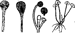
Figure 2.
Figure 3.
Figure 4. (see figure 2)—the causative agent of generalized mucormycosis, as well as pulmonary mycoses, etc.—is characterized by white mycelium, pear-shaped sporangia, 15 x 70 µ in diameter, and oval spores (2 x 3 µ). This fungus is poorly cultivated on liquid media, easily on solid media, especially on potatoes and carrots, at a temperature of 36–37°. The fungus is pathogenic to animals, especially rabbits, when administered intravenously. 3. Rhizomucor parasiticus (see figure 3)—the causative agent of pulmonary diseases, clinically similar to tuberculosis, but ending in recovery in a few months. This fungus is characterized by a mycelium that is first gray, then brownish in color, sporangia 35x80 µ in diameter, with a membrane studded with needle-like outgrowths, and round or oval spores 2x3 µ. The fungus is easily cultivated on solid media at a temperature of 38°; it is pathogenic to rabbits; upon intravenous administration, rabbits die in 3–7 days. Administration of the fungus under the skin and into the trachea gave a negative result. 4. Rhizopus (see figure 4) — the causative agent of a disease of the tongue (langue noire pileuse). Mycelium with numerous white rhizoids, with oblong black sporangia; oval, gray spores. To test the pathogenic properties of mucors, it is best to resort to experiments on rabbits (Barthelat). Upon intravenous administration, the animal dies on the 1st–6th day; upon intraperitoneal administration, death occurs later than upon intravenous administration; upon subcutaneous administration, a local process (abscess) is sometimes obtained. In experimental studies, the kidneys, striated muscles, liver, lungs, heart, spleen, and intestines are most frequently affected. Mucors are the causative agents of various mycoses (Verdun). Localization Clinical form Parasites Various organs Lungs Auditory passages Upper respiratory passages Generalized mucormycosis Pulmonary mucormycosis Otomucoromycosis Mucoromycosis (nasopharyngitis) Lichtheimia corymbifera Lichtheimia corymbifera, Rhizomucor parasiticus, Mucor mucedo Lichtheimia corymbifera, Lichtheimia ramosa, Rhizomucor septatus Lichtheimia corymbifera Of the Ascomycetes as human parasites, first, representatives of the family Aspergillaceae (Asp. fumigatus, flavus, gratioti, repens, glaucus; Sterigmatocystis nidulans; Penicillium) are of importance. They cause bronchomycoses, otomycoses, keratomycoses, lesions of the nose, skin, and nails; however, the clinical picture of mycoses caused by these molds presents nothing characteristic of them; the diagnosis is made on the basis of microscopic examination. Aspergillus is characterized by a strongly branching mycelium of varying width, straight unicellular conidiophores with a swelling at the end, from which chains of conidial spores depart. The size of the conidia varies depending on the species. Aspergilli also form asci with 4–8 spores.—Aspergillus fumigatus [see figure (v. II, art. 384)] is the cause of aspergillosis of the lungs and kidneys, as well as mycoses of the skin and auditory passages. It parasitizes humans, birds, horses, and cows. Aspergillus fumigatus is easily cultivated on general media, forms greenish-colored colonies on acidic media and brownish-black colonies on neutral media. Particularly rich growth is obtained on Raulin's medium. It curdles milk on the 12th day. It slowly liquefies gelatin. Susceptible animals are rabbits, guinea pigs, monkeys, pigeons. When the culture is introduced into a vein, the animal quickly dies from general septicemia with characteristic lesions of internal organs, especially the kidneys in rabbits and guinea pigs. When administered intraperitoneally, death occurs somewhat more slowly; local abscesses are observed upon subcutaneous injection of the culture. Infection of rabbits is also possible upon inhalation of spores, with a characteristic arrangement of the fungus in the bronchi.—Sterigmatocystis is characterized by conidiophores with a pear-shaped or oval swelling, covered with cylindrical basidia, from which 2 or more sterigmata with chains of conidia depart. The clinical picture of diseases caused by these fungi does not differ in any way from mycoses caused by aspergilli. Sterigmatocystis nidulans (see figure 5) was isolated by Eidam; optimum growth is 36–38°. It grows poorly on Raulin's medium, is cultivated on carrots, potatoes, and Sabouraud's medium. Inoculation of small amounts subcutaneously in rabbits and monkeys does not reveal pathogenic properties in the fungus. However, when administered intravenously in large amounts, the fungi are pathogenic also to rabbits. Penicillium [see separate table (art. 81–82), figures 4 and 5] forms a mycelium and conidiophores provided with transverse partitions; at the end they branch several times and terminate in chains of spores. Penicillium is widespread in nature as a saprophyte and only in exceptional cases leads a parasitic life. Among the species of Penicillium, Penicillium crustaceum (glaucum)—the causative agent of oto- and bronchomycoses—is found as a parasite; it has been known since 1763 (Linnaeus) and was isolated again in 1918 by Giordano from a case of pseudotuberculosis with a fatal outcome. The fungus is cultivated at 20–35°. Scopulariopsis, which is close to Penicillium, differs, however, in the character of its mycelium (irregular and short). Scopulariopsis brevicaulis (Brumpt and Langeron, 1910) was isolated from a case of onychomycosis. It is easily cultivated on ordinary media at 25°. A disease of the tongue caused by Scopulariopsis is known. Some Aspergillaceae (Aspergillus, Sterigmatocystis, Penicillium) are the causative agents of mycetoma. Besides them, this disease is caused by the fungus Madurella and Indiella (Hyphomycetes). Madurella mycetomi (see figure 6)—the causative agent of mycetoma (mycetomes à grains noirs—Madura foot)—forms a septate mycelium and oidia, which are formed by the division of mycelial segments into parts. The fungus forms sclerotia and sometimes secretes a brownish and black pigment. The fungus Madurella mycetomi is known, which forms sclerotia 0.5–1 mm in diameter in large numbers deep within the medium, and the fungus Madurella tozeuri, which sometimes forms sclerotia also on the surface of the medium (Pinoy). Indiella, the fungus causative agent of mycetoma (mycetomes à grains blancs), forms a septate mycelium from 1 to 5–10 µ in width, sometimes branching, and never forms pigment. The mycelium sometimes forms sclerotia characteristic of various species of this fungus (see figure 7). Yeast-like fungi, Saccharomyces, Cryptococcus, Endomyces, in the parasitology of humans and

Figure 5.
animals are usually united under the name of blastomycetes and are the causative agents of blastomycoses (see Blastomycetes). Blastomycetes sometimes also include from Fungi imperfecti: Cladosporium, Malassezia, Coccidioides, Oidium, Monilia. Definition
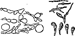
FIG. 6. Fig. 7. of the genus of budding fungi is based on their ability to form mycelium and asci (Castellani). Mycelium Asci 1. Cryptococcus . . 2. Saccharomyces . 3. Monilia..... 4. Endomyces . . . 5. Oidium..... No No Yes Yes No Yes Not Yes all form budding cells form mycelium dividing into quadrilateral and oval spores (oidia) Each of the indicated genera is divided into numerous species differing in their attitude toward carbohydrates and the character of growth on media. Of particular importance is the genus Monilia; it has been described in bronchomycoses, sprue, thrush; as a secondary infection—in cancers. Chronic diseases of mucous membranes (Macroglossia blastomycotica) caused by various species of Monilia, most often Monilia Pinoyi, are known. However, yeast-like fungi are often encountered as saprophytes as well.—To determine the pathogenic properties of fungi, experiments are resorted to on laboratory animals: rabbits, guinea pigs, white mice, and rats. When administered subcutaneously, local abscesses form; when administered into a vein and into the peritoneum, the animals die from septicemia with lesions of the kidneys, liver, and spleen. The speed with which sepsis develops depends on the species of the causative agent, its virulence, and the amount of culture introduced into the blood. Cultivation on media rapidly, within 2–3 weeks, weakens the virulence of the isolated fungus, and upon prolonged cultivation, the fungus completely loses its pathogenic properties. Upon passage through animals, however, the virulence of the fungus increases.—Cladosporium. This fungus forms straight yellow-green conidiophores, on which, most often at the end, an oval spore is formed, sometimes divided into two chambers. These spores, separated from each other by partitions, form moniliform chains, but never lie freely. Cladosporium madagascariense was isolated from a skin lesion (ulcerated tumor) on the leg. In tissues, the fungus forms egg-shaped or spindle-shaped masses 3–4 µ in length. Pathogenic for guinea pigs and white mice. Cladosporium Mansoni was isolated from scales in a skin disease, tinea nigra, resembling pityriasis versicolor. The fungus is easily cultivated on soli
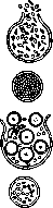
Figure 8.
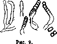
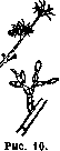
respiratory media. Growth is obtained after 2-4 days in the form of black colonies with a greenish tint; optimum growth temperature is 32°. Coccidioides (see Figure 8). The only species of this genus, Coccidioides immitis, occupies for now an intermediate position in classification between Saccharomyces and Monilia. It was discovered by Wernicke in 1892 in Brazil in a case of dermatomycosis (blastomycosis). The fungus also causes dermatitis coccidioides, or mycose de Posadas, quite frequently in the United States—a disease resembling cutaneous tuberculosis or mycosis fungoides. In tissues, the fungus forms large round cells up to 80 µ in diameter, with a membrane 3-6 µ thick. Spores are formed inside the cells. Fungal colonies in cultures are round, gray in color, transparent, and gradually spread from the depths of the medium over the entire surface, where they form mycelial threads and chlamydospores, sometimes lying in chains. Under anaerobic conditions, endogenous spores are produced, just as in the tissues of humans and animals. The fungus is pathogenic for rabbits, mice, guinea pigs, and monkeys. Mycelial forms of the fungus from culture, when inoculated into animals, cause abscesses in them, from which large round cells up to 34 µ in diameter, with endogenous spores, are subsequently isolated. Among typical Fungi imperfecti, the following should be noted: Hemispora—forms a fine, septate, branching mycelium. Conidiophores, usually branched at the base, form a conidium (so-called protoconidium) at the end by means of a circular constriction, separated by a thin, dense, brownish membrane. This first conidium then breaks down into a series of segments (so-called deuteroconidium). Hemispora stellata (see Figure 9) is extremely widespread in nature. As a parasite in humans, it was discovered by Vuillemin, then by Gougerot and Caraven in osteoperiostitis, and isolated from skin abscesses; it is easily cultured on ordinary media. Colonies are irregular in shape, wrinkled, initially brownish, then black. The fungus is pathogenic for rabbits. Inoculation of the fungus into the epiphysis of the tibia results in osteoperiostitis. — Hormodendron. Forms a branching septate mycelium; fruiting hyphae are straight, without swellings at the ends, divided by partitions, and sometimes branch. Spores are round or oval in shape, arranged in chains. Hormodendron Fontoynonti (see Figure 10) was isolated from cases of skin lesion known as "hodi-potsy," or parasitic depigmentation of the scalp and neck, in Madagascar. In skin scales, the fungus is found in large numbers in the form of: 1) oblong mycelium, cylindrical, swollen; 2) significant accumulations
of round spores. — Microsporon furfur, Achorion Schonleini, Trichophyton, Epidermophyton, Microsporon minutissimum, Trichosporon constitute the group of so-called dermatomycetes, or dermatophytes.
Oospora (Actinomyces, Cohnistreptothrix). Various species of lower fungi of the genus Oospora cause 1) actinomycosis (see) and 2) oosporosis. Their clinical picture is extremely diverse due to the fact that they affect almost all human organs—skin, lungs, pleura, kidneys, liver, eyes, lacrimal ducts, ears—sometimes causing general septicemia. Lung disease proceeds chronically and in its course resembles tuberculosis. Fungi of the genus Oospora are characterized by a thin, even mycelium 1 µ in diameter, branching, and in places divided into fragments resembling bacteria and cocci. Sometimes the branches of the mycelium gradually thicken and form chains of spores at the ends. The fungi stain easily with aniline dyes, are Gram-positive and acid-fast (like the tubercle bacillus). — Oosporoses are caused by fungi of Cohnistreptothrix. The fungi are difficult to culture and prefer anaerobic conditions. Known species include: Cohnistreptothrix bronchialis, isolated in 1921 by Castellani and others in bronchial disease; Cohnistr. Foersteri, isolated in 1874 from the lacrimal passages. Cohnistr. Israeli was isolated by Israel in 1896 in two cases (jaw and lung mycosis). The clinical picture of the diseases did not differ in any way from actinomycosis. The microscopic picture is also similar to that of actinomycosis. Cultures are obtained more readily in anaerobic than in aerobic conditions, on plain and sugar agar, by stab inoculation in the depth. Growth is also obtained on egg-white medium. In broth, it grows more easily under anaerobic conditions. On other media, there is almost no growth. Inoculation into animals is easily successful and produces typical lesions; rabbits and guinea pigs are suitable for experiment. Inoculation is performed subcutaneously and intraperitoneally. — Sporotrichons, see Sporotrichosis.
E. Plevako. 79
FUNGI
Related articles
Mentioned in
Cite this page
“Parasitic Fungi.” Soviet Medical Encyclopedia. English translation of Bolshaya Meditsinskaya Entsiklopediya, 1st ed. (Moscow, 1928–1936), ed. N. A. Semashko. https://sovietmedicalencyclopedia.pages.dev/article/parasitic-fungi/