Blastomycetes
Historical document, translated for reference. It reflects medical knowledge of the 1920s–30s and is not medical advice.
Summary
Blastomycetes are microscopic fungi including yeasts and similar organisms that reproduce by budding or division. Some species are pathogenic to humans and animals, causing blastomycosis, with different clinical manifestations including skin lesions and systemic disease.
Encyclopedia article (1928–1936)
Blastomycetes (Blastomycetes), a name used in medical literature to denote yeasts and certain fungi closely related to them. These are microscopic fungi represented by cells, either single or connected in chains, and sometimes even forming a primitive mycelium. The most characteristic feature of most is the budding of cells (see figure, 1); however, there are yeasts whose cells multiply by normal division. All true yeasts (see.), united in the family Saccharomycetaceae, form intracellular spores under certain conditions (see figure, 3), which determines their belonging to the simplest ascomycetes. Others (yeast-like fungi, or false yeasts) do not form such spores and resemble true yeasts only on the basis of reproduction by budding. Their systematic position is therefore unclear, all the more so since budding is characteristic of a whole range of other fungi at certain stages. Some species of both true and false yeasts can cause alcoholic fermentation; however, in most this ability is not expressed. B. are widely distributed in nature as saprophytes in soil, in sugary juice (nectar) of flowers, on the surface of fruits, etc. Others normally occur in connection with various organisms (bacteria or animals) as symbionts or parasites. Of the former, one can mention: 1) kefir yeasts (Torula kephir Nikolaewa and Torula ellipsoidea Nikolaewa); 2) Leben yeasts (Saccharomyces Lebenis Rist et Khoury and Mycoderma Lebenis Rist et Khoury); 3) ginger beer yeasts (Saccharomyces piriformis M. Ward); 4) the organism of "tea kvass" (Medusomyces Gisevii Lindau), etc. All these yeasts live in symbiosis with certain species of bacteria.
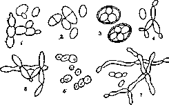
1 - Saccharomyces cerevisiae, budding; 2 - Schizosaccharomyces octosporus, division; 3 -Sacch. cerevisiae, endospores; i-Monospora cuspidata; S-Medusomyces Gisevii; в-Crypto-coccus linguae pilosae; 7-Sacch. Pastorianus, formation of primitive mycelium. Among the forms parasitic on animals, particularly interesting are: 1) Monospora cuspidata Metschnikoff-in the body cavity of the crustacean Daphnia; on this form, Metchnikoff discovered the phenomenon of phagocytosis; 2) Pseudo-saccharomyces applicatus parasiticus Kloker; this species and perhaps some related ones are found in the bodies of various insects, causing the formation of voluminous bodies composed of large cells filled with yeasts (so-called mycetomas). The latter are apparently rather symbionts of the insect and are passed from generation to generation through the eggs infected with yeast cells. Forms pathogenic to humans and generally warm-blooded animals are combined, mainly in the genus Cryptococcus (does not form spores-false yeasts). Several pathogenic forms are also indicated among the true yeasts, for example Schizosaccharomyces hominis Benedeck (see below). Close to blastomycetes is Endomyces albicans Vuill.-the causative agent of "thrush" in children.
l.
Kursanov. Blastomycosis, blastomycosis (from Greek blastos-bud, and mykes-fungus), synonyms: derm. blastomycotica, derm. coccidiodies, oidiomycosis, a rare skin lesion caused by yeast cells (budding blastomycetes). It has no definite geographical distribution, and if it was initially believed that American blastomycosis occurs in America and European blastomycosis in Europe, now, in view of descriptions of opposite cases, this view must be abandoned. The epidemiology has not been established. Statistics, due to the difficulty of recognition, are far from accurate. In the literature (up to 1927) apparently indisputable cases have been described. One of the first, Popov in 1872, proved the pathogenicity of yeast fungi. In 1891, Wernicke and Posadas described a case of blastomycosis, erroneously classifying the causative agent they found among animal parasites. In 1894, Busse, Buschke and Gilchrist, independently of each other, discovered and isolated pathogenic for animals yeast cells in skin diseases. The clinical picture and the isolated fungi in all cases described later by various authors turned out to be not quite homogeneous, since each new case could be classified with greater or lesser possibility under either the Busse-Buschke type or the Gilchrist type. The first type is the European deep blastomycosis, Busse-Buschke and the second type is the American blastomycosis, Gilchrist. The classification has been repeatedly revised and is now presented in the following form: I. Type Busse-Buschke (first type of the old classification), European form (in cultures-saccharomycetes), the rarest form, clinically expressed by the appearance of multiple gummas (rarely single), which, rather quickly disintegrating, form a large ulcer of irregular shape affecting the deep parts of the skin (see figure). The ulcer has undermined, pocket-like, somewhat infiltrated edges, and exudes a viscous, sometimes brown-red liquid. For this type, the process is characteristic of spreading to internal organs, where granulomatous nodules and nodules are formed from epithelioid and giant cells, which undergo suppuration, and the disease usually ends in death. Histologically in the skin, a defect of the epidermis is found at the site of the ulcer and slight acanthosis at the periphery. In the ulcer itself, Buschke distinguishes three zones: in the center-complete tissue decay, in the middle-proliferative-purulent foci, in the outer-individual granulomatous foci from epithelioid, plasma and giant cells.-II. Superficial blastomycosis, so-called cases of interdigital mycosis (cultures of fungi are not always the same); clinically the disease is characterized by the appearance of vesicles in the interdigital spaces of the hands and feet, at the periphery of the lesion. The skin is somewhat swollen and shows slight peeling.-III. American
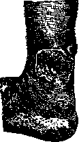
Blastomycosis of the skin.
blastomycosis, disease of Gilchrist, derm. blastomycotica, is characterized by the presence in cultures of yeast cells with mycelium; it initially develops not always uniformly - sometimes as a spot, sometimes as a nodule, sometimes as a blister, sometimes as a pustule. Later, all these elements merge, ulcerate, papillomatously grow, and according to their clinical picture most closely resemble either tuberculosis verrucosa cutis, or an ulcerated tuberculous syphilide, or cancer. For blastomycosis, small abscesses that secrete a sticky fluid are characteristic. Histologically, strong acanthosis, a large number of microabscesses, and a tuberculous structure of the tissue are found. Gilchrist's blastomycosis should be recognized as a rather local, benign disease; only in exceptional cases can it lead to metastases to internal organs. -IV. Transitional forms from the European to the American type. More often the clinical picture more closely approaches the American type, but from cultures fungi characteristic of the European type (saccharomyces) are isolated. Diagnosis in each individual case can present considerable difficulties due to clinical and histological similarity with syphilitic and tuberculous skin diseases. Since yeast cells as saprophytes are found in a number of other skin diseases, it is necessary, in addition to a positive bacterioscopic result, to also isolate pure cultures and prove their pathogenicity to animals. -Laboratory-experimental diagnosis. The main condition for the success of laboratory analysis for pathogenic blastomycetes is the sterile taking of material from an unopened focus (puncture, excision, scraping). Smears or sections prepared from pus or tissue of the focus are stained by Gram; under the microscope in the smears a small number of budding small, round yeast cells are found. Seeding of tissue or pus is done (aerobically and anaerobically-under a layer of sterile vaseline oil) on Sabouraud agar and slightly acidic potato medium. In the incubator, after 3-4 days, opaque fatty colonies, curd-like, white or pink in color, rarely with a brownish tint (American type) are visible; in smears from colonies gram-positive yeast cells are found, which in the Busse-Buschke type appear characteristic (round, slightly oval, small), in the American type there is a transition to Oidium (type Soor) - elongated cells with a tendency to form filamentous mycelium. For the precise establishment of the pathogenicity of yeast fungi, in each case clinically suspicious for blastomycosis, serological reactions are performed in the form of agglutination and complement fixation tests. In several cases (State Venereal Institute) clear agglutination of an emulsion of a 24-hour culture of blastomycetes was obtained when mixed with the blood serum of a blastomycosis patient (at a dilution of 1:200-400); the complement fixation reaction was not characteristic. It is also necessary to make sowings from the blood of patients (3-4 cubic cm of blood and 15-20 cubic cm of agar), since at a certain stage of the disease blastomycetes in humans, as in experiments on animals, penetrate into the blood. Autovaccines made from blastomycete cultures are highly toxic and can harm patients, causing further spread of the process. The pathogenic significance of the isolated cultures is also studied by injections (into the skin, under the skin, into the vein) in susceptible animals (mouse, rat, rabbit). Pathogenicity is sometimes very great: 1 loop kills in a period of several days to two weeks a rabbit weighing 2-2 1/2 kg, with symptoms of sepsis. With intracutaneous injection of old cultures, in rabbits a local process (hyperemia, infiltrate, tissue decay with suppuration) is observed, which lasts from one week to two months and to a certain degree resembles the course of American human blastomycosis; histologically, small-cell and neutrophilic infiltrations, cell decay in places, and epithelioid cells at the periphery are found; between cells there are sometimes in large numbers typical budding blastomycetes; the process ends in recovery with outcome in scarring. The rabbit's skin undergoes an allergic change in relation to the blastomycete fungus; immunity does not occur; superinfection is successful. The serum of a recovered rabbit has no therapeutic properties; agglutinating properties are also weakly expressed. -The prognosis varies: in cases where the clinical picture approaches the Busse-Buschke type, especially in the presence of metastases, it is very severe, even hopeless; on the contrary, in the so-called American (superficial) type, the prognosis is as a rule favorable, metastases to internal organs usually do not occur, and the process after a certain, sometimes long period ends in recovery. -Treatment in cases of the Busse-Buschke type is little reliable, although some have achieved recovery with huge doses of KI. In superficial blastomycosis, potassium iodide (up to 12.0 per day and even more) gives not bad results. Surgical intervention (curettage), X-ray therapy, radium, thermotherapy, salvarsan are also recommended; some also advise autovaccine. The experience of the State Venereological Institute (Moscow) showed that the latter should not be used.
3. Grasebin, Yu. Finkelstein.
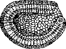
Figure 1. Blastopore of amphioxus.
Related articles
Mentioned in
Cite this page
“Blastomycetes.” Soviet Medical Encyclopedia. English translation of Bolshaya Meditsinskaya Entsiklopediya, 1st ed. (Moscow, 1928–1936), ed. N. A. Semashko. https://sovietmedicalencyclopedia.pages.dev/article/blastomycetes/