Pseudotuberculosis
Historical document, translated for reference. It reflects medical knowledge of the 1920s–30s and is not medical advice.
Summary
Pseudotuberculosis is a concept introduced in 1885 by Eberth that refers to a heterogeneous group of diseases characterized by the formation of tubercle-like lesions in organs, unrelated to tuberculosis. It is considered a form of septicemia, often observed in animals, caused by various bacteria including Yersinia pseudotuberculosis.
Encyclopedia article (1928–1936)
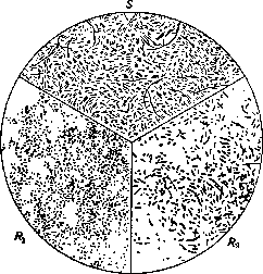
PSEUDOTUBERCULOSIS, a concept introduced in 1885 by Eberth, uniting a very heterogeneous etiologically group of diseases, for which the formation of tubercles in organs, externally resembling tuberculous ones, is characteristic. P. has no relation to tbc. Attempts have been made to replace the term P. with another for clarification. Thus, Baumgarten proposed instead of P. the designation 'injectiose miliare Leucocytombildung' or 'granulosis micrococcica', Pfeiffer - 'acute miliare Leucocytombildung', but nevertheless the name P. has been retained. It is more correct, as will be seen from the following, to consider P. a variety of septicemia, observed especially frequently in the animal kingdom. Cases of P. are distinguished, caused by bacteria, fungi (Blastomycetes, Streptothrix, Aspergillus, etc.), animal parasites (helminths) and even foreign bodies. The name P. is most commonly used in relation to processes of bacterial origin. Among the bacteria causing pseudotuberculous changes in animals and humans, one must name Bact. pseudo-tuberculosis rodentium (Pfeiffer), Bact. pseudo-tuberc. ovis (Preisz-Nocard), Bact. pseudotuberc-murium (Kutscher), etc. In individual cases of P., streptococci, paratyphoid bacteria, etc. have been found. The rodent P. bacillus (Bact. pseudotub. rodentium), which often causes severe epizootics among guinea pigs, rabbits, and other rodents; and has been isolated in human diseases, deserves the greatest attention. It is also interesting due to its similarity to the plague bacillus (Bact. pestis). This microbe was first isolated in 1883 (Malassez and Vignal); in 1889 it was described in detail by Pfeiffer. Bact. pseud. rodentium in smears from organs and from broth cultures is a short clumsy bipolar rod of ovoid shape, staining negatively by Gram. It is immobile, does not form capsules or spores, and grows on ordinary nutrient media. In liquid media it forms chains. According to the latest research, Bact. pseudotub. rod. gives a characteristic dissociation phenomenon, consisting in the disintegration of the culture into a series of variants: 1) 'smooth variant' - forms on agar smooth disk-like colonies with a shiny surface, with even edges, translucent with a bluish tint; the bacterial mass is viscous, emulsifies well; morphology of bacteria - thin rods 1.5-3μ in length, occasionally filaments (fig. 1 S). In broth - non-homogeneous turbidity. 2) 'Rough variant' - forms on agar colonies with a convex granular center, sharply delimited from a lace-like peripheral zone, turbid with a brownish tint; the bacterial mass is dense, does not give a homogeneous emulsion; morphology - shortened bipolar rods 0.8-1μ in length with an admixture of coarse forms (4-8μ in length) of bizarre outlines (fig. 1 R). In broth - flaky growth without turbidity. Besides the described extreme variants, a number of 'intermediate' forms are observed. The smooth variant is more virulent than the rough one. In laboratory cultures, the microbe is usually encountered in the form of the rough variant. - The biochemical properties of the variants are the same. The microbe does not liquefy gelatin; does not coagulate milk; does not form indole; decomposes glucose, levulose, maltose, and mannitol with acid formation without gas; does not change lactose and sucrose. The microbe's relation to some carbohydrates (rhamnose, adonite) and experimental infection of laboratory animals is of importance for distinguishing the P. bacillus from the plague bacillus (Bact. pestis). However, in practice differentiation may encounter considerable difficulties, and even immunoreactions in this case do not represent a completely reliable criterion. At present, one can rely on the following distinguishing features between these microbes: 1. White rats are resistant to infection with Bact. pseudotuberculosis while being very sensitive to Bact. pestis. This sign is quite reliable but relative. Cases of increased virulence of the P. bacillus with respect to white rats are known; the plague microbe, on the contrary, can lose its virulence when stored in the laboratory. 2. The characteristic difference is the unequal change in hydrogen ion concentration (pH) in cultures of these microbes: Bact. pseudotuberculosis alkalinizes the medium faster than Bact. pestis. 3. With respect to some carbohydrates, these microbes also show different activity, and the most reliable results are given by media with rhamnose and adonite, with respect to which the plague microbe is inactive in contrast to the P. bacillus. Epizootics of P. are most often observed among guinea pigs, less frequently rabbits are affected. Besides these rodents, according to a summary collected by Yurkevich, Bact. pseudotuberculosis was found several times in hares, then in cattle, pigs, horses, cats, chickens and songbirds. Cases of its detection in gray rats have also been described. The prevalence of this microbe in the external environment is noted - it has been found in drinking and waste water, in the air, in the soil, in spoiled oats. Infection under natural conditions apparently most often occurs through the digestive tract, as well as through the respiratory tract. The epidemiological role of insects has not been studied. Clinic. The disease in guinea pigs occurs in three forms. In the septicemic form, which kills the animal in 1-2 days, nodules do not have time to develop and phenomena of hemorrhagic septicemia (hemorrhages on serous membranes, etc.) come to the forefront. The most characteristic and common is the tuberculous-like form, lasting about 5-15 days and causing the death of the animal with phenomena of exhaustion with the picture described below. The third form is cachectic, in which the animal dies in 2-4 weeks without characteristic patho-anatomical changes. In experimental infection of guinea pigs, cases of recovery are sometimes observed (see Pathology of laboratory animals). - For the patho-anatomical picture of P. the following changes are characteristic: in the abdominal and chest cavities - semi-liquid viscous exudate; internal organs are hyperemic. The spleen is enlarged, dark red color, penetrated with grayish-white, slightly protruding above the surface of the organ nodules, ranging from millet seed to small pea size; nodules are also often found in the liver, lungs, omentum, intestine, and in the mesenteric lymph glands. The pseudotuberculous nodule (fig. 2)
Figure 1. Prints from colonies of variants of Bact. pseudotuberculosis rodentium. Morphology of bacteria of variants: smooth (S) and rough (R1 and R2). R1-periphery, R2-central colonies, where larger forms predominate.
Figure 2. Pseudotuberculous nodule of the liver. (According to Roger.)
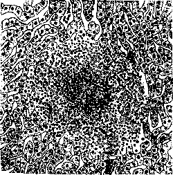
initially represents a miliary tissue necrosis (death of parenchymal cells around bacterial accumulations) and in acute course (which is generally observed in cases of P. in humans) reactive phenomena from the surrounding tissue are usually absent. In more prolonged course (more often in rodents) exudative processes develop due to lymphoid and polymorphonuclear cells. Giant and epithelioid cells are usually absent, although this by no means can be considered a rule. With the development of the process, the nodule consists mainly of caseous necrotic mass, often with softening in the center. No calcification of caseous masses is observed. For some rodents, characteristic features of the lesion are noted. Thus, for example, in rabbits, in addition to the above-described changes, characteristic changes in the vermiform appendix are observed. The appendix is enlarged, has the appearance of a dense tube, completely studded with nodules the size of a mustard seed. Bacteria in the developed nodules are rarely found microscopically (evidently as a result of loss of the ability to be stained). In more acute cases, on the contrary, accumulations of bacteria ('zoogloeae') are found, from which the French name 'tuberculose zoogleique' is derived. The culture of the causative agent, both from organs and from blood, is isolated without difficulty. From a differential-diagnostic point of view, it is important to mention that the similarity, more significant than with a tuberculous nodule, exists with nodules observed in glanders and in the subacute form of plague. It is also noteworthy to note that with intraperitoneal infection in guinea pigs, orchitis can develop, analogous to that of glanders (Ramon and others). Cases of bacterial P. in humans to the present time have been described in a very small number. A significant part of them should be considered doubtful, since the culture of the causative agent was not isolated directly from humans, but through inoculation to experimental animals, in which spontaneous P. cannot be excluded. In a review of the literature up to 1927, Poppe (Porre) cites 10 such doubtful cases and 11 cases, the etiology of which is undoubtedly associated with infection by Bac. pseudotuberculosis rodentium or closely related microbes. Diseases were observed in newborn children (in the first days of life) and in adults. The course is as a rule very acute, rapidly ending in death. In adults, a typhoid-like picture is observed, with high temperature, sometimes with jaundice. There are no characteristic diagnostic signs in the clinical picture. In the described cases, the diagnosis was made on the basis of bacteriological examination, and the culture of the causative agent was obtained either during life - from blood, or at autopsy - by sowing from internal organs affected by a characteristic patho-anatomical process. Several cases of localized lesions of a pseudotuberculous nature have also been described, which upon bacteriological examination gave cultures of microbes similar to the P. bacillus, for example, a case of inflammation of the middle ear, cases of conjunctivitis resembling Parino's disease, a case of appendicitis, in which the culture was obtained from ulcers and suppurating follicles of the removed segment of the intestine and lymph glands, and the isolated microbe agglutinated up to a dilution of 1:1280 with the patient's serum, etc. In newborn children, infection is assumed to occur during passage through the lower birth canal, and the infection apparently enters through the mouth, from where it enters the intestine and the portal vein system. Children die within 1-2 days. At autopsy, as a rule, lesions of the lymphatic apparatus of the intestine and mesenteric glands and multiple necrotic nodules in the liver and spleen are noted. Bacteriologically, among the strains of the P. bacillus isolated from humans, a division into two varieties is observed: one identical or almost identical with the microbe isolated from rodents, and the second, differing from it in its relation to Gram staining (Gram-positive). This second variety was as a rule observed in diseases of newborn children. Undoubtedly, the problem of P. deserves more attention than has been paid to it to date, and requires further study and accumulation of facts. This is especially true of the bacteriology, epidemiology, and clinic of bacterial P. in humans.
Related articles
Mentioned in
Cite this page
“Pseudotuberculosis.” Soviet Medical Encyclopedia. English translation of Bolshaya Meditsinskaya Entsiklopediya, 1st ed. (Moscow, 1928–1936), ed. N. A. Semashko. https://sovietmedicalencyclopedia.pages.dev/article/pseudotuberculosis/