MOLD
Historical document, translated for reference. It reflects medical knowledge of the 1920s–30s and is not medical advice.
Summary
Mold refers to fungi that appear in damp places and form colored growths on organic substances. The article describes different types of molds, their characteristics, reproduction methods, and their pathological significance in humans and animals.
Encyclopedia article (1928–1936)
MOLD, fungi (see illustration), appearing in damp places and forming on organic substances growths of various colors. Molds include fungi belonging to the Mucoraceae and Aspergillaceae families. Mucoraceae fungi, widely distributed in nature, have a branched mycelium without septa (sometimes septa are present, but not in large quantities) and reproduce by forming cnospores. The fertile hyphae bear at their ends thickenings in the form of sporangia heads, filled with spores; in some species there are aerial fertile hyphae, and at their base the mycelium forms outgrowths in the form of rootlets (rhizoides). There are species that reproduce by chlamydospores.
MOLD
408 Reproduction also occurs through the formation of zygospores. Inside the sporangia there is a columella, which is a spherical or pear-shaped outgrowth of the septum of the fertile hypha. Mucors include Mucor mucedo, Mucor pusillus (Jakowski), Mucor racemosus (Fresenius), Lichtheimia (mucor) corymbifera (Cohn), Rhizomucor septatus (Lucet and Constantin), etc. Different species of mucors differ in the presence or absence of rootlets (rhizoides), the shape of the columella, the presence of aerial hyphae, etc. The pathological significance of mucors is generally small. Mucor corymbifera has been found in inflammatory processes in the middle ear, in ulcerative processes in the nose; it was not found in sputum, but Fürbringer and Paltaluf (Furbringer, Paltauf) discovered it during autopsy in very dense, usually necrotic foci in the lungs. Mucor mucedo, Rhizomucor parasiticus in some cases cause prolonged lung diseases, occurring under the guise of tuberculosis (see Bronchomycoses, Pneumomycoses). Many mucors are pathogenic for animals.
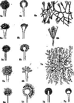
Mold fungi: 1a-1b-Aspergillus glaucus; 2a-2b-Aspergillus furnigalus; 3a-3b- Aspergillus flavus; 4a-4b-Aspergillus Wehmerl; 5a-6b-Sterigmatocystis nigra; 6a-6b-Penicillium glau-cum; 7a-7b-Mucor mucedo; 7c-Rhisopus.
The Aspergillaceae family includes Aspergillus, Penicillium and Sterigmatocystis. Aspergillus has a branched mycelium with septa; reproduction occurs by means of conidia. The fertile hyphae end with a thickening; on its outer surface sit unbranched conical outgrowths (basidia), which detach conidia from themselves. Penicillium has a branched mycelium with septa inside. The fertile hyphae branch at the ends; the branches end in flask-shaped outgrowths, bearing chains of conidia on themselves. Sterigmatocystis have a branched mycelium with septa. The fertile hyphae bear at the end a thickening of round or pear-shaped form; on its surface there are outgrowths-sterigmas; from them depart 2 or more secondary sterigmas, which detach conidia from themselves. Of all fungi belonging to the Aspergillaceae family, Aspergillus plays the greatest role in pathology; the lung and bronchial diseases caused by them often have a professional character, as they occur in persons dealing with moldy or dusty material, especially flour (wool sorters, sponge cleaners, rag pickers, etc.). The pulmonary mycosis caused by these fungi (aspergillosis) often occurs under the guise of tuberculosis (pseudotuberculosis aspergillina). Of the various species of Aspergillus, which differ mainly in the color of the pigment they form (furnigatus, flavus, niger, barbae, glaucus, etc.), Aspergillus furnigatus, forming a grayish-greenish mycelium, is of greatest importance. In sputum, this fungus is found either in grayish-white dense grains or in spherical brown-black foul-smelling lumps, sometimes reaching considerable size. In addition to lung lesions, Aspergillus (as well as other Aspergillaceae) also causes diseases of the middle ear (otomycosis), sometimes the cornea (keratomycosis), the nasal cavity (rhynomycosis), the trachea (tracheomycosis), the bronchi (bronchomycosis), the lungs (pneumomycosis), the skin (dermatomycosis), etc. [see separate "table (pp. 239-240), fig. 1]. In tropical countries, tumors (mycetomas) of the legs are found, caused by various species of fungi (Aspergillus, Sterigmatocystis) (see Madura foot). Fungi belonging to the Aspergillaceae family are particularly common in birds, for example in chickens. In France, a peculiar disease has been described (maladie des gaveurs de pigeons, Taubenzuchterkrankheit by German authors) (see Aspergillus, aspergilloses);
Mold fungi are easily stained by aniline dyes, not decolorized by Gram. Growth occurs on any nutrient media; an acidic reaction is preferable. For pathological products for the presence of pathogenic molds, it is recommended to examine them in the native state in a drop of water or glycerin; in such a preparation, the mycelium and spores of the fungi are clearly visible. For growing Aspergillus and other fungi, a protein-free Raulin medium is recommended (water-500.0; cane sugar-70.0; tartaric acid-4.0; ammonium nitrate-0.4; ammonium phosphate-0.1; potassium carbonate-0.6; magnesium carbonate-0.4; ammonium sulfate-0.25; iron sulfate-0.07; zinc sulfate-0.07; potassium silicate-0.07; manganese carbonate-0.07). When sowing particles of sputum containing Aspergillus into this medium, after a day at 37°, an abundant white velvety growth appears, which turns black after several days; a rabbit infected with the culture dies after 4-8 days; in its organs, inflammatory foci containing fungi are found. Natural nutrient media for mold fungi (as for fungi in general) are bread, potatoes, milk, etc.
V. Lyubarsky. 409
Related articles
Mentioned in
Cite this page
“MOLD.” Soviet Medical Encyclopedia. English translation of Bolshaya Meditsinskaya Entsiklopediya, 1st ed. (Moscow, 1928–1936), ed. N. A. Semashko. https://sovietmedicalencyclopedia.pages.dev/article/mold/