Fetal Position Anomalies
Historical document, translated for reference. It reflects medical knowledge of the 1920s–30s and is not medical advice.
Summary
This article describes various abnormal fetal positions during childbirth, including extended presentations, breech presentations, and transverse positions. It details the causes, diagnosis, and management approaches for each type of anomaly as understood in 1930s Soviet obstetrics.
Encyclopedia article (1928–1936)
FETAL POSITION ANOMALIES, deviations of the fetus from the position considered normal, i.e., from the longitudinal position with the head presentation, specifically the occipital part of the head, which occurs in 95% of all births. Deviations in the position of the fetus have numerous variants. From a practical point of view, all of them can be grouped into three categories: 1) extended presentations, 2) breech presentations, 3) transverse positions.
Extended presentations. Among extended head presentations, vertex, brow, and face presentations are distinguished. A number of factors—narrow pelvis, inelasticity of tissues of the lower segment, loss or perversion of the normal contractile ability of the uterus, furthermore—very large head, dolichocephaly, as well as deformities of the neck, especially in the area of the trachee—can cause delay in the downward movement of the occiput and may be the cause of varying degrees of head extension. In cases where head extension is moderate and the vertex enters the pelvic inlet, we speak of vertex presentations. With further extension, the brow will present (brow presentation). If extension reaches its maximum degree, then the face will present (face presentation). Brow and face presentations occur in 1-2% of all births. Diagnosis of these anomalies is established by external and internal examination; the latter is resorted to only in extreme cases under the current limitations of internal examination. Vertex presentation is typically found in flat pelves. The outcome of labor in this condition is largely related to the ability of the fetal head to mold and the degree of pelvic narrowing. With this anomaly, in cases of prolonged labor, it is necessary to terminate labor by operative means (forceps, perforation). Brow presentation belongs to the category of absolutely unfavorable conditions, and spontaneous labor with it occurs as an exception. For successful progress of labor with brow presentation, in case of timely diagnosis, one should facilitate the transition of brow presentation to occipital or face presentation by flexing or extending the head. If this cannot be achieved, then with intact membranes and sufficient dilation, labor can be terminated by external-internal version on the foot. If the mother absolutely wants to have a living child, abdominal cesarean section is indicated. Advanced brow presentation is resolved by perforation of the head. With this anomaly, ruptures of the soft birth passages are often observed, and children are born dead in 15-17% of cases. With face presentations, labor can end spontaneously only when the chin, which is the presenting point, comes under the pubic arch (anterior face presentation). If, however, the chin turns posteriorly, toward the promontory (posterior face presentation), then such a presentation is absolutely unfavorable and, like brow presentation, should be terminated either by version or cesarean section with timely diagnosis, or by perforation in advanced cases.
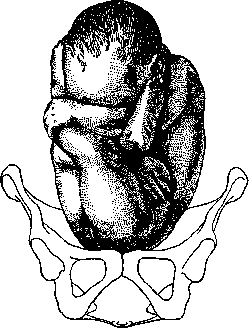
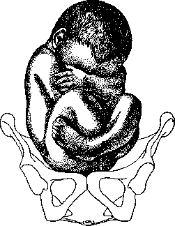
Breech presentations. With breech presentation, which occurs in 3% of all births, breech, knee, and foot presentations are distinguished. The etiology of breech presentations is related to a combination of various factors that interfere with the normal engagement of the head in the lower segment of the uterus. The main factor favoring the occurrence of such anomalies is excessive mobility of the fetus, which is more often observed with polyhydramnios, small fetuses, and twins. Deformities of the head with reduction of its weight, as occurs in underdevelopment of the skull, can also be the cause of breech presentation. Depending on the position of the fetus's legs, true breech and foot presentations are distinguished. Among breech presentations, simple or complete breech is distinguished when the fetus's legs are extended upward (see Figure 1), and incomplete breech when the legs are flexed and the feet are pressed against the buttocks (see Figure 2). In cases where with breech presentations the presenting part is the feet, we speak of complete foot presentation. If one leg prolapses while the other remains flexed at the hip joint, we speak of incomplete foot presentation. When the legs flexed at the knee joints become the presenting part, we speak of knee presentation. The diagnosis of breech presentation, except in rare individual cases, presents no difficulty. On external examination, the head is usually found in the upper part of the uterus, and fetal heart sounds are heard higher than with head presentations. Internal examination clarifies these signs by the morphological difference of the parts of the presenting breech. Labor with these anomalies is unfavorable for the mother due to its greater duration. As for the fetus, 15% are born in asphyxia or dead, since during breech labor difficulties may occur in the extraction of the aftercoming head, which causes compression of the umbilical cord.
Figure 1. Complete breech presentation (according to Bumm).
Transverse positions. In transverse positions, the axes of the fetus and uterus are perpendicular, forming an angle of varying degrees (see Figure 3). In transverse positions, the fetus lies such that one of its poles—the head—is usually located lower and is adjacent to one of the ribs of the uterus, while the other pole—the buttocks, being at the opposite rib of the uterus, stands higher, so that the fetus occupies an oblique position—the most common variety of transverse. Among transverse positions, left and right are distinguished depending on the location of the head. If it is turned to the left, we speak of the first or left transverse position; if to the right—of the second or right transverse position. In transverse positions, the shoulder most often presents, less frequently—the side of the fetus. Transverse positions occur in 0.5-0.75% of all births. In the etiology of transverse positions, excessive mobility of the fetus plays a role in cases of uterine atony and stretching in multiparous women, as well as with polyhydramnios, multiple pregnancies, and small fetuses. Placenta previa and tumors obstructing the pelvic inlet also contribute to the occurrence of transverse positions. Spontaneous labor with transverse positions is possible through spontaneous version, when with the onset of labor activity, oblique or transverse positions spontaneously transition to longitudinal. With small or macerated fetuses, or with a wide pelvis, labor can end spontaneously (under conditions of good labor activity) with the body folded in two. For the correction of transverse positions, external version is sometimes applied preventively before labor; with the onset of labor, with intact membranes and sufficient dilation, combined version on the foot is performed. With transverse positions, when the shoulder presents, in case of rupture of the membranes, prolapse of the hand occurs. The shoulder is impacted, the head and most of the body, strongly stretching the uterus, occupy both iliac fossae. The uterus compresses the fetus, a contraction ring may form, and advanced transverse position threatening uterine rupture occurs. In this case, labor requires immediate intervention by the operation of embryotomy (disarticulation of the fetus).
Figure 2. Incomplete breech presentation (according to Bumm).
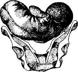
Figure 3. Transverse position; intact membranes.
In transverse positions, the fetus lies such that one of its poles—the head—is usually located lower and is adjacent to one of the ribs of the uterus, while the other pole—the buttocks, being at the opposite rib of the uterus, stands higher, so that the fetus occupies an oblique position—the most common variety of transverse. Among transverse positions, left and right are distinguished depending on the location of the head. If it is turned to the left, we speak of the first or left transverse position; if to the right—of the second or right transverse position. In transverse positions, the shoulder most often presents, less frequently—the side of the fetus. Transverse positions occur in 0.5-0.75% of all births. In the etiology of transverse positions, excessive mobility of the fetus plays a role in cases of uterine atony and stretching in multiparous women, as well as with polyhydramnios, multiple pregnancies, and small fetuses. Placenta previa and tumors obstructing the pelvic inlet also contribute to the occurrence of transverse positions. Spontaneous labor with transverse positions is possible through spontaneous version, when with the onset of labor activity, oblique or transverse positions spontaneously transition to longitudinal. With small or macerated fetuses, or with a wide pelvis, labor can end spontaneously (under conditions of good labor activity) with the body folded in two. For the correction of transverse positions, external version is sometimes applied preventively before labor; with the onset of labor, with intact membranes and sufficient dilation, combined version on the foot is performed. With transverse positions, when the shoulder presents, in case of rupture of the membranes, prolapse of the hand occurs. The shoulder is impacted, the head and most of the body, strongly stretching the uterus, occupy both iliac fossae. The uterus compresses the fetus, a contraction ring may form, and advanced transverse position threatening uterine rupture occurs. In this case, labor requires immediate intervention by the operation of embryotomy (disarticulation of the fetus).
Related articles
Cite this page
“Fetal Position Anomalies.” Soviet Medical Encyclopedia. English translation of Bolshaya Meditsinskaya Entsiklopediya, 1st ed. (Moscow, 1928–1936), ed. N. A. Semashko. https://sovietmedicalencyclopedia.pages.dev/article/fetal-position-anomalies/