Craniotomy
Historical document, translated for reference. It reflects medical knowledge of the 1920s–30s and is not medical advice.
Summary
Craniotomy is an ancient obstetric procedure involving reducing the volume of a fetal head by perforating the skull. The article describes the historical development of this operation and the various instruments used for perforation and extraction.
Encyclopedia article (1928–1936)
Craniotomy (from Greek cranion - skull and temno - I cut), reduction of the volume of the fetal head by perforating the skull. Craniotomy consists of two moments: 1) preparatory perforation of the skull and 2) the moment of delivery - extraction of the reduced volume head (cranioclasia). Both moments follow directly one after another; in cases not requiring special urgency, one can leave the expulsion of the perforated head to the forces of nature. Craniotomy belongs to the most ancient obstetric operations; in olden times, doctors unfamiliar with the mechanism of labor, forceps and version, always readily resorted in difficult births to reducing the fetus by one method or another (see Obstetric Operations). Thus, Craniotomy is mentioned already in the Indian Vedas (Ajurveda Susruta) 1,800 years before Christ, and its description is found in physicians of the Greek, Roman, and Arab schools, in Hippocrates, Paul of Aegina, Philomenus, Albucasis, etc. After the introduction into practice in the 16th century (1550) of version of the fetus (Ambroise Pare) and in the 18th century (1751-54) of forceps (Levret, Smellie), the number of Craniotomies began to decrease so much that the director of the Göttingen obstetric clinic Osiander (1830) considered it a merit that during 40 years of his activity he performed Craniotomy only once, and that in his youth. However, in the 18th, 19th and 20th centuries, many more perfected instruments for craniotomy appeared under various names, which by no means indicates a forgetting of this operation. At present, for perforating the head, instruments known as perforators are used, and for extracting the drilled head - so-called cranioclasts. Perforators are divided into trephine-like, scissors-like, and spear-like. Among the trephine-like are the perforators of Leisnig-Kivish, Braun, Martin, Guyon, etc. As can be seen from Fig. 1, a trephine-like perforator has a long sleeve, in which there is a rod with a handle at one end and a trephine drill and crown at the other. When rotating the handle, first the drill comes out and fixes the instrument, and then the teeth of the trephine come out, which first drill through the soft parts, and then the bone. Next come scissors-like perforators; of these, the oldest is Smellie, modified by Walbaum (1758), Naegele (see Obstetric Instruments, Fig. 21), Oldham, etc. Scissors-like perforators are arranged in such a way that first the head is drilled with the point, and then, by pressing on the handle, the ends of the perforator are spread apart and thus the opening in the skull is enlarged. Finally, there are spear-like perforators, of which the perforator of Blo (see Obstetric Instruments, Fig. 22) is both spear-like and scissors-like.
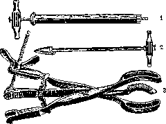
Figure 1. Trephine-like perforator. Figure 2. Phenomenov's perforator. Figure 3. Cranioclast Auvard's.
wedge-shaped.--Then come the spear-shaped perforators of Russian authors: the spear-shaped trephine of Phenomenov (figure 2), the wedge-shaped ones of Lazarevich, Shchetkin, Yastrebov, and others. With a low-lying head, it is more convenient to use a trephine perforator, with a high-lying head, a spear-shaped one. To extract the head after perforation, bone forceps of Menar-Stein, the brephotome of Lazarevich, and numerous other instruments known by the name tire-tetes (Mauriceau, Assalini, etc.) were initially used. Baudelocque (A. Baudelocque) proposed a special instrument - the cephalotribe; later modifications were proposed (Ritgen, Kilian, Busch, Scanzoni, Dubois, Braun, Etlinger, Lazarevich, etc.). The cephalotribe resembled ordinary obstetrical forceps, but its blades were longer, narrower, and had a slight cranial curvature. The ends of the blades almost touched. There was a compressing apparatus on the ends of the branches. Almost all these instruments strongly compressed (crushed) the head, increasing its anteroposterior dimension, which especially complicated the extraction of the head in a flat pelvis, and were very cumbersome. The original cephalotribe of Baudelocque weighed about 3.2 kg and reached a length of 53-54 cm. Instead of the cephalotribe, Simpson proposed for extracting the perforated head a special instrument, the cranioclast, representing forceps of Menar modified by him. This cranioclast was modified by Braun (Braun; 1868) in such a way that its branches were lengthened, the blades made thicker, and the lock, similar to the lock of Brueninghausen, in the forceps was replaced by a removable screw and a nut with wings (see Obstetrical Instrumentarium, figure 25). In England, Barnes as early as 1863 also modified Simpson's cranioclast, lengthening it, giving greater curvature to the inner blade, and arranging a movable screw at the end of the handle. It would therefore be more correct to call the modern cranioclast the Simpson-Barnes-Braun (see Obstetrical Instrumentarium). Auvard proposed an instrument consisting of three branches (figure 3; embryotome cephalique combine), connecting a solid piercing branch in the middle and two hollow branches at the ends on the sides. This is essentially a combination of a perforator, cranioclast, and cephalotribe. Auvard's combined head embryotome was later modified by Zweifel (see Obstetrical Instrumentarium, figures 26 and 27), Winter, and Walthard. In recent times, with the development of scientific obstetrics, new views on preserving the life of a mature intrauterine fetus, and the success of abdominal surgery, craniotomy has been applied less and less, and by the end of the first quarter of the 20th century, it was almost completely displaced in clinics and large maternity hospitals by cesarean section (see) and in rarer cases by pubiotomy. On the walls of the delivery room of the obstetric clinic of Pinard in Paris, there was even such an inscription: "La perforation sur l'enfant vivant est vecue". In Russia, Yastrebov as early as 1903 at the I Congress* of obstetricians and gynecologists proposed to strike perforation on a living fetus from the list of obstetric operations. Winter points out (1927) that perforation on a living fetus in clinics no longer has the right to exist and in his clinic it decreased from 0.9% (1908) to 0.19% (1922) due to better observation of labor and rapid intervention in cases threatening danger to the fetus. In the clinic for almost 20 years, in no case of a narrow pelvis was a living fetus perforated. Winter considers the current mortality rate in craniotomy to be 0.5%, while the morbidity rate is 15-20%. In Russia, the issue of limiting the application of craniotomy received considerable attention from Lebedev; Pobedinsky, however, at the V Congress of gynecologists, generally spoke that with greater care for newborns, not only the number of craniotomies but also preventive operations in general should be reduced. However, where the conditions of medical practice do not allow the creation of a corresponding aseptic environment for cesarean section, and further waiting during labor threatens the mother's life, craniotomy still takes place today, especially in cases of a dead fetus. As for the indications for this operation, first place should be given to all those dangerous complications during the act of labor that threaten the mother's life. This should first of all include significant spatial discrepancies between the presenting part of the fetus and the pelvic ring, i.e., narrowing of the pelvis from 6.5 cm to 7.5 cm conjugata vera, unsuccessful attempts at applying forceps, especially with a high-lying head and a stretched lower segment of the uterus, eclampsia (in a dead child), prolonged labor with the presence of endometritis septica, incorrect insertions of the head, for example in face presentation with the chin posterior, incomplete ruptures of the uterus, premature separation of the placenta with significant internal and external bleeding, narrowing of the birth canal (tumors, scar strictures). This operation can also be applied in diseases of the heart with disturbance of compensation, diseases of the lungs (tuberculosis, acute pneumonia), kidneys (nephritis with large edemas), and finally in hydrocephalus (puncture!). It is necessary to point out that at present, in most of the above-mentioned processes with a living fetus, except for cases with endometritis septica and unsuccessful application of forceps where infection is not excluded, cesarean section is performed in clinics and well-equipped obstetric institutions. With a dead fetus, the indications expand, however, one should not think that the death of the fetus itself gives the right to craniotomy to facilitate labor. Here too there must be definite indications, e.g. cases of prolonged labor in a narrow pelvis when the fetal heartbeats have ceased, with an umbilical cord prolapse with absence of pulsation in it - in the same pelvis, after decapitation, when the head separated from the umbilical cord meets an obstacle from the pelvis during manual extraction, during extraction of the fetus and inability to extract the head. The basis for such indications (in the expression of Schroder) is simple: "if the infant has died, then one must have in mind only the mother". Winter also raises the question whether it is permissible to take into account the social conditions of the parturient for establishing indications for craniotomy, and answers this question negatively. "Even in those cases where operations preserving the life of the fetus are associated with a certain danger to the mother, such as cesarean section and symphysiotomy, one should absolutely and not relatively value the life of the fetus as much as possible". The conditions for performing craniotomy should be: the waters have broken, the cervix is effaced, the dilation of the os is up to 3 fingers, and the head is immobile above the entrance or at the entrance to the pelvis. If the head has a tendency to move, the assistant should fix it as much as possible, since otherwise it is difficult to hold the perforator in a certain place. Technique of performing the operation. The parturient is placed on a transverse bed. The hair on the pubic region is shaved, and the latter is disinfected (best by smearing with Tincture of Iodine). In the presence of severe pain, it is desirable to disinfect the vagina with a solution of corrosive sublimate (1:2,000) or in diseases of the kidneys and anemia - with a solution of potassium permanganate. Then 2 or 4 fingers of the left hand are introduced into the vagina, and under their guidance and protection, the perforator is applied to the head; the trephine perforator held in the right hand is applied in the closed position; then, by turning the screw in the handle, the corkscrew is first introduced into the skull and then the trepan crown. Blo's perforator is inserted into the fontanelles or sutures and then opened and turned to one side and another. A cross-shaped opening is obtained. Both with Blo's perforator and other spear-shaped perforators, one has to press on one spot and drill; care is required so that the instrument does not slip off the convex parts of the skull and does not injure the soft parts of the birth canal or the hands of the operator itself. With a high-lying head, the handle of the perforator should be tilted downward and backward. Some obstetricians consider two acts in perforation: 1) formation of an opening in the skull and 2) removal of the brain - excerebratio. Skene (Alex. Skene) once proposed to perform perforation ad oculos (with the help of Sims' mirror). Indeed, it is more convenient to operate if the field of operation is visible. In this case, they proceed as follows: after exposure with mirrors, the skin on the fetus's head is grasped with bullet forceps, it is incised, and the perforator is applied to the exposed bone. Gorvitz used Ferguson's mirror in these cases. Phenomenov, in the absence of assistance and mirrors, advised grasping the head through the skin cover as firmly as possible with Muse's forceps, and, entrusting the assistant to hold them and pull them downward, to perform the puncture under the guidance of the hand with the help of a piercing instrument.
With occiput posterior presentation, the opening is made in the area of the small fontanelle; with vertex presentation, in the area of the large fontanelle; with oblique presentations of the head, e.g., in Naegele's and Litzmann's positions, in the area of the Gasserian fontanelles or the sagittal suture, which is more difficult; with face and brow presentations, in the area of the forehead, large fontanelle, and orbit; with perforation of the subsequent head through the mouth, in the area of the hard palate or in the area of the foramen occipitale magnum according to Michaelis' method in the space between the atlas and the posterior edge of the foramen magnum, or through the spinal cervical canal with removal of a portion of the vertebrae according to Kohnstein or by the method of L. Lickus (incising the skin to the bone, reaching the foramen occipitale magnum by a subcutaneous route). After an opening is made in the skull, a cranioclast is usually applied. When the head is extracted with it, the brain is expelled by itself, but as was stated above, exenteration—removal of the brain and washing it out with a physiological salt solution—may also be performed. For this purpose, e.g., Phenomenov's or Agafonov's spoon, connected with a catheter, is used, but in most cases this manipulation is unnecessary. When perforating the skull, it is necessary to destroy the medulla oblongata of the still living fetus with the centers of respiration and blood circulation using a perforator, because otherwise the child is extracted with signs of life, which produces a heavy impression on those present. Hammond cites a case where the extracted fetus cried after removal of about 60 g of brain. To stop the bleeding from the skull, tamponade was applied; the child breathed and died only 46 hours after the operation. Pernice reported on one child who survived the perforation and later became an idiot.--After the perforation is made, the cranioclast is applied to the head as follows: under the guidance of 4 fingers of the left hand, inserted into the vagina, the branch of the cranioclast with notches at the end is taken in the right hand and is passed into the opening in the skull so that the convexity of the spoon corresponds to the concavity of the other spoon applied from the outside to the head. The solid spoon must be inserted deeply enough so that its notches enter the cranial cavity; otherwise, when the instrument is closed and the bone is extracted, pieces of the skull are broken off, the points of application of force are reduced, which significantly complicates and prolongs the operation. After closing, a trial traction is first performed to determine how firmly the cranioclast is positioned. If it is not firmly positioned, it should be reapplied, preferably the hollow spoon to the face with a high-lying head, and traction should be performed downward and backward; as the skull progresses further, the spoons are gradually directed forwardward.
Related articles
Mentioned in
Cite this page
“Craniotomy.” Soviet Medical Encyclopedia. English translation of Bolshaya Meditsinskaya Entsiklopediya, 1st ed. (Moscow, 1928–1936), ed. N. A. Semashko. https://sovietmedicalencyclopedia.pages.dev/article/craniotomy/