Hip Joint
Historical document, translated for reference. It reflects medical knowledge of the 1920s–30s and is not medical advice.
Summary
The hip joint is a ball-and-socket joint formed by the head of the femur and the acetabulum of the pelvis. It supports the weight of the body and allows for various movements including flexion, extension, abduction, adduction, and rotation.
Encyclopedia article (1928–1936)
HIP JOINT, articulatio coxae (coxa, ae-archaic Latin word; French cuisse), term of Vesalius. The joint is formed by the head of the femur and the articular fossa (fossa acetabuli) of the hip bone. The head is considered spherical in shape, somewhat elongated in the frontal direction and flattened on the upper and anterior sides, which is why the hip joint is called nut-shaped (see Hip Bone). The heads of the femurs bear the load of the entire body transmitted by the pelvis, forming a parallel pair of connected levers of the first class. When standing, the posterior arm of the lever is balanced by the weight of the body, while the anterior arm is balanced by ligaments and the tone of the pelvic muscles. The pelvis can rest and be supported on one femoral head (when walking, when standing on one leg). The main movements of the hip joint are flexion and extension, i.e., rotation around a transverse axis passing through the centers of the heads. Flexion ends when the posterior muscles are stretched, extension-when the anterior ligament of the joint is stretched (see below). The range of flexion and extension in the hip joint is 90°, together with the pelvis-130°. Abduction and adduction occur within 60°, external rotation about 50°. The phylogenetic development of the hip joint is associated with the position of the body and the transition of its support to the lower extremities. In snakes and some lizards there are no lower extremities, in other cases there is a rudimentary pair in the form of small appendages located on the sides of the anus, and therefore there is no hip joint. A number of fossil (Saurischia) had well-developed hip joints-their bodies were held in a vertical or inclined position by the limbs and tail. In reptiles, in connection with the development of the pelvis (see.), there is a fusion of all three of its constituent bones and the formation of an articular cavity for the femur. In crocodiles, a separate cartilage (cartilago acetabularis) is isolated, displacing the pubic bone from participating in the formation of the articular cavity. In mammals, independently ossifying bones form the acetabulum, the acetabular bone of which (os acetabularae) is not homologous with the acetabular cartilage of crocodiles. In humans, the skeleton of the lower extremity appears in the mesenchymal basis in the form of a growing island. In the 2nd month of intrauterine life, the rudiment of the extremity is segmented with the formation of the hip joint and other joints. Preformation by cartilage begins with the girdle of the extremities. The cartilaginous rudiment of the hip bone consists of three separate parts. The epiphysis of the femoral cartilage is a wide plate-as the extremity grows in length, it gives the characteristic shape to the head of the femur. Ossification of the femur begins in the 6th week. In the 4th month, ossification islands appear in the acetabulum. The process proceeds very slowly, leaving for a long time a Y-shaped cartilage layer; final ossification occurs by 13-14 years, which has great practical importance. In the adult, the upper epiphysis develops into the head of the femur, the deviated part of the diaphysis forms the neck. In women, the neck is relatively shorter; the angle formed by the neck and body approaches a right angle. In children, the femoral head approaches a sphere, with age it becomes variable, with old age the upper and anterior sides of the head flatten-the place of greatest load, where pathological processes (malum coxae senile) occur. With age, the angle between the neck and body decreases, the bones become osteoporotic, the edges of the acetabulum become sharper and rougher. The acetabulum is formed at the junction of three bones: above-the ilium, laterally-the pubis, and below-the ischium. The acetabulum (acetabulum-vinegar cup) is bounded by a raised sharp edge, the upper, strongly protruding segment of which is called the 'eyebrow' of the acetabulum (supercilium acetabuli). Increasing the depth of the articular cavity, a so-called cartilaginous rim (labrum glenoidale) is attached along the entire bone edge, consisting of fibrous connective tissue. The width of the rim is 0.5 cm, in cross section it is triangular in shape, without sharp boundaries it passes into the cartilage of the articular cavity and into the fibers of the capsule of the hip joint. The anteroinferior edge of the acetabulum is interrupted by a notch (incisura acetabuli), which is compensated for by the transverse ligament of the acetabulum (lig. transversum acetabuli). On the bottom of the acetabulum there is a rough fossa (fossa acetabuli), surrounded in front, above and behind by a horseshoe-shaped, concave, cartilage-covered lunate surface (facies lunata). From the edges of the notch and the transverse ligament of the acetabulum, the round ligament (ligamentum teres femoris) goes, covered with a synovial layer and attached to the fossa of the femoral head. The joint capsule of the hip joint originates along the edge of the articular cavity, outward from the rim, due to which the latter is placed in the cavity of the joint. On the femur in front, the capsule attaches above the intertrochanteric line, behind it attaches medially from the intertrochanteric crest; therefore, most of the neck is included in the joint cavity. The joint capsule is strengthened by a number of interwoven ligaments (see Dislocations, fig. 9-11). The iliofemoral ligament (lig.iliofemorale, s. Bertini), embracing the joint in front, starting from the anteroinferior spine, attaches along the entire length of the intertrochanteric line. This ligament is the strongest in the human body (about 1 cm in thickness). On the inner-inferior semicircle of the hip joint is the pubocapsular ligament (lig. pubo-capsulare), going from the pubic bone to the superior semicircle of the greater trochanter. The posterior ischiocapsular ligament (lig. ischio-capsulare) starts from the ischium and passes with one part into the circular fibers of the capsule, the other attaches to the intertrochanteric fossa. Deep in the capsule, it is strengthened by the round ligament (zona orbicularis, s. lig. zonale femoris), which, like a ring, surrounds the middle of the femoral neck. The fibers of the three ligaments mentioned above, interweaving with the zona orbicularis, create support for the femoral neck. Between the ligaments there are weak points of the hip joint capsule, torn in dislocations (see). Between the iliofemoral and pubocapsular ligaments there is an anterior thinned place of the capsule. Between the iliofemoral and ischiocapsular ligaments there is a posterior thinned place of the capsule and between the pubocapsular and ischiocapsular ligaments there is an inferior one. Somewhat outward from the hip joint, between the iliofemoral ligament and the iliopsoas muscle, lies the iliopsoas bursa (bursa ilio-pectinea), sometimes communicating with the joint cavity, which has practical significance-the possibility of mutual transition of pus (see Muscles of man, fig. 10 and Bursa mucosa, fig. 4). Further fixation of the hip joint is accomplished by muscles that interweave with the capsule, pass over it and cause various movements of the joint. Flexion is produced by: iliopsoas (see Muscles of man, fig. 9-13), rectus femoris, tensor fasciae latae (partially), sartorius, gracilis, pectineus, adductor magnus (upper part), adductor brevis (when flexing the leg to 50°), gluteus maximus (with anterior fibers when rotating the thigh inward) (see Thigh, fig. 1). Extension-semimembranosus, semitendinosus, biceps femoris (long head), partly gluteus maximus, adductor magnus (when flexing the thigh beyond 50° with upper and middle parts), gluteus medius (posterior part) and others (see Thigh, fig. 2 and 3). Abduction-gluteus medius, gluteus maximus (middle part), gluteus minimus, piriformis, partly sartorius, tensor fasciae latae, and others (see Thigh, fig. 2 and 3). Adduction-adductor magnus, adductor longus, adductor brevis, gracilis, partly biceps femoris, semimembranosus, semitendinosus, pectineus and others. External rotation-quadratus femoris, external and internal obturator, piriformis, partially gluteus maximus (with extended leg), gluteus medius (posterior part), iliopsoas (simultaneously with flexion) (see Thigh, fig. 2 and 3). Internal rotation-gluteus medius (anterior part), gluteus maximus (with strong flexion) in combination with the action of adductor muscles (see Thigh, fig. 2 and 3). The blood supply to the hip joint is carried out by branches of the inferior gluteal (system a. iliaca externa) artery, surrounding the femur internally (a. circumflexa femoris med.-branch of the deep femoral artery) and obturator (a. obturatoria). From the latter, the artery of the acetabulum (a. acetabuli) departs, penetrating under the transverse ligament of the notch into the joint. Here it divides into two branches-to the acetabulum and for the head of the femur, into which it enters through the round ligament. Venous outflow from the hip joint goes along the systems of the deep femoral vein and the hypogastric vein with their numerous anastomoses. Lymphatic vessels of the hip joint pass through the obturator canal into the pelvis and flow into the hypogastric lymph nodes. Nerves to the anterior part of the hip joint capsule come from the femoral (n. femoralis) and obturator (n. obturatorius) nerves; to the posterior part of the capsule-from the sciatic nerve (n. ischiadicus). The topographic projection of the hip joint is determined as follows: through the middle of the line connecting the anterosuperior spine with the symphysis, a vertical is drawn, dividing the acetabulum and the head of the femur in half. In normal standing, the midpoints of the hip joint are located above the midpoints of the knee joints.
The deep position of the hip joint makes it impossible to palpate its contours; this is only possible with severe atrophy of the muscles and in dislocations, when the head exits the acetabulum, disrupting the Roser-Nelaton line relationship (see). The topography of the hip joint is clearly visible on frontal and horizontal sections. The figures show that the largest mass of muscles is located on the anterolateral and posterior sides of the hip joint. The neurovascular bundle of Lévi-Sit is on the anteromedial side, the sciatic nerve on the posteromedial side, etc., which has great practical importance for determining surgical approaches to the hip joint (see below).
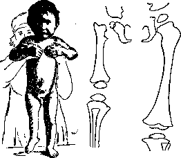
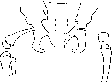
G. Richter. Pathology. Congenital changes in the hip joint. Congenital absence of the hip joint is observed together with cases of the very rare congenital absence of the femur (see Femur). Various degrees of this deformity may manifest either as absence of the proximal epiphysis or as insufficient or defective development of its elements. There is marked shortening of the thigh with coxa vara (see below), absence of a differentiated hip joint, and synostosis between the head of the femur and the acetabulum. These congenital deformities are extremely rare, and their origin has not yet received a fully precise explanation (fig. 1, 2, 3). This deformity is of the phocomelia type, in the origin of which several factors may play a role, for example, amniotic bands disrupting the vascularization of certain areas. This especially applies to the subtrochanteric area, which in terms of blood supply in the embryo depends on the diaphysis of the femur. There are no muscles here, and the blood supply to this area of bone depends on the aa. nutriciae, which run along the muscular attachments of the linea aspera femoris (Reiner). There are certain relationships between vascularization and ossification in the embryo. In some cases, obstruction of the feeding vessels from various causes can be assumed. Sometimes the possibility of a defect in the primary anlage or an intrauterine fracture cannot be excluded. Indications for treatment are either very shortening of the leg (of course in unilateral cases) or the presence of contracture accompanying coxa vara (see below), or a loose joint. Wearing a hip-leg splint extending in the form of
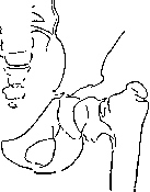
Figure 1. Congenital partial defect of the femur in a child (1 yr 10 mo), shortening 6 cm.
Figure 2. Schema of X-ray of the hips of the same child (see figure 1) with defect of the proximal end of the femur. a prosthesis can alleviate the distressing symptom of 'looseness' and compensate for the shortening, which in these cases can be quite significant. Surgical measures in the form of myotomies (sectioning of adductor muscles), osteotomies (see below) can sometimes correct the faulty position of the hip joint. The most common congenital disease of the hip joint is congenital dislocation and subluxation of it (see Dislocations). According to Isigkeit, 0.2% of all live births have this anomaly,
Figure 3. Bilateral defect of the femur in a 4-year-old
child together with absence of the fibula on the left. subluxations, however, according to Calot, are found upon careful study of X-ray films even more frequently. If these conditions are not recognized in a timely manner and not properly treated (bloodless reduction in unilateral dislocation in children under 7 years gives good joint function in more than 80%, in bilateral cases up to 5½ years - from 60% to 80%), then persistent disorders of hip joint function remain, significant disorders of statics in walking and standing. The essence of these disorders is that the thigh loses its support on the pelvis, because the head of the femur, not being in the acetabulum, during
loading freely slides along the lateral wall of the ilium, reaching in severe cases to the iliac crest itself. The passive apparatus for maintaining equilibrium of the trunk in relation to the thigh is lost when standing, because the ligamentous apparatus of the hip joint, decisive for this function, ceases to act, especially lig. ilio-femorale (see anatomy). Loss of function of m. glutaei medii and m. glutaei minimi, the points of origin and insertion of which are too close together, leads to loss of function of active maintenance of equilibrium of the pelvis in relation to the thigh during walking and standing. The first cause causes lordosis of the lumbar spine as a factor supporting passive equilibrium, the second cause gives patients the characteristic duck gait with the Trendelenburg phenomenon (see Dislocations).-The same picture, though in somewhat milder form, is also found in subluxations. There are a number of intermediate forms connecting dislocations and subluxations. Despite the smallness of the displacement of the head, sometimes measurable only in millimeters, the symptomatology in subluxation can strongly resemble that in dislocations. In subluxations, the acetabulum can be very flattened, the head of the femur can be flatter, valvular, mushroom-shaped (fig. 4). FLAT SHAPE OF THE HEAD
Figure 4. Congenital
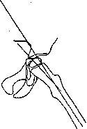
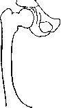
head of a girl. although this is difficult to prove. In most cases, however, this shape of the head of the femur is the outcome of pathological processes that were accompanied by softening of the head and subsequent deformation (Perthes' disease), osteomyelitis in early childhood, epiphysiolysis, deforming arthritis, etc. (coxa vara see below). Some authors (Calot, Waldenström and others) have attempted to isolate the flat head as a separate nosological unit - coxa plana, however most surgeons consider this deformation secondary. A very complex and heterogeneous group of pathological conditions, united under the name coxa vara and coxa valga, includes both congenital and acquired diseases. Under the name coxa vara is meant such a deformation in which the axis of the femoral neck with the axis of the femoral diaphysis forms an angle less than normal, in coxa valga - greater than normal. The size of this angle in normal fluctuates between 130° (in men) and 120° (in women). Pathological conditions are apparently determined primarily not by the size of the angle, not by morphology, but by functional disorders associated with this condition. For clinical purposes, the following definition is more suitable: coxa vara is such a morphological and functional deviation from norm in which the thigh in concentric standing in the acetabulum is in a state of adduction (fig. 5), in coxa valga - in a state of abduction (fig. 6) (Hagelund). Reduction of the angle can be given by almost all diseases of the hip joint. Due to the heterogeneity of diseases included in the coxa vara group, their classification was built by authors on the most various principles. By location: 1) coxa vara capitis, 2) coxa vara epiphysata, 3) coxa vara S29 trochanterica. By origin: 1) coxa vara congenita, 2) coxa vara rachitica, 3) coxa vara statica, or essentialis, or adolescent form, 4) traumatic coxa vara, 5) coxa vara after inflammatory diseases. Many'
Figure 5. Schematic drawing of coxa vara.
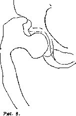
Figure 6.^ Schematic drawing of coxa valga. consider the classification of Hoffa and Drevermann (Hoffa, Drevermann) convenient: A. Congenital coxa vara. B. Acquired coxa vara: 1) symptomatic: "a) rachitic (fig. 7), b) osteomalacic (fig. 8), c) inflammatory, d) traumatic; 2) coxa vara essentialis (adolescentium, or static).
Figure 7. Coxa vara rachitica with rachitic curvature of the femur.
Coxa valga osteoma-lacica.
The mechanism of origin of coxa vara is simple in fractures of the femoral neck. The diaphysis is set at a more acute angle to the neck due to the traction of the adductor muscles. In other forms the mechanism is more complex. A gradually forming bend is assumed due to pathological softening of bone or decrease in its strength. Etiological factors: rickets, malaria, fibrous osteitis, inflammatory diseases, endocrinopathies. In the case of static coxa vara, a static-dynamic factor comes into play, which includes both abnormal load for this neck and abnormal traction of muscles (adductors). Patients suffering from static coxa vara (adolescent, essentialis), predominantly rural residents aged 12 to 18 years, attract attention with the bulkiness, massiveness, and cyanosis of their limbs. Bilateral affection is more common than unilateral. Gradually, pain radiating to the thigh, knee, and perineum becomes increasingly felt; pain increases with walking, subsides in
Figure 9. Angle of the base of the neck (98°) and the diaphyseal-epiphyseal angle (85°). In congenital coxa vara the angles may not coincide.
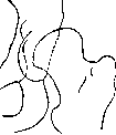
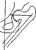
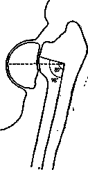
rest. Patients begin to limp. The leg assumes a position of adduction and external rotation. The greater trochanter rises above the Roser-Nelatonskaya line, and Trendelenburg's symptom appears. When rising to the knees, the patient's thighs cross. The thigh muscles atrophy somewhat. After 1-1½ years, the pains subside, but rapid fatigue when walking, shortening of the thigh, adduction, and external rotation remain, sometimes with hyperextension in the hip joint. Anatomically, this is coxa vara epiphysata. Radiographically, decalcification of the head and neck is found, and the gradual decrease of the collodiaphyseal angle. Subsequently, the epiphysis sinks more and more, resulting in a picture of epiphyseolysis (see below), the head of the femur flattens, and later becomes denser and takes on a mushroom-like shape. The epiphyseal cartilage ossifies (figures 9, 10). There are doubts about the possibility of diagnosing congenital coxa vara: in early childhood, X-rays are rarely taken, and the interpretation of an X-ray of the thigh in a newborn or infant presents the greatest difficulties. On the other hand, there are reasons to believe that some cases of static and rachitic coxa vara are congenital but only manifested later. The indication for treatment is the degree of functional disorders, not the morphological signs, no matter how pronounced they may be. Treatment in the initial acute stage—rest, constant traction with a weight of 2-3 kg, after the pain subsides, wearing a removable unloading apparatus for several months, massage and gymnastics in the lying position every evening, with the hip joint in a state of internal rotation, abduction, and flexion. In the completed process, if limitation of mobility, pain, fatigue of the leg, and Trendelenburg's symptom remain, surgical intervention is indicated. Redression, osteotomy of the neck, subtrochanteric osteotomy (see below) of various kinds, resection of the joint, arthroplasty are used; oblique subtrochanteric osteotomy is most frequently applied.
Coxa valga occurs less frequently and has relatively little clinical significance, but great biological interest. It can be a congenital disease. In muscular atrophies and paralysis of the adductor and extensor muscles, coxa valga is always observed, especially if the patient cannot use his limbs for a long time. The formation of the neck angle requires the proper play of muscles and load on the thigh; otherwise, coxa valga develops from inactivity. Coxa valga as a result of former inflammation or injury is a great rarity. Clinical picture: narrow thighs, the greater trochanter stands low and much more medially, limitation of adduction and increase in abduction of the thigh, an awkward gait with the foot turned inward. Sometimes coxa valga is combined with insufficient development of the acetabulum. In this case, the head of the femur is in a state of subluxation in relation to the acetabulum and easily dislocates, then giving a complete analogy with congenital dislocation of the hip. This picture is called coxa valga luxans. This latter form must be treated according to the principles of treating congenital dislocation of the hip; all other forms of coxa valga usually do not require treatment, occasionally giving rise to the fight against muscular contractures or to wearing orthopedic apparatus or to fixing operations in case of paralysis of the hip joint.
Figure 10. Coxa vara in an 8-year-old child with congenital changes in the epiphyseal nucleus.
Traumatic injuries, like other diseases of the hip joint, should be divided into intra-articular and extra-articular, open and closed. Fractures of the neck of the femur rank first in frequency as well as in their practical significance. According to Bruns, 6% of all fractures fall on the femur, of which 1.5% on the neck of the femur; after 70 years, all fractures fall on the neck of the femur. According to the anatomical sign, Matti distinguishes the following types: 1) fracture of the head, 2) subcapital or medial fracture of the neck, 3) intermediate fracture of the neck, 4) lateral fracture of the neck, 5) fractura colli femoris intertrochanterica, 6) fractura pertrochanterica, 7) fractura subtrochanterica, 8) isolated avulsions of the trochanters (figure 11). For the clinician, the important fact is that the first 3 groups are intra-articular injuries. Lateral fractures of the neck can sometimes be intra-articular, and finally the last 4 groups are always extra-articular injuries. Fractures of the head of the femur, its cartilaginous part, occur relatively rarely. They are difficult to diagnose both clinically and radiographically. Even Dupuytrene asserted that when falling on the leg or on the greater trochanter, compression fractures of the head of the femur often occur, which are not recognized and are considered bruises of the hip joint. Interpretation of the X-ray in this case is a difficult task, and damage to the head is often overlooked. A longer-lasting tumor, persistent pain on percussion along the axis of the diaphysis or neck of the femur, prolonged disturbances of motor function when loading the hip joint make one suspect a fracture of the head. Treatment. In the first period, unloading with the help of traction for up to 2 weeks. After absorption of the hemorrhage—movements, application of heat in the form of hot and dry-air baths
Figure 11. Classification of fractures of the neck of the femur. (According to Matti.)
according to Bier. The prognosis quo ad functionem is always serious, as secondary severe deformative changes in the joint may arise. These deforming arthritics sometimes indicate secondary operations: resection of the joint of arthroplastic type in young people and arthrodesis in old age. Intra-articular fractures of the neck represent a disease occurring somewhat more often in women and characteristic of the age over 50. In youth and sometimes in children, medial fracture of the neck occurs almost exclusively in the form of a rupture of the epiphyseal cartilage and traumatic avulsion of the epiphysis. Kocher, Volkovich, and others established that the mechanism of fracture is usually the following: falling on the greater trochanter, with the impact more often on the posterior-lateral surface of the trochanter major, along the axis of the neck of the femur; the force of impact is directed from behind and below upward and forward; from this compression along the neck, a compression fracture occurs at the weakest place, just under the head. The central fragment is fixed by the edges of the acetabulum, while the peripheral fragment begins to slide forward and upward. The significantly protruding intertrochanteric crest presses from behind on the neck, causing the fragments to move forward and the thigh to rotate outward. In its forward movement, the neck abuts against the immobile ligamentum Bertini, and the further action of the force creates conditions for a fracture from bending. In this case, the posterior edge of the peripheral fragment can be embedded in the spongy substance of the central fragment and sometimes give the so-called impacted fracture. When falling on the sole, when the force is directed along the axis of the femur, a pure form of fracture from shifting of the peripheral fragment, not reinforced, onto the fragment with the head, well fixed in the joint, can be obtained. A pure form of fracture from external rotation can occur when falling on the side, if the leg is fixed in this case and the body turned inward. Epiphyseolysis in children is a complete analogy to the subcapital fracture of the elderly. It can be the result of a very strong simultaneous trauma, sometimes repeated. First, there is a loosening of the epiphyseal connection, and then its complete separation. There are reasons to believe that in many cases the trauma affects a pathologically changed growth cartilage, and epiphyseolysis easily joins additionally here. Such traumatic separation of the epiphysis is more often observed in children with a habitus of the hypophyseal type (dystrophia adiposogenitalis). The displacement of the fragments of the neck of the femur corresponds to the mechanism of fracture. Usually the central fragment is in the position of abduction and rotation inward, the lateral fragment—in the rotated inward and adducted position.
Medial fractures resulting from falling on the back are almost always adductive with a high-lying trochanter and a strongly adducted thigh. Clinical picture: the leg is turned outward, spontaneous pains and on palpation of the inguinal region and the area of the hip joint, radiating sometimes to the knee, sometimes to all sides of the thigh. A blow to the greater trochanter, along the axis of the femur, sometimes causes significant pain. In most cases, one or another shortening of the thigh (up to 2 cm) can be established. The most constant sign is disturbance of function. Patients cannot actively adduct and abduct the thigh and lift it. Passive movements are possible, but are still accompanied by pain. The greater trochanter stands above the Roser-Nelatonskaya line. The distance from the spina iliaca anterior superior to the apex of the greater trochanter is shorter on the affected side. The triangle of Brian on the affected side ceases
Figure 12. Triangle of Brian on the healthy side.
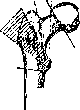


The triangle becomes isosceles - the vertical leg decreases, the horizontal one lengthens (fig. 12, 13). An X-ray can be difficult to interpret in cases of incomplete fracture, impacted fracture, and in very rare cases in pathological fractures. In such patients, only repeated structural X-rays clarify the situation and allow differentiation from a contusion or sprain of the hip joint. The prognosis for medial fractures must be made with great caution, since in most cases we are dealing with elderly people, for whom prolonged immobilizing treatment carries a number of dangers in the form of exacerbation of purulent bronchitis, hypostatic pneumonia, thrombosis with subsequent embolism, urinary retention with subsequent cystitis, bedsores, weakening of cardiac activity. To achieve complete bony union and restoration of function in these patients appears to be an exceptionally difficult task. Kocher speaks of this as an exception, but according to Hubner, 74.8% of fractures have poor union. Complete consolidation of the fracture sometimes occurs only after 8-10 months, or even after a year. Causes of poor healing: 1) senile osteoporosis, 2) atrophy and resorption of the crushed neck, 3) absence of periosteum (bone-forming) on the neck, 4) interposition of parts of the hip joint capsule, 5) unfavorable vascular anatomy. In fractures of the neck with complete rupture of the joint capsule, the central fragment can be completely deprived of its blood vessels and undergo aseptic necrosis. Under such conditions, callus forms only on the peripheral fragment. If at least half of the capsule is intact, the blood supply is sufficient. A very important factor is inadequate reduction and fixation of fragments, and finally early weight-bearing. Recent studies indicate that with proper treatment technique, the percentage of successful cases can be brought to 80-90, while in the remaining cases pseudarthrosis results. Under general anesthesia, the fragments are reduced. This is achieved by turning the leg, i.e., the peripheral fragment, inward, followed by abduction of the leg and slight flexion. Only in this position does contact occur with the central fragment, which is in the position of abduction and inward rotation. At the same time, the capsule is stretched and stabilizes the fragments. The fragments are fixed in the achieved position either with a plaster cast or with traction. At present, the Whitman plaster cast, applied from the axillary region to the toes of the foot, is used. The cast is kept for 2-3 months. After this, active and passive gymnastics in bed are begun, with the leg constantly in a state of abduction and inward rotation. Weight-bearing not earlier than 1/2 year. Sometimes in this cast the patient can move around on crutches.
Figure 13. Brian's triangle on the affected side.
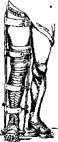
The treatment of medial fractures of the neck is in most cases conservative. Large statistics show that surgical treatment gives only 40-50% success. The latest data (Bohler, Smith-Petersen) give somewhat better figures. Under local anesthesia or in the above-mentioned position of the leg, fixation can also be achieved with skeletal or adhesive traction. Fixation continues until the patient can actively lift the leg. During the entire bed rest, the patient does gymnastics, moves as much as possible, and participates in physical exercises. Weight-bearing also not earlier than 1/2 year, when an X-ray will indicate bony union of the fragments. This method has recently been successfully applied in the Sklifosovsky Institute of Traumatology and Emergency Aid in Moscow. Lofberg, after correctly positioning the fragments with a hammer blow (of his own design), impacts the fragments and fixes them in this position. Reduction of fragments sometimes encounters difficulties: greater traction (up to 10 kg) must be applied, and the Tavernier method (fig. 14) and others must be used for reduction. All the described methods are not applicable in very old, obese, emphysematous, prostatic patients, etc. In them, the primary concern is function, not the correct anatomical positioning of the fragments. They cannot be kept in bed for long due to the danger of various complications and marasmus. Walking on crutches in the Volovich apparatus, Thomas splint (fig. 15) and other devices gives the patient a tolerable existence.
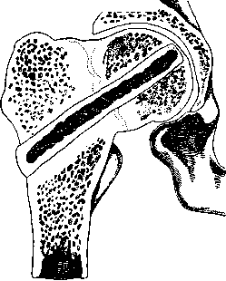
As for surgical treatment, its purpose, after exposing the fracture site, is to join the fragments by one method or another. Wire sutures, screws passed through the entire neck and both fragments, all kinds of clips (Bolzung) from autogenous material (tibial plates), heteroplastic material (plates from bovine bone), all kinds of metal plates and screws are proposed. Kocher, believing that bony union cannot be achieved at all in medial fractures, removed the head and inserted the neck into the acetabulum even in fresh cases. Large statistics show that these surgical measures in fresh cases give worse results (no more than 40% success) than conservative methods (up to 80%). Causes: significant rarefaction of bones due to the presence of a foreign body, as a result of which fixation of the fragments weakens, then secondary fracture of the transplant, suppuration of the wound, poor regenerative capacity of bones in the elderly. Despite the authority of Lexer, Delbet, Henderson, etc. (Lexer, Delbet, Hen- Fig. 16. Smith-Petersen's incision: Leg after fracture of the femoral neck. 1-spina ili ant. superior; 2-line of incision; 3-m. tensor fasciae latae; 4-greater trochanter; 5-lower end of incision, CTBO intersection of tractus ilio-ti- In fresh biahs, the fracture of the femoral neck recedes into the background before conservative methods. However, recently many authors (Smith-Petersen, Bohler, Tavernier, Hotz) have again raised the question of surgical treatment of femoral neck fracture. After reduction of the fragments, a specially constructed nail by Smith-Petersen is driven into the area of the greater trochanter. This nail consists of 3 faces. Each face represents a thin and sharp plate. Such a nail is easily inserted and firmly fixes both fragments, preventing them from shifting. The Smith-Petersen incision (fig. 16) begins immediately below the anterior superior iliac spine, extends downward and somewhat绕过绕过 the base of the greater trochanter; after opening the fascia, the m. tensor fasciae latae is bluntly separated and retracted outward, the m. rectus femoris and m. sartorius outward, after which the anterior capsule of the hip joint is exposed (fig. 17). After opening the joint, the fragments are reduced (fig. 18). From this same incision, into the subtrochanteric fossa, under visual control, the Smith-Petersen nail (fig. 19) is inserted, which passes through both fragments (fig. 20, 21). The correctness of the operation is controlled by the restoration of passive movements in the hip joint. Postoperative period 7-10 days. Short plaster cast, after which walking on crutches sometimes even without any cast. According to Hotz, from a small incision below the greater trochanter after reduction of the fragments, a screw (10-12 cm long) is passed through the neck of the head and acetabulum. The direction and depth of insertion are determined by X-ray with the help of lines previously marked on the skin. A control X-ray must be taken immediately. In case of correct fixation of the femur to the pelvis (fig. 22), walking can be allowed as early as the 2nd day. The above-mentioned reduction method by Tavernier is also supplemented by fixation of the fragments with a special screw under the control of a portable X-ray apparatus. Traumatic epiphyseal separations in children are treated the same as medial fractures. In severe cases with complete separation of the epiphysis and significant displacement, surgical treatment with possible positioning of the fragments in the correct position and preservation of the growth cartilage. In this position, it is fixed for a month.
Figure 18. The next stage according to Smith-Petersen.
After opening the joint, reduction of the fragments of the femoral neck.
Figure 19. Smith-Petersen's nail.
Figure 20. The fragments are joined in the correct position with a special instrument consisting of a protector, head holder, fragment fixator, and special pusher for the nail.
Lateral and intertrochanteric fractures of the femoral neck have the same clinical picture, prognosis, and therapy. The fracture results from a fall on the greater trochanter, sometimes with its comminution and avulsion of the lesser trochanter. A fracture can also occur from a fall on the leg with force along the axis of the femur (flexion fracture). The clinical picture on Figure 21. The nail is passed through both fragments. Figure 22. Correct fixation of the femoral neck with the Smith-Petersen nail.
Figure 22. Correct fixation of the femoral neck with the Smith-Petersen nail.
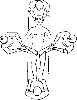

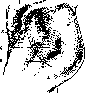
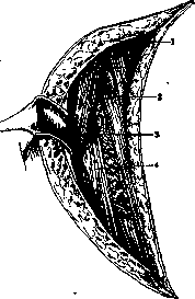
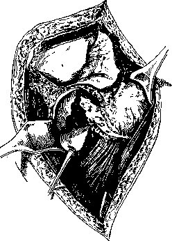
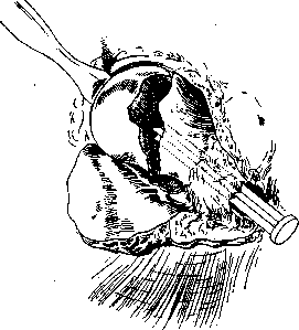
resembles a medial fracture, only the outward rotation of the thigh is less, while the shortening of the limb is usually greater (up to 4 cm). The angle of inclination of the neck, i.e., traumatic coxa vara, is always more pronounced. The conditions for fracture healing are significantly simpler than with medial fractures, and a strong bony callus can be formed in a relatively short period. A very unpleasant complication with these fractures is extensive crushing of the greater trochanter and surrounding bone, which later form large callous masses extending to the capsule of the hip joint. This results in prolonged and persistent limitations of mobility and pain with movements in the hip joint. When occurring with occasional intra-articular fractures, the course of the disease can be as severe as with medial fractures of the neck with poor consolidation and scanty callus formation. Impacted fractures in this area may sometimes not be recognized in time, are treated as bruises, and with early weight-bearing and movements lead to very severe forms of traumatic coxa vara or traumatic coxa adducta. These patients sometimes require subsequent surgical intervention (see below, osteotomy).-Treatment. Under local or spinal anesthesia, rarely under general anesthesia, traction is applied to the fractured limb. If the neck angle is greatly reduced, skeletal traction on the thigh or upper end of the tibia can achieve very strong abduction of the thigh-up to 50°. With minor changes in the neck angle, traction through soft tissues is recommended-flexion in the hip joint not more than 30°. The outward rotation of the thigh is corrected either by special traction turning the thigh outward, or by a Volkmann splint, or by fixing the lower leg and foot. Traction is applied for 2-2½ months, followed by at least a month of wearing a Thomas splint or plaster cast, under constant monitoring of the neck angle. In very elderly people, a light plaster cast can be applied immediately in the abducted position and they can be put on crutches. In rare cases where it is impossible to properly reduce the fragments using various traction methods, surgical treatment is resorted to. Through an incision in the area of the greater trochanter, open reduction of the fragments is performed. After this, they are fixed with a Lambotte screw or secured with wire or other material wrapped around them (fig. 23, 24).
Figure 22. Method of fixing fragments together with the pelvis according to Hotz with a long screw. 83»
Figure 23.
Figure 24. Pertrochanteric fractures most often occur from sudden hyperextension of the leg when falling backward and from forced rotation and adduction of the thigh when falling to the opposite side. The peripheral fragment is usually displaced forward and inward. In the clinical picture, the prominent position of the greater trochanter is striking, its immobility during rotation of the femoral diaphysis, often a significantly displaced forward peripheral fragment can be palpated; shortening of the leg by 3-5 cm, outward rotation of the thigh. Treatment of these fractures requires marked flexion of the hip and knee joints; otherwise, they are treated the same as intertrochanteric fractures. Isolated fractures of the greater trochanter are extremely rare. These are usually avulsion fractures from uncoordinated action of the m. glutaeus medius and other muscles, which, when contracting, can displace the fragment significantly upward and backward. Without significant displacement of the fragment, treatment is conservative. With wide separation, the fragments must be sutured or screwed together. Duration of treatment is 5-6 weeks until recovery of the impaired abduction movements and outward rotation of the thigh. An isolated fracture of the lesser trochanter is equally rare and represents an avulsion fracture from forced contraction of the m. ilio-psoas. Characteristic symptoms are limitation of outward rotation of the thigh and inability to lift the thigh in a sitting position with the knee joint extended (Ludloff's symptom). This phenomenon is explained by the fact that lifting the thigh in this position is possible only with the help of the m. ilio-psoas, since the m. rectus femoris cannot function under these conditions, while the activity of the m. ilio-psoas has ceased due to avulsion of its attachment point. Treatment is usually conservative for 2-6 weeks. Subtrochanteric fractures, in terms of mechanism and course of the disease, have much in common with fractures of the upper third of the thigh. Complex pictures of combinations of fractures at multiple levels of the neck are encountered. Despite continuous improvement in the technique of treating femoral neck fractures, especially in recent years, world statistics indicate that at least 20% of femoral neck fractures do not unite. According to Anschiutz, of this number, about 20% die from the consequences of the fracture or complications (pneumonia, bedsores, marasmus). In approximately 30% of patients of small weight and not stout, these fractures proceed well with the most conservative methods. In these cases, although there is no bony union, the peripheral end with its processes and edge projections sinks into the lesser trochanter or other part of the central fragment and thus provides solid support for the leg, and the patient does not need serious assistance. 50% of patients require surgical intervention. Indications for surgery: constant pain and a pronounced Trendelenburg symptom, i.e., complete inability to bear weight on the leg.
Figure 25. Valgization according to Lorenz; oblique osteotomy in the sagittal plane: 1-capsule of the head on the ilium; 2-acetabulum.
Operations are bloodless and bloody. Bloodless operations aim to place the distal fragment in a position where it has support. Inversion, according to Lorenz, is placement in a position of inward rotation, abduction, and extension, i.e., placement in the opposite position and fixation in this position; transposition, according to Drevermann, is placement of the fragment in the anterior position for support on the pelvic bone. Finally, repeated application of a Whitman plaster cast after reduction of the fragments.-Bloody operations. There are various methods: extra-articular and articular (in the area of the fracture), preserving and not preserving movement (creating only support), preserving normal anatomy and disfiguring (arthroplasty). Of extra-articular methods, subtrochanteric transverse or oblique wedge osteotomies are often used recently. The distal fragment is usually abducted by 30-35°, placed in the correct position, and the leg is fixed with a plaster cast or traction for 1½-2 months. Subsequent treatment includes massage and gymnastics. In 1918, Lorenz proposed for old congenital dislocations of the hip an oblique osteotomy that provides solid support for the leg in the normal position in the area of the acetabulum. The bone is cut in the sagittal plane at the level of the trochanteric fossa, the lower fragment is inserted into the fossa, and the upper fragment apposes it laterally, forming an angle. After bony union, the proximal end of the femur resembles a fork with two prongs, on which the pelvis firmly rests (fig. 25). Hass suggests making the bone cut in the frontal plane to avoid anterior displacement of the central fragment due to traction from the m. ilio-psoas (fig. 26). This operation is called valgization or forking (according to Lorenz, Hass, Kirmisson, Baire) and is also valuable in the treatment of pseudarthrosis of the femoral neck; in this case, the fork embraces the central fragment of the neck with the head, pressing against it (fig. 27). An incision is made on the outer side of the thigh from the greater trochanter downward. After cutting the bone, downward traction and abduction up to 40-45°; after the lower fragment has displaced to the desired position under the femoral head, fixation for 2-3 months. Schanz's osteotomy also provides support and eliminates adduction of the thigh, but is based on a different principle. A deep transverse osteotomy of the femur is performed at the level of the ischium. An angle is formed between the fragments, open outward. The central fragment approaches the pelvis. After union of the fragments
Figure 26. Valgization according to Hass; osteotomy in the frontal plane.
the Trendelenburg phenomenon disappears, as the pelvis can no longer drop to the healthy side, resting on the diseased thigh. Before cutting the bone, 2 special screws (fig. 28) are inserted above and below the fracture site, parallel to each other. After osteotomy, they are set at the desired angle (35-40°) (fig. 29), their protruding ends are secured, and a plaster cast is applied to the entire limb to the toes. After a month, the screws are removed by making a window in the plaster. Another month and a half later, the plaster cast is removed. Schanz's operation provides
Figure 28.
Figure 29. weaker support but shortens the leg less and provides greater mobility than Lorenz's operation (fig. 30).-Similar to Schanz's method is Pauwel's operation, applied by several authors for the treatment of pseudarthrosis of the neck (fig. 31). After wedge osteotomy and positioning of the fragments at an angle,
Figure 30. Condition of the hip joint after Schanz's operation.
the Trendelenburg phenomenon disappears, as the pelvis can no longer drop to the healthy side, resting on the diseased thigh. Before cutting the bone, 2 special screws (fig. 28) are inserted above and below the fracture site, parallel to each other. After osteotomy, they are set at the desired angle (35-40°) (fig. 29), their protruding ends are secured, and a plaster cast is applied to the entire limb to the toes. After a month, the screws are removed by making a window in the plaster. Another month and a half later, the plaster cast is removed. Schanz's operation provides
Figure 28.
Figure 29. weaker support but shortens the leg less and provides greater mobility than Lorenz's operation (fig. 30).-Similar to Schanz's method is Pauwel's operation, applied by several authors for the treatment of pseudarthrosis of the neck (fig. 31). After wedge osteotomy and positioning of the fragments at an angle,
Figure 30. Condition of the hip joint after Schanz's operation.
the Trendelenburg phenomenon disappears, as the pelvis can no longer drop to the healthy side, resting on the diseased thigh. Before cutting the bone, 2 special screws (fig. 28) are inserted above and below the fracture site, parallel to each other. After osteotomy, they are set at the desired angle (35-40°) (fig. 29), their protruding ends are secured, and a plaster cast is applied to the entire limb to the toes. After a month, the screws are removed by making a window in the plaster. Another month and a half later, the plaster cast is removed. Schanz's operation provides
Figure 28.
Figure 29. weaker support but shortens the leg less and provides greater mobility than Lorenz's operation (fig. 30).-Similar to Schanz's method is Pauwel's operation, applied by several authors for the treatment of pseudarthrosis of the neck (fig. 31). After wedge osteotomy and positioning of the fragments at an angle,
Figure 30. Condition of the hip joint after Schanz's operation.
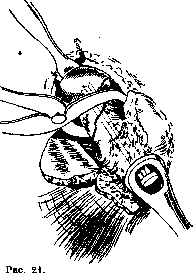
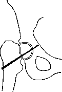


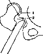
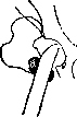
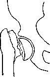
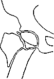
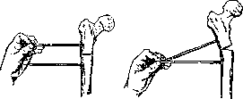
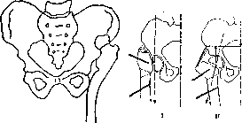
Figure 31. I-before osteotomy according to Pauwel'y; II-after the operation. When opened outward, the pseudarthrosis line becomes almost horizontal and thus the force of gravity compresses the fragments of the femoral neck rather than separating them, which eliminates painful phenomena. To restore function to the weakened mm. glutaeus medius and minimus, the greater trochanter with attached muscles is detached and transplanted to the diaphysis more peripherally, to return proper function to the muscle deprived of its normal attachment point. This operation (Veau-Lamy) as a standalone procedure is unreliable, but when combined with other operations it secures their success. Good functional results are given by operations in which the freely lying head is removed and the neck is inserted into the acetabulum (fig. 32, 33).-If the neck is short, different methods have been proposed for its lengthening: part of the greater trochanter is detached, both trochanters are moved peripherally, various wedge-shaped osteotomies are added (Schlegel, Golyanitsky and others). König, Borchard, Schmieden and others insert the greater trochanter into the acetabulum. The leg is fixed in a plaster cast for 2½-3 months in an abducted position. With these methods, ankylosis of the hip joint is expected, i.e., restoration only of the supporting function.
Figure 33. A-plane of detachment of the greater trochanter; B-transplanted greater trochanter; C-joint capsule.
Lexer's plastic operation provides not only support but also restores movement in the joint (fig. 34). Incision of skin and fascia (see below-operation of Murphy, Lexer), after opening the joint-removal of the head; by free transplantation of a piece of bone taken from the removable head and placed subperiosteally, the overhang in the area of the acetabulum edge is increased; then the neck is shaped, wrapped with a flap of fascia with fat taken from the thigh, and inserted into the acetabulum; layered sutures on the wound. After a month of fixation, active and passive movements. Lexer's results are good.-If the head is not necrotic and the patient is not old, there are indications for bone synthesis with complete restoration of both function and anatomy of the femoral neck. Fragments of the femoral neck in pseudarthrosis are fixed with the help of an inserted wedge (Bolzune) into the acetabulum with interposition of fascia and fat between the head and acetabulum; the greater trochanter is transplanted lower according to Veau-Lamy; A-freely transplanted piece of head for increasing the overhang above the head.

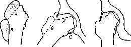
Figure 34. Plastic operation of Lexer for pseudarthrosis; i-before the operation; ii-after removal of the head; the neck is inserted into the acetabular cavity with interposition of fascia and fat between the head and acetabulum; the greater trochanter is transplanted lower; A-freely transplanted piece of head for increasing the overhang above the head.
If the head is not necrotic and the patient is not old, there are indications for bone synthesis with complete restoration of both function and anatomy of the femoral neck. Fragments of the femoral neck in pseudarthrosis are fixed with the help of an inserted wedge (Bolzune) into the acetabulum with interposition of fascia and fat between the head and acetabulum; the greater trochanter is transplanted lower according to Veau-Lamy; A-freely transplanted piece of head for increasing the overhang above the head. Heteroplastic (metallic nails, screws and others). A wedge can be introduced either extra-articularly from the area of the greater trochanter (Delbet, Lexer and others) or using the incision for arthrotomy of the hip joint (see above incision of Smith-Petersen, Goetz) (fig. 35). The negative side of the osteosynthetic method is indicated above. Braytsev succeeded in obtaining a good long-term result by using the following method: he scraped out the removed head and left only the shell of cartilage and a thin layer of bone and fitted it onto the neck. A short neck covered with articular cartilage resulted. The theoretical basis: the established fact that articular cartilage is nourished by the synovial fluid. One can speak of pseudarthrosis no earlier than a year, before this period one may be dealing with an unhealed fracture of the neck. Closed injuries include contusion and sprain (distorsion) of the hip joint. By contusion is meant injury to the internal parts of the joint, mainly the synovial membrane, by direct violence followed by effusion of blood into the joint. The articular cartilage can sometimes be damaged, torn, detached. Usually there is also contusion of the para-articular soft parts of the skin, a hematoma that gives a crunch on palpation. Movements, standing and walking are painful but possible. After hemarthrosis, hydrops of the joint with its instability often persists for a long time. X-ray film excludes fractures, with large hemorrhage it gives diffuse darkening. The greater trochanter is not displaced.-Distorsion of the hip joint is the result of indirect force, it is the result of action on the long lever arm, as in dislocation. In this case, the joint capsule can rupture, ruptures of the reinforcing ligaments (lig. Bertini and others) occur, para-articular large hemorrhages, sometimes also hemarthrosis. Diagnosis: tenderness on palpation of lig. Bertini or other torn ligament and with certain movements of the joint when the ligaments are tensed. In general, joint function is not impaired. The skin is usually unchanged.-Open injuries to the soft parts of the hip joint area are significant when the joint or bones or nearby pelvic organs are simultaneously injured, especially if the wounds are infected. The very deep position of the joint often makes correct diagnosis difficult.-Gunshot wound to only the soft tissues of the hip joint is conceivable only as a tangential wound. In all other cases, either the bone or, more rarely, only the joint capsule is injured. Injuries to bones vary in degree-from detachment of a small piece of the head to its complete comminution and of the pelvic bones with simultaneous injury to the perineum, pelvic organs, nerves and vessels. In gunshot wounds with a small zone of injury, diagnosis of injury to the hip joint is difficult. Cases without suppuration leave no consequences. Cases with suppuration give an extremely severe picture of suppurative coxitis; very severe pain, the joint capsule is stretched by purulent content but meets resistance in the huge muscular mass of the hip joint, hence high fever and pain. Langenbeck's symptom appears (the a. femoralis on the affected side is better palpated than on the healthy side, due to its elevation by the stretched hip joint). The prognosis is severe. The American war of 1866 gave 93% mortality, the Franco-Prussian-74.7%, the last imperialist war also a large number, although exact data are lacking. Long-term results (Erlacher) also indicate the severity of the disease: out of 100 cases-65 ankyloses of the joint, 14 unstable joints, 12 severe limitations of mobility, 7 minor limitations of mobility, 2 normal. For contaminated wounds, active intervention is necessary: primary treatment, resection of the joint, in more rare, very extensive injuries, disarticulation in the hip joint. In subsequent treatment, timely additional arthrotomies, counter-incisions, drainage which have to be kept for a long time. Fixation with splint, plaster cast or traction. Sometimes infection becomes noticeable only from the 5-6th day.
Figure 35.
Non-traumatic diseases of the hip joint. Acquired extra-articular diseases of the hip joint can involve the skin, subcutaneous tissue, fascia, muscles, vessels, nerves, mucous bursae and bones. Of the large number of mucous bursae in the hip joint area (from 14 to 21), the large bursa trochanterica under the tendon attachment of m. glutaei maximus, on the lateral posterior surface of the greater trochanter, bursa ilio-pectinea in front on lig. ilio-femorale under m. ilio-pectineus, often communicating with the joint, and bursa at the attachment of t. ilio-psoas to trochanter minor have great practical importance. Into these bursae hemorrhages, effusions can occur, they can be in a state of inflammation, arising both hematogenously and by transition from adjacent tissues. Hygromas, tuberculous, gonorrheic, purulent bursitis are observed. For differential diagnosis with lesions of the hip joint, strictly localized local tenderness, limitation only of those movements in the hip joint in which the bursa is compressed, but other movements in the hip joint remain free, e.g., in bursitis trochanterica-limitation of inward rotation, in burs, ilio-pectinea-limitation of extension, in burs, troch. minor (see above)-Ludloff's symptom. Treatment depending on etiology.-Of diseases of the fascia, the so-called snapping hip joint (hanche a ressort, schnappende Hüfte) deserves attention. The snapping phenomenon is clearly perceived by touch, and sometimes even by the ear, in a certain position of the thigh or with certain movements. At this, a jumping of a strong cord over the posterior edge of the greater trochanter is noticeable, accompanied by pain. The mechanism of this phenomenon depends on the shortening of tractus iliotibialis fasciae latae, to which m. tensor fasciae latae and m. glutaeus maximus are attached, or on the thickening of the greater trochanter. Thus the etiological factors are different: scar contraction of tractus Maissiatius, changes in the gluteus maximus muscle and m. tensor fasciae latae, periostitis, tumors in the area of the greater trochanter, bursitis of the greater trochanter bursa and others. The essence is that the thick fascial band with difficulty jumps over the posterior edge of the
the greater trochanter. The snapping phenomenon is clearly perceived by touch, and sometimes even by the ear, in a certain position of the thigh or with certain movements. At this, a jumping of a strong cord over the posterior edge of the greater trochanter is noticeable, accompanied by pain. The mechanism of this phenomenon depends on the shortening of tractus iliotibialis fasciae latae, to which m. tensor fasciae latae and m. glutaeus maximus are attached, or on the thickening of the greater trochanter. Thus the etiological factors are different: scar contraction of tractus Maissiatius, changes in the gluteus maximus muscle and m. tensor fasciae latae, periostitis, tumors in the area of the greater trochanter, bursitis of the greater trochanter bursa and others. The essence is that the thick fascial band with difficulty jumps over the posterior edge of the
greater trochanter. The snapping phenomenon is clearly perceived by touch, and sometimes even by the ear, in a certain position of the thigh or with certain movements. At this, a jumping of a strong cord over the posterior edge of the greater trochanter is noticeable, accompanied by pain. The mechanism of this phenomenon depends on the shortening of tractus iliotibialis fasciae latae, to which m. tensor fasciae latae and m. glutaeus maximus are attached, or on the thickening of the greater trochanter. Thus the etiological factors are different: scar contraction of tractus Maissiatius, changes in the gluteus maximus muscle and m. tensor fasciae latae, periostitis, tumors in the area of the greater trochanter, bursitis of the greater trochanter bursa and others. The essence is that the thick fascial band with difficulty jumps over the posterior edge of the
greater trochanter. The snapping phenomenon is clearly perceived by touch, and sometimes even by the ear, in a certain position of the thigh or with certain movements. At this, a jumping of a strong cord over the posterior edge of the greater trochanter is noticeable, accompanied by pain. The mechanism of this phenomenon depends on the shortening of tractus iliotibialis fasciae latae, to which m. tensor fasciae latae and m. glutaeus maximus are attached, or on the thickening of the greater trochanter. Thus the etiological factors are different: scar contraction of tractus Maissiatius, changes in the gluteus maximus muscle and m. tensor fasciae latae, periostitis, tumors in the area of the greater trochanter, bursitis of the greater trochanter bursa and others. The essence is that the thick fascial band with difficulty jumps over the posterior edge of the
of the trochanter and gives the phenomenon of clicking. There are people who can cause this phenomenon voluntarily and without any pain, so the presence of the phenomenon does not yet indicate disease. Treatment depending on etiology. In surgical intervention, it is sometimes necessary to perform 'tractotomy,' chiseling off the protruding part of the greater trochanter, suturing the fascia to the greater trochanter and aponeurosis of m. vasti lateralis, etc. Intra-articular diseases (see Arthritis, Coxitis, Perthes' disease and Joints). Among non-infectious diseases, deforming arthritis (see) is relatively common, for which a more correct name, arthrosis deformans, has recently been proposed. This is a chronic degenerative process, which is characterized by a combination of atrophic and proliferative processes in the bones and articular cartilage, thickening and overgrowth of the articular villi, and the formation of marginal protrusions, cartilaginous and bony. Due to impaired nutrition, the cartilage softens and splits into separate fibers at the sites of greatest stress, the bone is polished and sclerosed. Individual ulcers and tubercles appear on the articular surface. Ultimately, the shape of the head changes sharply, taking on the most bizarre outlines. When this disease develops after 40 years, it is considered the result of tissue wear and is called malum coxae senile. However, similar forms are also found in significantly younger ages. The onset resembles chronic joint rheumatism, it develops (sometimes bilateral, often in people engaged in heavy physical work and exposed to cooling, more often in full, massive people. Pain appears, sensitivity to
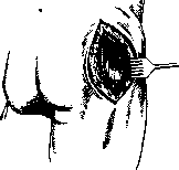
Figure 37. Langenbeck's incision-spreading of muscles.
cooling, to load. Sometimes at the beginning of the disease there are no symptoms except very severe pain, which leads to recognizing neuralgia of the joint, coxalgia (coxodynia, coxagra) and differentiating it from sciatica. Later, limitations of movement join-first rotation outward and abduction, and then
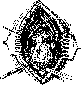
Figure 38. Exposure of the greater trochanter for subsequent temporary detachment with a chisel; according to Langenbeck, the trochanter is not severed.
flexion. The patient develops a jumping g; walking with small steps. X-ray film reveals changes in the shape of the head and marginal protrusions. Treatment: unloading and enhancement of local tissue nutrition. Hot sulfur baths, mud therapy, iodine; for unloading and rest-splint apparatus, cane,
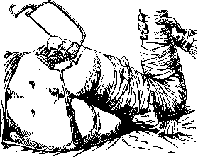
Figure 39. Operation according to Langenbeck-filing of the femoral head.
rubber soles, moderate mechanotherapy without load. In severe cases-arthrodesis or arthroplasty of the hip joint. Deforming arthroses of neurogenic origin in the most severe form occur as arthropathies, more often in tabes, less often in syringomyelia. _ Here a most varied picture of atrophic and hypertrophic processes is observed
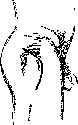
Figure 40. Kocher's incision.
with displacement of the acetabulum, spontaneous dislocation and fractures, disappearance of the head and neck. New bone formation occurs not only in the joint but also on the articular capsule and surrounding ligaments and muscles. Clinical picture: extensive exudate, range of motion exceeding normal, movements are accompanied by clicking and crepitation with complete absence of pain. The patient walks freely but limps. X-ray film gives a typical picture of disfigured articular ends with extensive free bodies and disappeared areas of bone. A rare finding in the hip joint-osteochondritis dissecans (see Osteochondritis), and the diagnosis is complicated by the fact that it is very difficult to radiologically establish the joint mouse in the hip joint. The pathological processes listed above, both infectious and non-infectious, often result in either significant limitation of mobility or complete ankylosis, sometimes a sharply painful, non-functioning hip joint, sometimes a loose one. Often with this, there is also a faulty position of the thigh-adduction and flexion. A number of operations necessary to correct the position of the thigh are listed above (see above osteotomy). In a sharply painful as well as a loose joint, extra-articular arthrodesis (see Coxitis) or resection of the joint is often indicated. Technique of hip joint resection. The most common lateral and posterior incisions. The lateral incision of Langenbeck is made
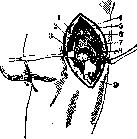
Figure 41. Surgical anatomy, modified anatomy during the incision described by König: before dislocation of the head: 1-mm. gemelli and obturator internus; 2-piriformis; 3-posterior part of the joint capsule; 4-t. piriformis and spina iliaca posterior superior; 5-m. glutaeus medius and minimus; 6-m. glutaeus maximus; 7-m. glutaeus medius; 8-anterior part of the joint capsule; 9-trochanter.
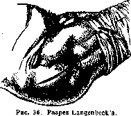
15 cm long, on the middle line of the thigh, and penetrates to the bone (fig. 36). With wide hooks, the muscles are spread apart, the anterior and posterior edges of the greater trochanter with the muscles attached to it are chiseled off (fig. 37, 38, 39). If the remaining part of the greater trochanter is removed, wide exposure of the joint is obtained, the head is dislocated and sawed off. Langenbeck does not sever the greater trochanter. Wreden's incision begins slightly below spina iliaca anterior superior, runs arcuoid to the posterior edge of the greater trochanter and bends slightly
downward. The exposed trochanter is driven upward and inward and is turned upward with the muscles. At the end of the operation, the greater trochanter is fixed in place with a nail or periosteal sutures.
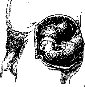
Kocher's method. Arcuate incision with convexity forward around the greater trochanter,
Figure 42. Hip joint exposed by incision according to Sprengel.
its upper half runs along the fibers of m. glutaei max. (fig. 40), the lower one along the axis of the thigh. M. glutaeus max. is split along the fibers, the greater trochanter is exposed. To the neck, one approaches in the interval between m. piriformis and m. glutaeus med. (fig. 41), the capsule is opened and the head is removed after dislocation.-Anterior incision of Sprengel-from spina iliaca posterior (fig. 42) along crista ilii to spina iliaca anterior, from here it turns at a right angle and ends at the base of the greater trochanter; after exposure of crista ilii, all muscles attached to os ilii are stripped off, and going along its surface, the acetabulum is opened after pulling the entire flap downward. Clapp (Klapp) makes the incision forward from the middle of crista ilii, 2 cm below it, the vertical part of the incision is carried around the trochanter
posteriorly, cuts through m. glutaeus med. and minim to the bone and strips it downward, and between the anterior edge of m. glutaei med. and tensor fasciae latae goes, bluntly separating, to the acetabulum and joint capsule. In bilateral ankylosis of the hip joint or multiple joint involvement in immobility of non-infectious origin, arthroplasty may be indicated, i.e., obtaining a movable
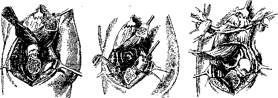
Fig. 45. Figure 46: Arthroplasty according to Murphy; cutting out a flap of fascia with fat from the skin flap on a pedicle.
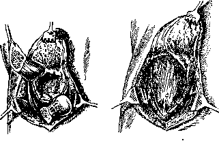
Figure 47
Figure 48
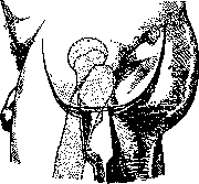
Figure 49. Arthroplasty according to Lexer;
skin incision and removal of the greater trochanter.
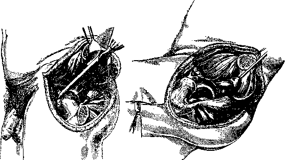
Fig 50
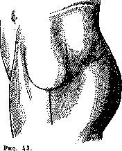
Figure 51. joint. Contraindications are usually tuberculosis and fresh purulent processes. Technique of the operation according to Murphy. ^The patient lies on the healthy side. A Y-shaped incision goes around the greater trochanter downward 5 cm, forward 4 cm and backward 2½ cm,
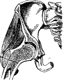
Figure 52. Arthroplasty according to Lexer; diagram of the final result of the operation. "vertically goes upward; from the lowest point of the U-shaped flap, a longitudinal incision along the thigh (fig. 43). The wound is spread with hooks; with a Gigli saw the greater trochanter is sawed off with attached muscles, the muscles are freed and the large flap is turned upward. Then with a wide chisel the ankylosis is separated, the head is dislocated (fig. 44). With the help of special instruments-a spherical trepan and a hollow rasp-the articular ends are modeled and smoothed (fig. 45). Then from the skin flap a flap of fascia with fat on a pedicle is cut out (fig. 46), placed in the newly formed acetabular cavity and sutured at the edges (fig. 47). The head is placed in the acetabulum, the greater trochanter is fixed in place. Layered sutures on the wound (fig. 48^. Plaster cast for 2½ weeks, then traction 3-4 weeks. Passive and active gymnastics.-Technique of arthroplasty according to Lexer. U-shaped incision (fig. 49, 50, 51, 52). The greater trochanter is severed with a chisel and turned upward with the muscles. Then, according to Murphy, only with free transplantation of fascia with fat. Postoperative treatment without immobilization, early movements. The above-mentioned incisions of Sprengel,
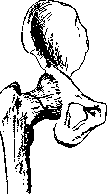
Figure 53. Saddle-shaped osteotomy of the femoral neck according to Rauh.
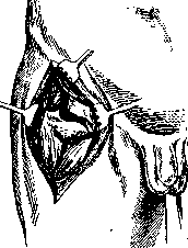
Figure 54. Saddle-shaped osteotomy according to Rauh.
Smith-Petersen and Clapp are also convenient for arthroplasty, allow for good inspection of the wound and are less traumatic. Patients are operated on in the supine position. In order to obtain slight flexion and extension of the hip joint, Paire (Raug) proposed (Figs. 53, 54) a simpler operation for ankylosis—a saddle-shaped osteotomy of the neck with a fascial graft between the fragments. Tumors. Intra-articular primary tumors of the hip joint are extremely rare, while para-articular ones are comparatively more common. On the bone in the area of the greater trochanter, enchondromas, chondrofibromas, myelogenous sarcomas, and exostoses are observed. From the soft parts, tumors of the skin, lymph glands, metastases of cancer of the uterus, penis, etc., angiomas, and neurofibromas may originate. Relatively often in the area of the greater trochanter, metastatic cancers originating from cancer of the breast, hypernephromas, and the thyroid gland are encountered. They begin with severe tearing pains when neither clinically nor radiographically can a tumor be established, and they quickly lead to spontaneous fractures, which clarify the diagnosis.
a. shnee.
Related articles
Mentioned in
Cite this page
“Hip Joint.” Soviet Medical Encyclopedia. English translation of Bolshaya Meditsinskaya Entsiklopediya, 1st ed. (Moscow, 1928–1936), ed. N. A. Semashko. https://sovietmedicalencyclopedia.pages.dev/article/hip-joint/