Arthrodesis
Historical document, translated for reference. It reflects medical knowledge of the 1920s–30s and is not medical advice.
Summary
Arthrodesis is a surgical procedure to immobilize a joint by fusing the articular ends, primarily indicated for flail joints following poliomyelitis when active mobility is lost. The technique involves removing articular cartilage and fixing the joint ends in a specific position, with various methods including intra-articular and para-articular approaches.
Encyclopedia article (1928–1936)
ARTHRODESIS (from Greek arthron-joint and desis-binding), a name proposed in 1878 by the Vienna surgeon Albert for an operation to secure the joint ends with the aim of eliminating mobility in the articulation. The indication for arthrodesis is most often flail joints following previous poliomyelitis, when active mobility has been completely lost and passive mobility is increased. Arthrodesis is contraindicated: 1) in such extensive lesions that even after ankylosis of several joints the wearing of an orthopedic apparatus is inevitable; 2) if after the operation there is a possibility of worsening the function of adjacent, less affected joints; therefore it is desirable to first test by means of a plaster cast the effect of the proposed arthrodesis on the statics and dynamics of the entire limb; 3) in children under 10 years of age, to avoid subsequent deformations from curved epiphyseal growth. Technique. The classic method consists of decartilagination: the articular cartilage is scraped, cut away, or removed with a chisel (see figure); the thus refreshed joint ends are fixed for several months in a specific position by means of an immovable bandage. Consequently, an economical resection (in a deformed joint-plastic) is performed without excision of the capsule; in other methods of arthrodesis, instead of or in addition to cartilage removal, the joint ends are secured with bolts, staples, or struts from autogenous or other material. Depending on how the fastenings are arranged, intra-articular and para-articular (paraarticular) arthrodeses are distinguished. An example of the first modification is Lexer's (Bolzung) method, in which from the plantar side, through the calcaneus and talus bones, centrally penetrating the ankle joint, a bone nail is driven into the epiphysis of the tibia. The second
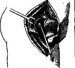
Arthrodesis of the right hip joint according to Vulpius. The articular cartilage of the femoral head has been removed. The sharp spoon is used to scrape out the cartilage of the acetabulum. modification is represented by a very large number of variants of para-articular arthrofixation by means of struts or splints applied from one side or both sides. For the take of a transplant, it is better if the splint not only bears with its ends but also adheres throughout its length to the corresponding parts of the skeleton. This requirement is satisfied by the splint method, for example, for the foot-a subperiosteally laid splint from the tibia (Cramer, Vreden) or a piece of rib (Kornev). The merit of the method is determined, besides considerations of the mechanical strength of the fixation, by the successful use of biological properties of bone tissue and the consistency of the method with the functional requirements of the given case. Metal screws and staples, which hold firmly at first, later cause around themselves rarifying osteitis and become loose. Only in the case where the operative trauma and subsequent functional irritation stimulate osteogenic abilities in the bone bed, does the primary mechanical connection of the joint ends turn into organic fusion. From this point of view, autogenous material deserves unconditional preference over heteroplastic. But obtaining the first is associated with an additional operation, and a heterotransplant can be ready in any size, shape and strength. Therefore, the indications for the use of such material as ox bone remain valid, which fixes well, irritates little and is slowly resorbed. For the technique of osteoplastic arthrodeses, it is necessary to take into account the following (proven by experiments of W. Muller) peculiar biological reaction. If, by closing the joint para-articularly, the synovial cavity is not eliminated, then the organism strives by all means to restore the articulation. The bone bridge thrown across the joint melts, and even without an external cause for fracture, a pseudoarthrosis forms in the transplant. Therefore, osteoplastic arthrodesis must be accompanied by decartilagination of the joint. Finally, mention should also be made of the danger of recurrence or subsequent curvature if arthrodesis is performed in incomplete paralysis and unilateral traction of the remaining muscle is not taken into account. As for the joints individually, arthrodesis of the foot is most often practiced. All articular surfaces around the talus bone are to be closed. This is most radically achieved by decartilagination after temporary removal of the talus (Tosetti-Lorthioir method). However, in children under 12 years of age, the talus contains so much cartilage and so little bone substance that the small remainder of it, preserved after the operation, can later be completely resorbed without a trace when placed back in its place. Arthrodesis of the knee is indicated in an overextended and lax joint in the lateral direction. The simplest and surest method consists of economical resection and securing the joint ends by means of the patella, the refreshed surface of which is placed in the hollows carved out on the front side of the thigh and tibia. Arthrodesis of the hip joint is necessary in those extremely severe cases of childhood paralysis when the balance of the trunk in the sagittal plane is lost. Arthrodesis of the hand, in a position of slight dorsal hyperextension, by means of para-articular transplantation of a bone strut, significantly improves the function of the fingers in radial nerve paralysis. Arthrodesis of the elbow is performed extremely rarely and is not indicated when the anatomical integrity of the hinge is preserved. Arthrodesis of the shoulder, in the absence of material for muscle plastic surgery, deserves wide application. The shoulder secured in an abducted position to the horizontal is actively elevated by contraction of the m. serrati.
Related articles
Mentioned in
Cite this page
“Arthrodesis.” Soviet Medical Encyclopedia. English translation of Bolshaya Meditsinskaya Entsiklopediya, 1st ed. (Moscow, 1928–1936), ed. N. A. Semashko. https://sovietmedicalencyclopedia.pages.dev/article/arthrodesis/