Arthrotomy
Historical document, translated for reference. It reflects medical knowledge of the 1920s–30s and is not medical advice.
Summary
This 1930s encyclopedia article details the surgical procedure of arthrotomy, outlining indications and various surgical approaches for the hip and knee joints.
Encyclopedia article (1928–1936)
ARTHROTOMY (from the Greek a'rthron - joint and tome - cutting), the surgical opening of a joint, providing a more or less free access to it, is used: a) for the removal of purulent exudates, loose joint mice, and foreign bodies, b) for the reduction of old or irreducible congenital dislocations, c) for performing joint resection due to tumors, tuberculosis, or infectious foci, d) for arthroplasty (arthrolysis) in cases of ankylosis. Incisions for arthrotomy have a typical direction and are named after their authors. Hip joint. The methods of surgical access to the joint vary depending on the purpose of the operation, and accordingly are divided into anterior, lateral, posterior, and superior. - Anterior methods (see figure 1): 1. Lücke and Schede method: with the patient in the supine position, the skin incision is made one finger below the anterior superior iliac spine straight down along the lateral edge of the sartorius muscle; after cutting the aponeurosis, it is easy to penetrate bluntly into the intermuscular space between the sartorius and rectus femoris muscles (laterally) and the iliopsoas muscle (medially); deep down, the joint capsule is exposed and incised; small spatial relations hinder further work within the joint itself. 2. Tilling method: a longitudinal incision, 10 cm long, along the anterior edge of the greater trochanter, which is exposed, chipped off with a chisel along with the muscles attached to it and
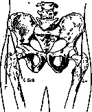
Figure 1. Anterior methods: T - Tilling; L Sch - Lücke and Schede; Lo - Lorenz.
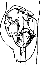
Figure 2. Lateral methods: B1 - Vreden I; B2 - Vreden II; L - Langenbeck.
pulled backward; then the thigh is rotated medially, the lesser trochanter is felt and also chipped off with a chisel, thereby providing wide access to the neck and joint. 3. Hoff and Lorenz method - for blood-reduced reduction of congenital hip dislocations; skin incision from the anterior superior iliac spine down along the outer edge of the tensor fasciae latae muscle. 4. Ludloff method - the same indications; with the thigh abducted at a right angle, a skin incision is made along the lateral edge of the adductor magnus muscle. - Lateral methods (see figure 2): 1. Classical Langenbeck method with König's modification; with the thigh flexed at an angle of 45°, an incision is made with a resection knife down to the bone, starting from the midpoint of the distance between the posterior superior iliac spine and the greater trochanter, rectilinearly downward through the apex of the trochanter and along the upper third of the thigh; with a periosteal elevator, the middle gluteal, piriformis, internal and external obturator muscles are separated subperiosteally and pulled backward, while the minimum gluteal and vastus lateralis muscles are separated from the anteromedial surface and pulled forward. The capsule is opened, the thigh is brought medially, the head is dislocated, and the round ligament is cut. According to König's modification, the muscle attachments are separated with a chisel along with thin bone plates, and the remaining trochanteric prominence is finally resected and removed. 2. Vreden method: an arcuate skin incision from the anterior superior iliac spine (with convexity forward and downward), crossing the lateral surface of the thigh four fingers below the greater trochanter, then upward to the posterior superior iliac spine; the greater trochanter is chipped off with a wide chisel from the outside and back, upward and inward, and pulled upward together with the muscles, which exposes the femoral neck and, consequently, access to the joint. In conclusion, the greater trochanter is fixed in its former place with a nail or periosteal sutures. 3. Vreden II method (for arthroplasties); the incision starts from the middle of the iliac crest 1 cm below it, runs parallel to it to the anterior superior iliac spine, from here it turns vertically downward along the middle of the anterior surface of the thigh to half its length and at a right angle turns to the lateral surface to its middle; the gluteal musculature is dissected away from the lateral wall of the pelvis without damaging the periosteum to the edge of the acetabulum. 4. Kofman method: vertical incision from the apex of the greater trochanter along the upper third of the thigh; perpendicular to it, with a slight
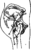
Figure 3. Posterior and superior methods: Sp - Sprengel; K - Kocher; W - White; O - Oschman.
convexity downward above the trochanter, a second incision is made, twice as short as the first; continuation of the operation according to Langenbeck. - Posterior methods (see figure 3): 1. Kocher method: an angular incision runs from bottom to top from the posterior surface of the base of the greater trochanter to its apex, then upward and backward along the fibers of the gluteus maximus muscle; subcutaneous fatty tissue, deep fascia, and, longitudinally, the fibers of the gluteus maximus are incised, and the attachment of the gluteus medius to the trochanter is exposed; deep down they penetrate into the space between the gluteus medius and minimus (in front) and the tendon of the piriformis (behind); the attachment site of the gluteus medius and minimus muscles, innervated by the superior gluteal nerve, is separated subperiosteally from the greater trochanter and pulled forward with a hook; when the flexed thigh is turned outward, the iliofemoral ligament is separated, the capsule is incised, the subperiosteal attachments of the piriformis, obturator internus, gemelli, and obturator externus muscles, innervated by the inferior gluteal nerve, are separated and pulled backward with a hook; the ligamentum teres is severed from behind and below the head with the thigh strongly adducted inward and flexed, and the head is dislocated backward. 2. White method: a posterior, bone-penetrating, semi-circular incision that starts from the midpoint between the anterior superior iliac spine and the apex of the greater trochanter, curves around the femur posteriorly one finger away from its edge, and ends four fingers below the base of the greater trochanter; the capsule is incised along the edge of the acetabulum, and the head is dislocated. The method is very traumatic, but provides good drainage in severe purulent inflammation. 3. Oschman method: an arcuate incision from the anterior superior iliac spine past the apex of the greater trochanter posteriorly to the ischial tuberosity; the greater trochanter is chipped off with a chisel and pulled forward together with the muscles, exposing access to the joint. 4. Superior method (see figure 3) by Sprengel: an incision from the posterior superior iliac spine along the iliac crest to the anterior superior spine, from where it turns down to the greater trochanter; an incision through the fascia and the attachment of the gluteus medius and minimus along the crest, and the separation by an elevator of all muscles with the periosteum from the lateral wall of the pelvis to the acetabulum. Knee joint. In the absence of a universal method that, with minimal trauma to functionally important tissues, would provide perfect access to all sections of the joint, there are many methods that can be successfully used depending on the purpose of the operation. Methods are divided into longitudinal, transverse, and flap methods. - Longitudinal methods (see figure 4): 1. Median incision by Park, as well as Billroth-Ollier-Starkov, starts from the upper border of the suprapatellar pouch, goes down through the patella to the lower edge of the tibial tuberosity; with the knee flexed at a right angle, the patella is sawed through, the tendon of the quadriceps muscle and the patellar ligament are incised longitudinally. The tibial tuberosity is also split with a chisel along the midline and pulled to the sides along with the overlying edges of the incision. 2. External lateral incision by Chassaignac along the outer edge of the patella and tendons. 3. External arcuate Kocher incision runs along the outer surface of the thigh from the border of the upper edge of the pouch and the lateral edge of the vastus externus muscle down one finger outward from the patella and curves medially below the tibial tuberosity. After incision of the skin, subcutaneous tissue, fascia of the thigh, and the lower edge of the vastus externus muscle, the upper pouch of the knee is opened; the tibial tuberosity is knocked off with a chisel along with the periosteal lining of the tibia and the entire flap is reflected medially. Subsequently, for complete exposure of the joint, the intercondylar eminence is chipped off with a chisel along with the cruciate ligaments attached here, after which the articular end can be easily dislocated anteriorly and raised, making it possible to inspect the posterior pouches of the joint. At the end of the operation, the intercondylar eminence and tibial tuberosity are fitted into their places, fixed anteriorly with catgut sutures, the capsule is sutured, the entire flap is turned laterally, and the skin wound is sutured tightly. 4. Internal lateral Langenbeck incision along the inner edge of the patella and tendons. 5. Bilateral incision (Jeffray and Sedillot); two semi-lunar incisions on both sides of the patella, which begin at the upper border of the upper pouch on both sides of the tendon, then the inner incision goes down in an arc, curving toward the inner edge of the patellar ligament, and the outer incision with its lower end curves backward in the direction of the head of the fibula, without reaching its posterior surface, to avoid wounding the professional peroneal nerve; incision of the aponeurosis and capsule. - Transverse methods: 1. Volkmann method - skin incision between both femoral condyles
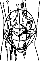
Figure 4. Longitudinal methods: B.O.S. - Billroth-Ollier-Starkov; Ch - Chassaignac; K - Kocher; L - Langenbeck. Transverse method: V - Volkmann. Flap methods: Te - Textor; H - Hahn; Ti - Tilling.
through the middle of the patella, which is sawn transversely; the lateral and cruciate ligaments are divided. 2. Moreau adds two longitudinal incisions at the ends of Volkmann's transverse incision in the shape of the letter H. - Flap methods: 1. Textor: with the knee bent at a right angle, a semilunar incision is made from the posterior edge of one femoral condyle to the posterior edge of the other, with its convexity facing downwards, passing between the apex of the tibial tuberosity and the patella; the patellar ligament, lateral ligaments, and capsule are severed, the flap is reflected upwards, the intercondylar eminence is chipped off with a chisel, together with the attachment of the cruciate ligaments. Subsequently - fixation of the intercondylar eminence in its place with a single suture, suturing of the severed ligaments, bursa, and skin. 2. Hahn: an arcuate incision with its convexity upwards between both condyles around the patella, transection of the tendon of the quadriceps femoris muscle and reflection of the flap downwards. 3. Tilling: an arcuate incision with its convexity downwards between the anterior edges of both condyles, passing below the tibial tuberosity, which is detached with a chisel from bottom to top and inwards and, together with the patellar ligament and the flap, is reflected upwards; upon incision of the capsule, the medial and lateral femoral condyles with their attached ligaments are similarly detached with a chisel; the cruciate ligaments are severed, and the end of the femur is dislocated anteriorly. Ankle joint. 1. Kocher's method. An external semilunar incision, which begins at the outer edge of the tendon of the extensor digitorum communis muscle, runs below the tip of the malleolus and arches upwards for 10 cm along the posterior edge of the fibula to the outer edge of the Achilles tendon; the exposed tendons of the peroneal muscles and the lateral ligaments of the malleolus are transected transversely, the periosteum is separated from the lateral surface of the malleolus, talus, and tibia up to the medial malleolus, the foot is dislocated medially, and the joint is opened. In conclusion, the foot is brought into the normal position, the tendons and wound edges are sutured. 2. König's method. Two anterior incisions: the medial one begins 2 cm above the joint line, is directed along the anterior edge of the tibia downwards through the joint to the tuberosity of the navicular bone; the lateral incision, parallel and equal in length to the first, begins at the same height near the anterior edge of the lateral malleolus. The resulting flap is separated subperiosteally from the bones of the leg and the talus together with the tendons; the joint is opened. Shoulder joint. 1. Langenbeck's anterior method: a straight incision runs from the anterior edge of the acromion process downwards along the intertubercular sulcus and along the anterior side of the deltoid muscle, ending slightly above the attachment of this muscle. The fibers of the muscle are separated bluntly, the tendon of the biceps brachii muscle is exposed and retracted laterally; a longitudinal incision of the periosteum is made along the intertubercular sulcus, which is separated on both sides from both tubercles together with the muscle attachments, while to facilitate this the shoulder is rotated sideways. Upon separation of the periosteum, the head is easily dislocated into the wound. 2. Kocher's posterior method: the incision begins at the acromioclavicular articulation along the upper edge of the spine of the scapula to its middle, then curves arcuately downwards toward the axillary fossa, ending at a distance of two fingers from its posterior edge; the acromioclavicular articulation is opened, the attachment of the trapezius muscle and the overlying part of the deltoid muscle are divided along the upper edge of the spine of the scapula, and are bluntly separated from the underlying muscles. The acromion process is cut off obliquely in the direction of the neck of the scapula and, together with the deltoid muscle, is retracted anteriorly across the head of the humerus, exposing the posterolateral surface of the shoulder joint and the head covered by the tendons of the muscles that rotate the shoulder outwardly (supraspinatus, infraspinatus, teres minor muscles). From the intertubercular sulcus, first the greater tubercle is severed, then the lesser tubercle, which are retracted posteriorly and anteriorly together with the muscles, after which the head can be dislocated posteriorly. Elbow joint. 1. Langenbeck's method. An incision along the medial edge of the olecranon, 10 cm long, down to the bone, divides the triceps muscle longitudinally at the top, as well as the periosteum of the humerus and ulna; separation with a raspatory of the periosteum, together with the attachment of the triceps muscle, first laterally, then medially (together with the ulnar nerve); upon strong flexion at the elbow, the ends of the bones are dislocated into the wound. 2. Kocher's method. An incision from the lateral side 5 cm above the joint, then straight down over the head of the radius, curving arcuately along the edge of the anconeus muscle to the edge of the ulna. The incision penetrates between the triceps muscle (posteriorly) and the brachioradialis and extensor carpi radialis muscles (anteriorly); lower down - between the anconeus muscle (posteriorly) and the extensor carpi ulnaris muscle (anteriorly); the periosteum is separated first in the medial direction, together with the triceps muscle on the arm and the anconeus muscle on the elbow, the entire flap is reflected medially, then the periosteum is separated together with the capsule, lateral ligaments, and the attachment of the extensor tendons, in the lateral direction; the bones are easily dislocated into the wound. Wrist joint. 1. Langenbeck's method. A dorsoradial incision, starting from the middle of the ulnar edge of the second metacarpal bone upwards onto the forearm through the wrist joint and 5 cm above it; the aponeurosis of the dorsal carpal ligament and the periosteum together with the capsule are divided between the tendons of the following muscles: on the ulnar side - the proper extensor of the index finger, extensor digitorum, extensor digiti minimi muscles, which, together with the separated periosteum, are reflected in the same ulnar direction; on the radial side - the extensor carpi radialis brevis and longus, and long abductor of the thumb muscles - are reflected together with the periosteum toward the radius. Upon flexion of the hand, the ends of the bones are dislocated dorsally into the wound. 2. Kocher's method. A dorsoulnar incision, starting from the middle of the radial edge of the fifth metacarpal bone and upwards onto the forearm, through the middle of the wrist; the dorsal carpal ligament is divided, the tendon of the extensor digiti minimi muscle is retracted in the radial direction; the joint is opened, the capsular ligament together with the attachment of the extensor carpi ulnaris muscle is displaced with a raspatory to the ulnar side, and to the radial side - together with the extensor tendons.
A. Kozlovsky.
ARTHROTOMY, see Mastoiditis, Highmoriantris.
Related articles
Mentioned in
Cite this page
“Arthrotomy.” Soviet Medical Encyclopedia. English translation of Bolshaya Meditsinskaya Entsiklopediya, 1st ed. (Moscow, 1928–1936), ed. N. A. Semashko. https://sovietmedicalencyclopedia.pages.dev/article/arthrotomy/