Kronlein Operation
Historical document, translated for reference. It reflects medical knowledge of the 1920s–30s and is not medical advice.
Summary
The Kronlein operation, introduced in 1886, involves temporary resection of the outer bony wall of the orbit to provide wider access. It is used for various orbital conditions including tumor removal, foreign body extraction, and treatment of retrobulbar hemorrhages while preserving the eyeball.
Encyclopedia article (1928–1936)
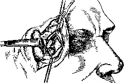
KRONLEIN OPERATION (Kronlein), proposed by K. in 1886 and described by him in 1888, consists of the temporary resection of the outer bony wall of the orbit with the aim of providing wider access into it. First performed by K. for the removal of a dermoid cyst of the orbit, it has found application in various cases: for diagnostic purposes in exophthalmos of unknown causes; to facilitate operations in the posterior part of the eyeball and in the optic nerve (e.g., removal of a subretinal cysticercus, opening of the epineural space of the optic nerve in papilledema); it is also used in pulsating exophthalmos for ligation and removal of pulsating dilated veins and aneurysms; in removal of tumors of the optic nerve with preservation of the eyeball; for the removal of orbital tumors in general, especially if they are located in the depth of the orbit—near its apex, as well as foreign bodies that have penetrated deeply into the orbit or become wedged in its bony walls; sometimes-for finding hidden foci of chronic inflammation, for removal of retrobulbar hemorrhages. In all these cases, the main aim is the preservation of the eyeball and, as far as possible, the associated soft parts (nerves, vessels, muscles) necessary for its normal functioning. Technique of the operation. 1. Incision of soft tissues in the anterior temporal region, arc-shaped, convex forward, 6-8 cm in length. The beginning of the incision is at the top where the easily palpable linea semicircularis of the frontal bone intersects with the horizontal line passing above the eyebrow parallel to the upper orbital edge 1-1.5 cm above it. The incision goes downward in an arc, and its middle, the most convex part of the arc-is at the lateral canthal ligament. From here the incision goes downward and backward to the zygomatic arch, where it ends at the midpoint of a straight line connecting the lateral angle of the palpebral fissure with the tragus of the ear. The incision in the upper and lower thirds includes skin, muscle, and fascia, while in the middle third it penetrates to the bone at the outer edge of the orbit. 2. With a sharp elevator, the periosteum (periorbita) is stripped on the inner surface of the outer wall of the orbit, starting 1 cm above the sutura zygomatico-frontalis and continuing to the fissura orbitalis inferior. 3. Protecting the soft tissues of the orbit with a wide plate, resection of the bony wall is performed with a sharp thin chisel. First, the zygomatic process of the frontal bone is cut at its base, the chisel is turned, and from the depth of this horizontal section, the outer wall of the orbit is pierced along an oblique line backward and downward to the inferior orbital fissure at its midpoint. Then, with a lower horizontal section, the frontal process of the zygomatic bone is cut, and continuing this resection, the beginning of the fissurae orbit. inferior is reached. Thus, a wedge-shaped piece of the outer wall of the orbit (fig. 1) is selected and turned outward, remaining in connection with the temporal muscle and fascia. 4. The exposed periorbita is opened horizontally along the lateral rectus muscle, is spread upward and downward; then a wide access into the retrobulbar space is opened for its examination and the application of appropriate surgical techniques, depending on the given case (fig. 2). To facilitate access into the muscular cone, the lateral rectus muscle is sometimes also cut. At the end of the operation in the orbit, this muscle is sutured again, and the bony piece is fitted in place and fixed in the normal position with catgut sutures on the periosteum.
Figure 1. Position and size of the bony wedge.
Finally, the skin wound is sutured. Kronlein himself performed the operation under general anesthesia, but most surgeons use local anesthesia. The operation has gained general recognition and wide application in retrobulbar surgery. Damage to the n. facialis, ptosis is rarely observed after it; more often-limitation of mobility of the lateral rectus muscle. The eyeball is not damaged at all by the operation, and the cosmetic result after it is satisfactory. K. o. has caused many modifications, expressed mainly in the making of a better incision of the soft tissues [such are the modifications of the bony parts, revealing still wider access into the orbit. In general, the original skin incision of Kronlein has not been preserved and has been replaced by more convenient ones in terms of access to the orbit and integrity of the muscle-fascial formations. The incision of S. S. Golovin (orbitotomia externa simplex) is especially convenient also in that it allows the orbital operation to be started with a simpler technique and only if necessary, after a small additional incision, to proceed to the resection of the outer wall.

Related articles
Cite this page
“Kronlein Operation.” Soviet Medical Encyclopedia. English translation of Bolshaya Meditsinskaya Entsiklopediya, 1st ed. (Moscow, 1928–1936), ed. N. A. Semashko. https://sovietmedicalencyclopedia.pages.dev/article/kronlein-operation/